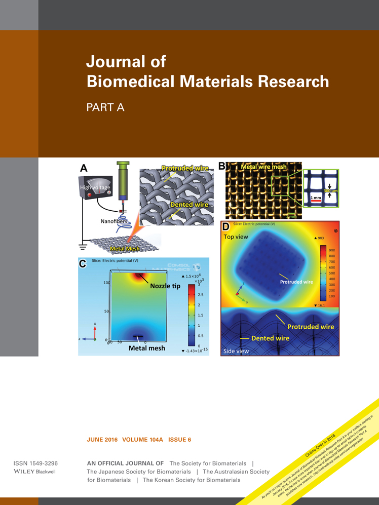
A hybrid microfluidic system for regulation of neural differentiation in induced pluripotent stem cells
Zahra Hesari
Deparmentof Pharmaceutics, Faculty of Pharmacy, Tehran University of Medical Sciences, Tehran, Iran
Nanotechnology Research Centre, Faculty of Pharmacy, Tehran University of Medical Sciences, Tehran, Iran
Search for more papers by this authorMassoud Soleimani
Department of Hematology and Blood Banking, Faculty of Medicine, Tarbiat Modaress University, Tehran, Iran
Search for more papers by this authorFatemeh Atyabi
Deparmentof Pharmaceutics, Faculty of Pharmacy, Tehran University of Medical Sciences, Tehran, Iran
Nanotechnology Research Centre, Faculty of Pharmacy, Tehran University of Medical Sciences, Tehran, Iran
Search for more papers by this authorMeysam Sharifdini
Department of Medical Microbiology, School of Medicine, Guilan University of Medical Sciences, Rasht, Iran
Search for more papers by this authorSamad Nadri
Medical Biotechnology and Nanotechnology Department, Faculty of Medicine, Zanjan University of Medical Science, Zanjan, Iran
Search for more papers by this authorMajid Ebrahimi Warkiani
School of Mechanical and Manufacturing Engineering, Australian Centre for NanoMedicine, University of New South Wales, Sydney, Australia
Search for more papers by this authorCorresponding Author
Mehrak Zare
Skin and Stemcell Research Center, Tehran University of Medical Sciences, Tehran, Iran
Correspondence to: R. Dinarvand; e-mail: [email protected]Search for more papers by this authorCorresponding Author
Rassoul Dinarvand
Deparmentof Pharmaceutics, Faculty of Pharmacy, Tehran University of Medical Sciences, Tehran, Iran
Nanotechnology Research Centre, Faculty of Pharmacy, Tehran University of Medical Sciences, Tehran, Iran
Correspondence to: R. Dinarvand; e-mail: [email protected]Search for more papers by this authorZahra Hesari
Deparmentof Pharmaceutics, Faculty of Pharmacy, Tehran University of Medical Sciences, Tehran, Iran
Nanotechnology Research Centre, Faculty of Pharmacy, Tehran University of Medical Sciences, Tehran, Iran
Search for more papers by this authorMassoud Soleimani
Department of Hematology and Blood Banking, Faculty of Medicine, Tarbiat Modaress University, Tehran, Iran
Search for more papers by this authorFatemeh Atyabi
Deparmentof Pharmaceutics, Faculty of Pharmacy, Tehran University of Medical Sciences, Tehran, Iran
Nanotechnology Research Centre, Faculty of Pharmacy, Tehran University of Medical Sciences, Tehran, Iran
Search for more papers by this authorMeysam Sharifdini
Department of Medical Microbiology, School of Medicine, Guilan University of Medical Sciences, Rasht, Iran
Search for more papers by this authorSamad Nadri
Medical Biotechnology and Nanotechnology Department, Faculty of Medicine, Zanjan University of Medical Science, Zanjan, Iran
Search for more papers by this authorMajid Ebrahimi Warkiani
School of Mechanical and Manufacturing Engineering, Australian Centre for NanoMedicine, University of New South Wales, Sydney, Australia
Search for more papers by this authorCorresponding Author
Mehrak Zare
Skin and Stemcell Research Center, Tehran University of Medical Sciences, Tehran, Iran
Correspondence to: R. Dinarvand; e-mail: [email protected]Search for more papers by this authorCorresponding Author
Rassoul Dinarvand
Deparmentof Pharmaceutics, Faculty of Pharmacy, Tehran University of Medical Sciences, Tehran, Iran
Nanotechnology Research Centre, Faculty of Pharmacy, Tehran University of Medical Sciences, Tehran, Iran
Correspondence to: R. Dinarvand; e-mail: [email protected]Search for more papers by this authorAbstract
Controlling cellular orientation, proliferation, and differentiation is valuable in designing organ replacements and directing tissue regeneration. In the present study, we developed a hybrid microfluidic system to produce a dynamic microenvironment by placing aligned PDMS microgrooves on surface of biodegradable polymers as physical guidance cues for controlling the neural differentiation of human induced pluripotent stem cells (hiPSCs). The neuronal differentiation capacity of cultured hiPSCs in the microfluidic system and other control groups was investigated using quantitative real time PCR (qPCR) and immunocytochemistry. The functionally of differentiated hiPSCs inside hybrid system's scaffolds was also evaluated on the rat hemisected spinal cord in acute phase. Implanted cell's fate was examined using tissue freeze section and the functional recovery was evaluated according to the Basso, Beattie, and Bresnahan (BBB) locomotor rating scale. Our results confirmed the differentiation of hiPSCs to neuronal cells on the microfluidic device where the expression of neuronal-specific genes was significantly higher compared to those cultured on the other systems such as plain tissue culture dishes and scaffolds without fluidic channels. Although survival and integration of implanted hiPSCs did not lead to a significant functional recovery, we believe that combination of fluidic channels with nanofiber scaffolds provides a great microenvironment for neural tissue engineering, and can be used as a powerful tool for in situ monitoring of differentiation potential of various kinds of stem cells. © 2016 Wiley Periodicals, Inc. J Biomed Mater Res Part A: 104A: 1534–1543, 2016.
Supporting Information
Additional Supporting Information may be found in the online version of this article.
| Filename | Description |
|---|---|
| jbma35689-sup-0001-suppinfo.docx1.2 MB |
Supporting Information |
| jbma35689-sup-0002-suppinfogfs1.tif3.7 MB |
Supporting Information Figure S1 |
| jbma35689-sup-0003-suppinfofs2.tif3.3 MB |
Supporting Information Figure S2 |
| jbma35689-sup-0004-suppinfofs3.tif5.6 MB |
Supporting Information Figure S3 |
Please note: The publisher is not responsible for the content or functionality of any supporting information supplied by the authors. Any queries (other than missing content) should be directed to the corresponding author for the article.
REFERENCES
- 1 Lai BQ, Wang JM, Ling EA, Wu JL, Zeng YS. Graft of a tissue-engineered neural scaffold serves as a promising strategy to restore myelination after rat spinal cord transection. Stem Cells Dev 2013; 23: 910–921.
- 2 Anderson KD, Sharp KG, Steward O. Bilateral cervical contusion spinal cord injury in rats. Exp Neurol 2009; 220: 9–22.
- 3 Zamani F, Amani-Tehran M, Latifi M, Shokrgozar MA, Zaminy A. Promotion of spinal cord axon regeneration by 3D nanofibrous coresheath scaffolds. J Biomed Mater Res Part A 2014; 102A: 506–513.
- 4 Segers VFM, Lee RT. Stem-cell therapy for cardiac disease. Nature 2008; 451: 937–942.
- 5 Raasch K, Malecki E, Siemann M, Martinez MM, Heinisch JJ, Bakota J, Kaltschmidt C, Kaltschmidt B, Rosmeyer H, Brandt R. Identification of nucleoside analogs as inducers of neuronal differentiation in a human reporter cell line and adult stem cells. Chem Biol Drug Design 2015; 86. PUB-ID: 2713693
- 6 Temple S. The development of neural stem cells. Nature 2001; 414: 112–117.
- 7 Gage FH. Mammalian neural stem cells. Science 2000; 287: 1433–1438.
- 8 Nadri S, Kazemi B, Eslaminejad MB, Yazdani S, Soleimani M. High yield of cells committed to the photoreceptor-like cells from conjunctiva mesenchymal stem cells on nanofibrous scaffolds. Mol Biol Rep 2013; 40: 3883–3890.
- 9 Takahashi K, Tanabe K, Ohnuki M, Narita M, Ichisaka T, Tomoda K, Yamanaka S. Induction of pluripotent stem cells from adult human fibroblasts by defined factors. Cell 2007; 131: 861–872.
- 10 Park IH, Zhao R, West JA, Yabuuchi A, Huo H, Ince TA, Lerou PH, Lensch MW, Daley GQ, Reprogramming of human somatic cells to pluripotency with defined factors. Nature 2008; 451: 141–146.
- 11 Lowry WE, Richter L, Yachechko R, Pyle AD, Tchieu J, Sridharan R, Clark AT, Plath K. Generation of human induced pluripotent stem cells from dermal fibroblasts. Proc Natl Acad Sci USA 2008; 105: 2883–2888.
- 12 Okada M, Oka M, Yoneda Y. Effective culture conditions for the induction of pluripotent stem cells. Biochim Biophys Acta 2010; 1800: 956–963.
- 13 Mahairaki V, Ryu J, Peters A, Chang Q, Li T, Park TS, Burridge PW, Talbot CC, Asnaghi L, Martin LJ, Zambidis ET, Koliatsos VE. Induced pluripotent stem cells from familial Alzheimer's disease patients differentiate into mature neurons with amyloidogenic properties. Stem Cells Dev 2014; 23: 2996–3010.
- 14 Hanna J, Wernig M, Markoulaki S, Sun CW, Meissner A, Cassady JP, Beard C, Brambrink T, Wu LC, Townes TM, Jaenisch R. Treatment of sickle cell anemia mouse model with iPS cells generated from autologous skin. Science 2007; 318: 1920–1923.
- 15 Xu CY, Inai R, Kotaki M, Ramakrishna S. Aligned biodegradable nanofibrous structure: A potential scaffold for blood vessel engineering. Biomaterials 2004; 25: 877–886.
- 16 Bilousova G, Jun du H, King KB, De Langhe S, Chick WS, Torchia EC, Chow KS, Klemm DJ, Roop DR, Majka SM. Osteoblasts derived from induced pluripotent stem cells form calcified structures in scaffolds both in vitro and in vivo. Stem Cells 2011; 29: 206–216.
- 17 Nelson T, Martinez-Fernandez A, Yamada S, Perez-Terzic C, Ikeda Y, Terzic A. Repair of acute myocardial infarction by human stemness factors induced pluripotent stem cells. Circulation 2009; 120: 408–416.
- 18 Wernig M, Zhao JP, Pruszak J, Hedlund E, Fu D, Soldner F, Broccoli V, Constantine-Paton M, Isacson O, Jaenisch R. Neurons derived from reprogrammed fibroblasts functionally integrate into the fetal brain and improve symptoms of rats with Parkinson's disease. Proc Natl Acad Sci USA 2008; 105: 5856–5861.
- 19 Zhang N, An MC, Montoro D, Ellerby LM. Characterization of human Huntington's disease cell model from induced pluripotent stem cells. PLoS Curr 2010; 2: RRN1193.
- 20 Song B, Sun G, Herszfeld D, Sylvain A, Campanale NV, Hirst CE, Caine S, Parkington HC, Tonta MA, Coleman HA, Short M, Ricardo SD, Reubinoff B, Bernard CC. Neural differentiation of patient specific iPS cells as a novel approach to study the pathophysiology of multiple sclerosis. Stem Cell Res 2012; 8: 259–273.
- 21 Tsuji O, Miura K, Okada Y, Fujiyoshi K, Mukaino M, Nagoshi N, Kitamura K, Kumagai G, Nishino M, Tomisato S, Higashi H, Nagai T, Katoh H, Kohda K, Matsuzaki Y, Yuzaki M, Ikeda E, Toyama Y, Nakamura M, Yamanaka S, Okano H. Therapeutic potential of appropriately evaluated safe-induced pluripotent stem cells for spinal cord injury. Proc Natl Acad Sci USA 2010; 107: 12704–12709.
- 22 Wang Y, Lee WC, Manga KK, Ang PK, Lu J, Liu YP, Lim CT, Loh KP. Fluorinated graphene for promoting neuro-induction of stem cells. Adv Mater 2012; 24: 4285–4290.
- 23 Zhang J, Li L. Stem cell niche: Microenvironment and beyond. J Biol Chem 2008; 283: 9499–9503.
- 24 Kshitiz, Park J, Kim P, Helen W, Engler AJ, Levchenko A, Kim DH. Control of stem cell fate and function by engineering physical microenvironments. Integr Biol 2012; 4: 1008–1018.
- 25 Warkiani EM, Lim CT. Microfluidic Platforms for Human Disease Cell Mechanics Studies. Materiomics: Multiscale Mechanics of Biological Materials and Structures. Springer Vienna 2013; 107–119.
- 26 Conover JC, Notti RQ. The neural stem cell niche. Cell Tissue Res 2008; 331: 211–224.
- 27 Polacheck WJ, Li R, Uzel SGM, Kamm RD. Microfluidic platforms for mechanobiology. Lab Chip 2013; 13: 2252–2267.
- 28 Chaudhuri PK, Warkiani ME, Jing T, Lim CT. Microfluidics for research and applications in oncology. Analyst 2016; 141: 504–524.
- 29 Shemesh J, Jalilian I, Shi A, Yeoh GH, Knothe Tate ML, Warkiani ME. Flow-induced stress on adherent cells in microfluidic devices. Lab Chip 2015; 15: 4114–4127.
- 30 Eglitis MA, Mezey E. Hematopoietic cells differentiate into both microglia and macroglia in the brains of adult mice. Proc Natl Acad Sci USA 1997; 94: 4080–4085.
- 31 Warkiani ME, Lou CP, Liu HB, Gong HQ. A high-flux isopore micro-fabricated membrane for effective concentration and recovering of waterborne pathogens. Biomed Microdev 2012; 14: 669–677.
- 32 Ardeshirylajimi A, Soleimani M, Hosseinkhani S, Parivar K, Yaghmaei P. A comparative study of osteogenic differentiation human induced pluripotent stem cells and adipose tissue derived mesenchymal stem cells. Cell J 2014; 16: 235–244.
- 33 Taniyama Y, Tachibana K, Hiraoka K, Aoki M, Yamamoto S, Matsumoto K, Nakamura T, Ogihara T, KanedaY, Morishita R. Development of safe and efficient novel nonviral gene transfer using ultrasound: Enhancement of transfection efficiency of naked plasmid DNA in skeletal muscle. Gene Ther 2002; 9: 372–380.
- 34 Doetsch F. A niche for adult neural stem cells. Curr Opin Genet Dev 2003; 13: 543–550.
- 35 Panchision DM, McKay RD. The control of neural stem cells by morphogenic signals. Curr Opin Genet Dev 2002; 12: 478–487.
- 36 Wilson CJ, Clegg RE, Leavesley DI, Pearcy MJ. Mediation of biomaterial–cell interactions by adsorbed proteins: A review. Tissue Eng 2005; 11: 1–18.
- 37 Bunge MB. Bridging areas of injury in the spinal cord. Neuroscientist 2001; 7: 325–339.
- 38 Hayman MW, Smith KH, Cameron NR, Przyborski SA. Growth of human stem cell-derived neurons on solid three-dimensional polymers. J Biochem Biophys Methods 2005; 62: 231–240.
- 39 Teng YD, Lavik EB, Qu X, Park KI, Ourednik J, Zurakowski D, Langer R, Snyder EY. Functional recovery following traumatic spinal cord injury mediated by a unique polymer scaffold seeded with neural stem cells. Proc Natl Acad Sci USA 2002; 99: 3024–3029.
- 40 Yamada KM, Cukierman E. Modeling tissue morphogenesis and cancer in 3D. Cell 2007; 130: 601–610.
- 41 Kim DH, Beebe DJ, Levchenko A. Micro-and nanoengineering for stem cell biology: The promise with a caution. Trends Biotechnol 2011; 29: 399–408.
- 42 Ding T, Lu WW, Zheng Y, Li Zy, Pan Hb, Luo Z. Rapid repair of rat sciatic nerve injury using a nanosilver-embedded collagen scaffold coated with laminin and fibronectin. Regen Med 2011; 6: 437–447.
- 43 Chen CS, Mrksich M, Huang S, Whitesides GM, Ingber DE. Geometric control of cell life and death. Science 1997; 276: 1425–1428.
- 44 Patel S, Kurpinski K, Quigley RJ, Gao H, Hsiao BS, Poo MM. Bioactive nanofibers: Synergistic effects of nanotopography and chemical signaling on cell guidance. Nano Lett 2007; 7: 2122–2128.
- 45 Nori S, Okadaa Y, Yasudaa A, Tsuji O, Takahashia Y, Kobayashia Y, Fujiyoshib K, Koiked M, Uchiyamad Y, Ikedae E, Toyama Y, Yamanakag S, Nakamurab M, Okano H. Grafted human-induced pluripotent stem-cell-derived neurospheres promote motor functional recovery after spinal cord injury in mice. Proc Natl Acad Sci USA 2011; 108: 16825–16830.
- 46 Nutt SE, Chang EA, Suhr ST, Schlosser LO, Mondello SE, Moritz CT, Cibelli JB, Horner PJ. Caudalized human iPSC-derived neural progenitor cells produce neurons and glia but fail to restore function in an early chronic spinal cord injury model. Exp Neurol 2013; 248: 491–503.
- 47 Li X, Xiao Z, Han J, Chen L, Xiao H, Ma F, Hou X, Li X, Sun J, Ding W, Zhao Y, Chen B, Dai J. Promotion of neuronal differentiation of neural progenitor cells by using EGFR antibody functionalized collagen scaffolds for spinal cord injury repair. Biomaterials 2013; 34: 5107–5116.


