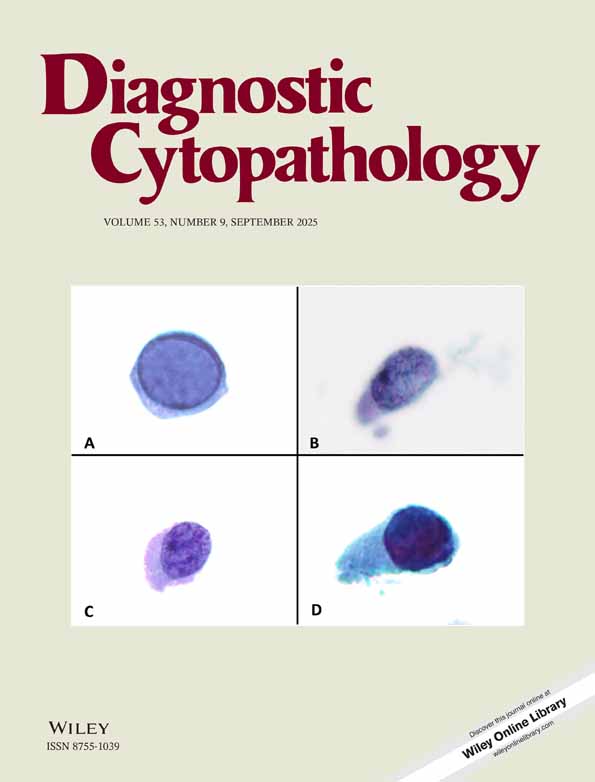
Mesothelial hyperplasia with reactive atypia: Diagnostic pitfalls and role of immunohistochemical studies—a case report
Abstract
The cytomorphologic features of highly reactive mesothelial cells can be difficult to distinguish from malignant cells. We report on an unusual case of mesothelial hyperplasia in a pericardial effusion. The specimen contained bizarre-shaped cells and large tissue fragments in a patient with a history of lung carcinoma. The atypical cells were negative for CEA and LeuM-1 and positive for cytokeratins (AE1/3) and HBME-1. Strong HBME-1 positivity supported a mesothelial origin of the atypical cells and led to the diagnosis of reactive mesothelium. While HBME-1 cannot be used as the sole marker to establish an mesothelial origin; its use in a immunohistochemistry panel may be useful in individual cases to distinguish reactive mesothelial cells from carcinoma in effusion cytology. Diagn. Cytopathol. 2000;22:113–116. © 2000 Wiley-Liss, Inc.


