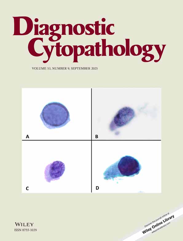
Rare epithelial sheets with “cancer-like” nuclei presenting against a background of cytologically normal-appearing endometrial epithelium in endometrial brush samplings: Establishing a differential diagnosis
Abstract
Liquid-fixed Tao brush samples showing small quantities (less than 10%) of endometrial epithelial sheets with cancer-like nuclei that are admixed with benign endometrium raise a concern about false-positive cytology interpretations. This paper details 7 cases along with the outcomes of three prior papers that touch on the differential diagnosis of “small amounts of atypical epithelium with cancer-like nuclei.” Liquid-fixed, cytocentrifuge-processed Tao brush endometrial samples from 7 women showing “small amounts of atypical epithelium with cancer-like nuclei” were followed up by hysterectomy. Clinical presentations, diagnostic tests, surgical procedures, and tissue outcomes are detailed. Four women had hyperplastic polyps (three with focal atypical complex hyperplasia, and one with focal atypical simple hyperplasia). One had endometrial microcarcinoma. One had p53-positive endometrial intraepithelial carcinoma (EIC). One had endometrial intraepithelial neoplasia (EIN). The differential diagnosis of the index cytological finding, “small quantities of endometrial epithelial sheets with cancer-like nuclei admixed with benign endometrium,” includes hyperplastic polyp, EIC, microcarcinoma/EIN, and carcinoma metastatic to the endometrium. Combining endometrial brushing, endovaginal sonography/sonohysterography, and selective immunostaining may be useful for both detecting and sorting out these lesions. Diagn. Cytopathol. 1999;21:378–386. © 1999 Wiley-Liss, Inc.


