Epidemic of cutaneous fowlpox in a naïve population of chickens and turkeys in Austria: Detailed phylogenetic analysis indicates co-evolution of fowlpox virus with reticuloendotheliosis virus
Abstract
Cutaneous fowlpox is a disease of chickens and turkeys caused by the fowlpox virus (FWPV), characterized by the development of proliferative lesions and scabs on unfeathered areas. FWPVs regularly carry an integrated, active copy of the reticuloendotheliosis virus (REV), and it has been hypothesized that such FWPVs are more problematic in the field. Extensive outbreaks are usually observed in tropical and sub-tropical climates, where biting insects are more difficult to control. Here, we report an epidemic of 65 cutaneous fowlpox cases in Austria in layer chickens (91% of the cases) and broiler breeders and turkeys, all of them unvaccinated against the disease, from October 2018 to February 2020. The field data revealed appearance in flocks of different sizes ranging from less than 5000 birds up to more than 20,000 animals, with the majority raised indoors in a barn system. The clinical presentation was characterized by typical epithelial lesions on the head of the affected birds, with an average decrease of 6% in egg production and an average weekly mortality of 1.2% being observed in the flocks. A real-time multiplex polymerase chain reaction (PCR) confirmed the presence of FWPV-REV DNA, not only in the lesions but also in the environmental dust from the poultry houses. The integration of the REV provirus into the FWPV genome was confirmed by PCR, and revealed different FWPV genome populations carrying either the REV long terminal repeats (LTRs) or the full-length REV genome, reiterating the instability of the inserted REV. Two selected samples were fully sequenced by next generation sequencing (NGS), and the whole genome phylogenetic analysis revealed a regional clustering of the FWPV genomes. The extensive nature of these outbreaks in host populations naïve for the virus is a remarkable feature of the present report, highlighting new challenges associated with FWPV infections that need to be considered.
1 INTRODUCTION
Fowlpox is a worldwide distributed, slow spreading disease of gallinaceous birds with substantial economic impact on commercial poultry production (Tripathy, 2018). The causative agent of fowlpox is a brick-shaped double-stranded DNA virus named fowlpox virus (FWPV), which is the type species of the genus Avipoxvirus within the family Poxviridae (Skinner et al., 2012). FWPVs are epitheliotropic, with transient epithelial hyperplasia, inflammation and necrosis being typical features of FWPV infections. Furthermore, large, cytoplasmic and eosinophilic inclusions can be observed in poxvirus-infected cells, a characteristic aspect observed early on by Bollinger (1877). Clinically, fowlpox can be presented in two forms: (a) cutaneous form, which is characterized by development of proliferative lesions and scabs on unfeathered areas and (b) diphtheritic form, characterized by the development of diphtheritic lesions on the upper parts of the digestive and respiratory tracts (Tripathy, 2018). The disease occurs either by direct contact of birds or by inhalation of dust and aerosols generated by contaminated feathers and scabs, or mechanically by biting insects (Giotis & Skinner, 2019). Nonetheless, the onset of clinical signs is preceded by an incubation period that can vary between 4 and 10 days (Tripathy & Reed, 2020). Vaccination is a critical intervention to control the disease in the field, especially in tropical and sub-tropical areas where biting insects play an active role in the spreading of the disease, or in certain areas where the disease is endemic (Giotis & Skinner, 2019). For that, chicken embryo origin (CEO) and tissue culture origin (TCO) vaccines are available, with the latter being more attenuated. Live non-attenuated pigeonpox viral strains, which are less pathogenic in chickens and turkeys, can also be used to vaccinate these birds. Alternatively, recombinant fowlpox vaccines are also accessible, which provide additional protection to heterologous pathogens.
One feature of FWPVs that remains to be fully understood is the presence of an integrated, nearly intact provirus copy of reticuloendotheliosis virus (REV) in most field strains. Differently, vaccine strains seem to carry only REV long terminal repeat (LTR) remnants in their genome (García et al., 2003; Moore et al., 2000; Singh et al., 2000, 2003). Some preliminary investigations suggest that the REV integration in FWPV field strains increases their pathogenicity (Singh et al., 2005; Zhao et al., 2014), but the complete mechanism behind it was not elucidated, although immunosuppression by released REV has been suggested (Wang et al., 2006).
Fowlpox outbreaks are considered limited in temperate climates and, thus, of moderate importance. However, here we report a series of outbreaks of cutaneous fowlpox in fowlpox-unvaccinated layer and broiler breeder chickens and turkey flocks in Austria, between 2018 and 2020, with significant economic impact in the Austrian poultry industry.
2 MATERIALS AND METHODS
2.1 Outbreak description, flock data and sampling
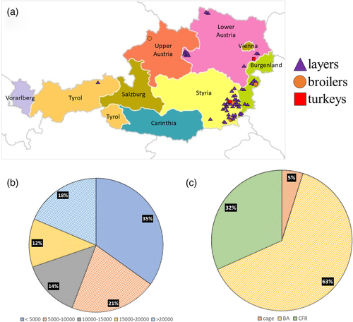
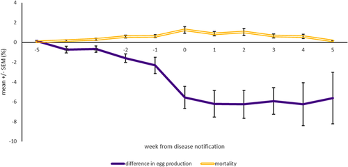
From October 2018 to February 2020, 65 outbreaks of cutaneous fowlpox were observed in poultry farms in Austria, housing exclusively non-vaccinated birds against the disease. Most of the affected poultry were layer chickens (91% of the cases), with outbreaks being also recorded in broiler breeder flocks (6%) and turkey flocks (3%). Most cases were reported from the federal state of Styria (58%), followed by the federal states of Burgenland (20%), Lower Austria (19%), with one case being recorded in Upper Austria and Tyrol (1.5%) (Figure 1a). In total, 63% and 32% of the affected flocks were raised in a barn or conventional free-range system, respectively, with 35% of outbreaks taking place in flocks with less than 5000 birds (figure 1b,c). The age of birds varied between 11 and 75 weeks when they developed clinical signs. Additionally, red mites were present in most of the affected layer chicken flocks, regardless of the husbandry system. Available production data from 13 affected flocks revealed that these flocks were experiencing an average drop in egg production of 6% and an average increased weekly mortality of 1.2%, when the disease was notified (Figure 2). The veterinarian intervention consisted of a supportive treatment of vitamins to improve the recovery of the affected flocks. Samples from each outbreak, consisting either of dead birds with lesions or just skin tissue samples from birds presenting lesions, were submitted to the Clinic for Poultry Medicine at the University of Veterinary Medicine Vienna, for diagnostic confirmation and further investigations. Additionally, dust samples from the poultry houses were also submitted to study the presence of the virus in the environment.
2.2 Histopathology
Processing of skin samples for histology investigation initiated with a fixation step in a 4% neutral buffered formaldehyde solution (SAV LP GmbH, Flintsbach, Germany), followed by dehydration and embedding in paraffin. Next, the samples were cut into 4-μm-thick sections in a microtome (Microm HM 360; Microm Laborgerate GmbH, Walldorf, Germany), and the generated tissue sections were mounted on glass slides and stained with haematoxylin and eosin (H&E) for microscopic assessment.
2.3 DNA extraction and multiplex real-time polymerase chain reaction
Viral DNA was extracted from 25 mg skin samples or 0.2 cm3 dust samples using the DNeasy Blood and Tissue Kit (QIAGEN, Vienna, Austria) according to the manufacturer's instructions with slight modifications for the dust samples, as described before (Sulejmanovic et al., 2019). The presence and the threshold cycle (CT) detection of FWPV and REV DNA in the samples were determined in a multiplex real-time polymerase chain reaction (PCR), using primers and probes targeting the FWPV 4b core protein and the REV gag genes, as previously described by Hauck et al. (2009). The multiplex real-time PCR was carried out in an Agilent AriaMx (Agilent Technologies, Vienna, Austria) using a Brilliant III Ultra-Fast QPCR Master Mix with Low ROX kit (Agilent Technologies). Each 20-μl reaction mixture consisted of 2-μl template DNA, 500 nM and 400 nM (final concentration) of each primer (forward and reverse) for FWPV and REV, respectively, and 150 nM and 250 nM of probe for FWPV and REV, respectively. Cyclic conditions included initial denaturation at 95°C for 3 min, 40 cycles of denaturation at 95°C for 5 s and combined annealing/extension step at 60°C for 10 s. The fluorescence data were collected during the latter step. Data analysis was performed using Agilent AriaMx software version 1.7 (Agilent Technologies) by setting the threshold automatically. A negative extraction and a no template control (NTC) were used in each run to confirm the absence of contamination in the assay.
2.4 Generation of Illumina reads
Two selected samples—PA18/24608 and PA19/1236—were sequenced using the NextSeq platform (Illumina) to obtain complete FWPV genomes. For this purpose, 100 ng of total DNA from each sample was used to prepare the next generation sequencing (NGS)-libraries by using NEBNext Ultra II FS DNA Library Prep Kit (New England BioLabs GmbH, Frankfurt am Main, Germany). Following the adaptor-ligation, DNA fragments were amplified by PCR using a set of Dual Index Primers (NEBNext Multiplex Oligos for Illumina; New England BioLabs GmbH). The size and distribution of each of the generated NGS-libraries was confirmed with the Agilent High Sensitivity DNA Kit (Agilent Technologies) and the Agilent Bioanalyzer 2100 (Agilent Technologies). Each NGS-library with a size of approximately 300 bp was sequenced using 150 bp paired-end read-mode on the NextSeq platform (Illumina) at Vienna BioCenter Core Facilities GmbH, Next Generation Sequencing Facility, Vienna, Austria.
2.5 Generation of Sanger sequences
A preliminary assembly from the Illumina reads was generated using the CLC De Novo Metagenome Assembly module (QIAGEN) to assess whether additional sequences were needed to obtain good quality viral genome assemblies. There, it was observed that the REV genome reads formed a separate contig independent of FWPV. Thus, a set of PCRs was established in order to amplify the integration site. PCR primers amplifying left hand (FPV-201-F: 5′-TGTGCGACGATAAATACTACG-3′; REV-gag-R: 5′-CCACCCTAAAGTCTAGAGTCC-3′) and right hand (REV-env-F: 5′-CATACTGGCATCAATCGTACC-3′; FPV-203-R: 5′-AAAAGTCTTCAAACTGACGGG-3′) integration site were designed to determine the exact REV proviral integration position for each sample. All PCRs were performed using Hot Start Master Mix Kit (QIAGEN) and 10 pmol of each primer. Thermal cycling conditions were as follows: initial denaturation at 95°C for 15 min, 35 cycles of 95°C for 30 s, 55°C for 30 s and 72°C for 2 min and a final amplification step at 72°C for 10 min. The PCR products were analyzed by gel electrophoresis in a 1% (w/v) Tris acetate–EDTA–agarose gel at 100 V for 60 min, stained with GelRed (Biotium, Vienna, Austria) and visualized under ultraviolet light (BioRad Universal Hood II; Bio-Rad Laboratories, Hercules, CA, USA). Correct size PCR products (1347 and 1200 bp for left- and right-hand integration site, respectively) were purified from agarose gel by using QIAquick Gel Extraction Kit (QIAGEN) and subsequently cloned into pCR4-TOPO vector by using the TOPO TA Cloning Kit for sequencing (Invitrogen, Life Technologies, Vienna, Austria) according to manufacturer's instructions. Three positive clones per sample were sequenced by Sanger sequencing using M13 primers (LGC Genomics, Berlin, Germany) with each clone being sequenced in both directions. Assembly and analyses of sequences were performed with Accelrys Gene, version 2.5 (Accelrys, San Diego, CA) software.
2.6 Assembly of FWPV genomes
A visual representation of the whole bioinformatics pipeline is illustrated in Figure S1. The Illumina reads were adaptor trimmed using cutadapt (Martin, 2011) (parameters -a AGATCGGAAGAGC -A AGATCGGAAGAGC -m 20) followed by assembly with the De Novo Metagenome Assembly module from CLC Genomics Workbench 20 (QIAGEN). The generated metagenomics contigs were analyzed with the CLC Taxonomic Profiling module (QIAGEN) against a database of 12 complete FWPV genomes downloaded from the NCBI Assembly databank, and contigs matching to FWPV genomes were retained. The resulting contigs (unordered contigs) were further improved with the aid of Sanger sequences generated in the previous section, using the CLC De Novo Assembly module. To perform scaffolding, whole genome alignment was constructed with the CLC Whole Genome Alignment tool including the 12 FWPV genomes from NCBI (see Section 2.7) together with our two FWPV strains. In this step, the CLC Whole Genome Alignment tool also reorders the contigs of the two strains based on the multi-genome alignment and generates two sets of ordered contigs, one for each strain. Annotation of both strains was also performed with the CLC Whole Genome Alignment tool using the protein sequences from strain US-FWPV (GenBank accession: AF198100) as a reference. The two final assemblies were submitted to NCBI under the accessions PA18-24608 for strain PA18/24608 and PA19-1236 for strain PA19/1236.
2.7 Phylogenetic analyses
Complete sequences of 12 FWPV genomes (Table S1) together with the outgroup Flamingopox virus genome (MF678796) were retrieved from GenBank (26 July 2021) to confirm the phylogenetic relationship of the newly generated Austrian FWPV strains. The final assemblies of our two strains were artificially concatenated and aligned together with the 12 FWPV genomes from NCBI and with the Flamingopox virus genome using the CLC Whole Genome Alignment tool followed by selection of conserved blocks using the online tool GBlocks. In order to determine the phylogenetic relationship of the two REV genomes integrated into the Austrian FWPV strains, a total of 33 REV complete genomes were also retrieved from GenBank (15 November 2021) and aligned with the CLC Whole Genome Alignment tool followed by selection of conserved blocks using the online tool GBlocks (https://ngphylogeny.fr/tools/tool/276/form) (Talavera & Castresana, 2007). Phylogenetic analyses for both FWPV and REV genomes were performed by a standard maximum likelihood approach using a General Time Reversible model with Γ-distribution (four parameters) (Nei & Kumar, 2000), implemented in the MEGA 11 software (Tamura et al., 2021) with a support generated by a 500-replicate bootstrap.
2.8 Confirmation of REV integration into the FWPV genome
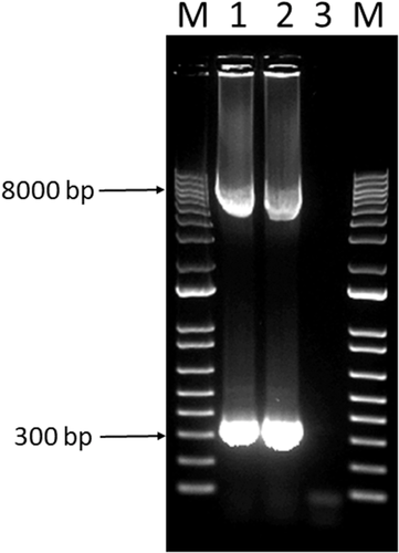
The integration of the REV provirus into the FWPV genome was further assessed in the fully sequenced samples PA18/24608 and PA19/1236 by a PCR targeting sequences that flank the REV integration site. For that reason, the UltraRun LongRange PCR Kit (QIAGEN) was used together with a previously published primer pair (Joshi et al., 2019). The PCR amplification consisted of a 25-μl reaction containing 10 μl of UltraRun LongRange PCR Master Mix, 500 nM of each primer, 11-μl nuclease-free water and 1.5 μl of template DNA. After initial denaturation for 3 min at 93°C, the target DNA was amplified for 35 cycles, which consisted of denaturation for 30 s at 93°C, annealing for 30 s at 56°C and extension for 4 min at 68°C, with a final extension step of 10 min at 72°C. The PCR products were analyzed by gel electrophoresis in a 1% (w/v) Tris acetate–EDTA–agarose gel at 100 V for 60 min, stained with GelRed (Biotium) and visualized under ultraviolet light (Biorad Universal Hood II; Bio-Rad Laboratories). Fragment sizes were determined with reference to a 1-kb ladder (Invitrogen, Life Technologies).
2.9 Virus isolation from environmental samples
The viral infectivity in environmental samples was investigated by inoculation of the chorio-allantoic membrane (CAM) of chicken embryos. Thus, the dust samples PA19/8161, PA19/27661 and PA20/2004, which had the lowest CTs for FWPV in the real-time PCR, were homogenized by vortexing in phosphate buffered saline (PBS) (20% wt/vol), containing 1 mg/ml streptomycin and 100,000 IU/ml penicillin. Then, the homogenates were clarified by centrifugation at 2000 × g for 15 min. The supernatant was collected, and 200 μl of the sample material was inoculated on the CAM of 11-day-old specific-pathogen-free chicken embryos (VALO Biomedia GmbH, Osterholz-Scharmbeck, Germany) according to a protocol of Spackman and Stephens (2016), in a total of five embryos per sample. An additional group of embryos was inoculated with PBS and kept as a negative control. Embryos were candled and monitored for mortality on a daily basis and, at 6 days of post-inoculation (dpi), CAMs were harvested and examined for pock lesions, with the procedure being repeated for three passages in total. In the absence of lesions, the inoculated CAMs were investigated by real-time PCR.
3 RESULTS
3.1 Gross pathology and histopathology findings
During the post-mortem investigation, birds presented nodular lesions that varied from small, focal and yellowish eruptions to spherical wart-like masses on unfeathered areas of the skin such as the comb, wattle, eyelids and the snood (turkeys) (Figure 3a,b). The histology assessment of the lesions revealed marked hyperplasia and ballooning of the epidermal cells with the consequent cell degeneration (Figure 3c). Additionally, epidermal cells presented the characteristic eosinophilic inclusion bodies in the cytoplasm associated with fowlpox infections. Lymphocytic infiltration was commonly observed in association with the lesions.

3.2 Multiplex real-time PCR investigations and viral infectivity in dust samples
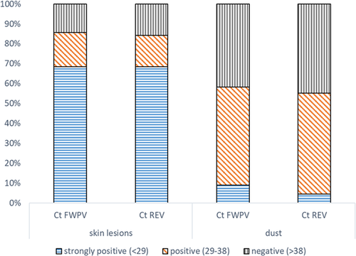
The results of the multiplex real-time PCR investigations were divided into three categories according to the CT: strongly positive (CT < 29), positive (CT 29–38) and negative (CT > 38) (Figure 4). Based on this demarcation, 69% of the assessed skin lesions were strongly positive for both FWPV and REV, with 17% being positive and 14% being negative. Regarding the dust samples, only 5%–9% were strongly positive for FWPV and REV, with the majority of samples being just positive (50%). In total, 41%–45% of dust samples turned out negative in the real-time PCR for FWPV and REV.
The CAMs of chicken embryo inoculated with selected dust samples presented no changes, such as focal pock lesions or generalized thickening. Real-time PCR investigations of inoculated CAMs confirmed the negative result.
3.3 Genome and phylogenetic analysis
Two real-time PCR positive samples—PA18/24608 and PA19/1236—were sequenced on NextSeq Illumina platform with the aim to obtain whole FWPV genomes responsible for skin lesions in affected birds. Through integration of Illumina and Sanger sequences, two high-quality assemblies were produced with total lengths of 262,783 bp for PA18/24608 and 282,351 bp for PA19/1236. A multiple genome alignment between the two strains highlighted two 10 kb regions (Figure S2) that are present in PA19/1236 but absent in PA18/24608, which explain the length discrepancy between the two genomes. However, both regions were located at contig borders in PA18/24608 and were conserved in other sequenced strains from NCBI, implying that they were simply not sequenced in PA18/24608 and cannot be regarded as true indels. Phylogenetically, both genomes were assigned to the species fowlpox virus, in virtue of their clustering with other FWPV strains (Figure 5a). Including the country metadata in the tree for all the FWPV genomes (Figure 5b, Table S1), a clustering based on country is apparent, with both Austrian strains joined in the same group.
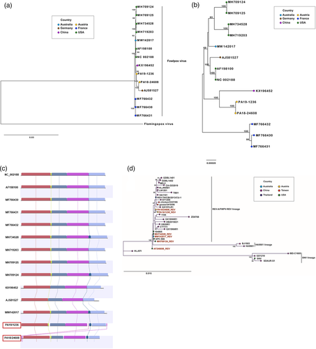
Both our strains incorporated a near-full-length REV provirus integration (Figure 5c), similar to the US strains MH709125 and MH734528, and the Australian strain MW142017. The three strains from France, however, also included a full REV integration, but the corresponding sequence data are missing from the NCBI entries for those strains. The remaining strains in the NCBI database only enclosed a LTR fragment with homology to all other REV provirus integrations. Interestingly, a detailed examination of different metadata revealed that FWPV genomes with the full REV integration are associated with samples derived from animal tissue, while FWPV genomes without the full REV-integration originate from samples derived from cell culture (Table S1).
The REV provirus was located between the FVP201 and FVP203 genes of the FWPV genome, a feature reported previously for REV integrations, and consisted of the complete 5′-LTR, the gag, pol and env genes, and a partial 3′LTR. A multi-genome alignment for the REV provirus region, including all available strains, was constructed to understand the evolutionary dynamics of the REV integration within the FWPV genome (Figure 5d, Table S2). This analysis displayed three main lineages within the REV provirus phylogeny: (i) the REV-A/FWPV-REV lineage, which is the largest one and includes all the FWPV-integrated REV genomes, (ii) the HA9901 lineage and (iii) the SNV lineage. The Austrian proviral REV genomes reported here formed a small separate subcluster within the REV-A/FWPV-REV lineage (Figure 5d).
3.4 Integration of REV provirus into the FWPV genome
A PCR amplification was performed on samples PA18/24608 and PA19/1236 using primers flanking the REV insertion in the FWPV genome. Such investigation aimed to characterize the REV insertion into the FWPV as full, partial or absent. The PCR amplification exposed two amplicons for each sample, corresponding to different lengths of the REV provirus: a large amplicon of around 8 kb, corresponding to the nearly full length of the REV provirus, and a shorter amplicon of around 300 bp (Figure 6). The results suggest the presence of different FWPV genome populations in the investigated skin samples.
4 DISCUSSION
Fowlpox is one of the earliest described diseases in poultry, being already thoroughly studied in the 1870s by von Bollinger, who performed microscopical observations of the cytoplasmatic eosinophilic inclusion bodies (von Bollinger, 1873, 1877). Since then, a vast body of knowledge on fowlpox and FWPVs was developed and, consequently, at present there is a great number of options to prevent and control the disease. Nonetheless, new challenges have arisen that need to be resolved, such as the increasing number of outbreaks on previously vaccinated farms, the upsurge of reports of atypical or severe disease presentation and the lack of understanding on the interplay between FWPV and the inserted REV provirus (Chacón et al., 2020; Giotis & Skinner, 2019; Mirzazadeh et al., 2021; Tripathy & Reed, 2020; Zhao et al., 2014).
In the present study, we report the occurrence of an epidemic of cutaneous fowlpox in Austria, mainly in layer chickens, but also in broiler breeders and turkeys, all of them unvaccinated against the disease, which lasted for a year and a half. Likewise, a re-emergence of fowlpox outbreaks was observed in Germany, from 1999 onwards, which was attributed to the lack of prophylactic vaccination together with the increase of alternative rearing systems (Lüschow et al., 2012). In the present epidemic, vaccination was used as an intervention strategy and started to be implemented in succeeding flocks of 18 farms diagnosed positive, nine of which were multi-age, which might have contributed to the phasing out of the outbreaks.
The geographical distribution, the husbandry system and the size of the affected flocks showed a predominance of outbreaks in the federal state of Styria, in flocks with less than 5000 birds, which were raised in a barn system (deep litter). Considering the different housing systems, indoor or outdoor, the epidemic mirrored the husbandry of Austrian layer flocks (Zloch et al., 2018). The clinical presentation of the outbreaks was characterized by typical epithelial lesions on the head of the affected birds, corresponding to the characteristic epithelial hyperplasia with cell ballooning, degeneration and cytoplasmatic inclusion bodies (Bollinger bodies), as previously reported for cutaneous fowlpox (Tripathy & Reed, 2020). Additionally, a slight increase of 1.2% in mortality and an average drop of 6% in egg production were observed in flocks for which data were available. High variances were recorded for the egg production, reflecting the different age of affected flocks. Similarly, a slight increase in mortality and drop in egg production have been previously described in flocks suffering from fowlpox (Tripathy & Reed, 2020). Reported values vary from 0.03% increase of mortality per day and 0.7% decrease of egg production in milder cases to 8% increase in mortality and 10% reduction of egg production in birds more severely affected (Chacón et al., 2020; Fallah Mehrabadi et al., 2020; Tripathy & Hanson, 1978), hence identical to the values observed in the present study.
One of the most remarkable features of the present report is the high number of fowlpox outbreaks that took place over the period of a year and a half. More extensive outbreaks are usually observed in tropical and sub-tropical climes, where biting insects are harder to control (Giotis & Skinner, 2019). In the present epidemic, nonetheless, the length of the epidemic curtails the seasonal influence, thus downplaying the role of mosquitoes. Interestingly, however, there is an overlap between denser poultry areas in Austria—southeast of Austria—and areas invaded by alien mosquito species in the last decade (Schoener et al., 2019; Seidel et al., 2016), which could introduce new dynamics in the epidemiology of certain poultry diseases, such as fowlpox. Alternatively, red mites, which are highly prevalent in layer farms throughout Europe (George et al., 2015; Moro et al., 2009; Sparagano et al., 2009; Waap et al., 2019; Zloch et al., 2018), were present in the majority of affected chicken flocks of the present study, and are important vectors for FWPV (Huong et al., 2014; Oh et al., 2020; Shirnov et al., 1972). Despite not being subjected to analysis in our investigation, they possibly played an important role in spreading FWPV in the affected flocks and in the persistence of FWPV in the farms.
In the present report, we used a previously established duplex real-time PCR (Hauck et al., 2009) to detect both FWPV and REV provirus genomic sequences in skin samples from affected birds and dust from the poultry house since it is a sensitive, specific, quick and simple detection method. The majority of skin samples were strongly positive for both FWPV and REV, with Q-PCR CT values of less than 29. Interestingly, 13% of the tested skin samples turned out to be negative for both FWPV and REV. Presumably, the high incidence of cutaneous fowlpox cases lead to misdiagnoses in the field, with lesions on the comb of the birds, such as scratches, being confounded with early cutaneous lesions of fowlpox. Real-time PCR results for dust samples showed higher CT values (30–38), indicating a lower viral load in the dust compared to the skin lesions. Presumably, one might expect a certain dilution of the viral DNA in the environment, which could hamper the conclusions of such comparison. Nonetheless, inhalation or ingestion of dust has been linked to the spread of fowlpox (Giotis & Skinner, 2019), and therefore, the detection of FWPV-REV DNA in dust confirms the potential risk of the environmental contamination in the prevalence and spread of fowlpox in poultry farms. Hereof, in a previous report of cutaneous fowlpox in commercial turkeys in Austria, the share of labour and farm equipment was found to play an important role in the introduction of the virus from affected layer chickens in the vicinity of the turkey farm (Hess et al., 2011). Interestingly, the last field outbreaks in the present study took place right before the mandatory COVID-19 lockdown in March 2020, ordered by the Federal Government of Austria, which severely restricted the movement of people, and therefore, might have limited the transmission of fowlpox between farms.
The viral infectivity in the dust samples was investigated by inoculation of the CAM of chicken embryos, but no live virus was isolated. Such finding might be a consequence of either a dilution of the original material, as previously reported (Hess et al., 2011), or that dust is a suboptimal specimen for isolating viruses in chicken embryos due to the presence of other contaminant agents, or rather due to an absence of viral infectivity. Similar results were recently reported for the infectious laryngotracheitis virus (ILTV) in which the virus could not be retrieved after chicken embryo inoculation with ILTV-DNA positive field dust, a result attributed to inactivating properties of the dust such as desiccation (Bindari et al., 2020). In future studies, the infectivity of FWPV in the dust might be investigated by in vivo transmission studies, as recently reported for ILTV (Yegoraw et al., 2021).
It has been hypothesized that FWPVs containing an integrated copy of REV in their genome are more problematic in the field (Giotis & Skinner, 2019); hence, the screening for the REV integration in our samples was of major concern in the study. The primers and probe used for the detection of REV-provirus specific DNA were based on the sequence of the group-specific antigen gene (gag), which do not comprise the integration site of REV into the FWPV genome, and thus, an exogenous origin of REV cannot be totally excluded by this method. However, the detection of REV DNA in skin samples, which is not the ideal replication site for REV (Zavala & Nair, 2020), and in dust, with the CT values for REV and FWPV DNA matching in both sample types, strongly suggest that the REV genome detected by multiplex real-time PCR corresponds to the integrated REV provirus. Yet, we further investigated the integration of REV by PCR amplifications using primers specific for FWPV sequences flanking the REV insertion sites. Notably, the strains investigated revealed a short REV amplicon, likely corresponding to the LTRs, and an additional long amplicon, which shall correspond to the nearly full-length REV genome. Likewise, the co-existence of different FWPV genome populations carrying either the REV LTRs or the full-length REV genome in infected cells was reported recently (Chacón et al., 2020; Joshi et al., 2019; Mirzazadeh et al., 2021). This is a reflection of the instability of the REV proviral genome due to homologous recombination between the REV LTRs during FWPV replication, resulting in the loss of the provirus (Hertig et al., 1997; Niewiadomska & Gifford, 2013). Nevertheless, the role of the integrated REV provirus in the present epidemic is inconclusive. It has been suggested that the REV provirus might increase the FWPV virulence and/or fitness (Giotis & Skinner, 2019). Still, in the present study, no cases of severe fowlpox were observed, and the virus fitness in the field seems to have a multi-factorial basis, as previously discussed.
The whole FWPV genome sequences from two outbreaks revealed almost no variation between FWPV-epidemics strains in Austria. In general, the variation among FWPV strains, based on available full genomes or either polymerase or P4b sequences, seems indeed very low, but clear differences between strains, independent of REV integrations, could be seen. Such differences were prominent when metadata were added to the phylogenetic tree, with the FWPV genomes appearing organized in regional clusters, revealing a geographic evolutionary dynamic of FWPVs. Surprisingly, the AF198100/NC002188 strain does not cluster with other US isolates (MH709124, MH709125, MH719203 and MH734528), but with AJ581527 isolated in Germany. Although this might be accounted for by commonality in passage history (Afonso et al., 2000; Laidlaw & Skinner, 2004; Mayr & Malicki, 1966), poorly documented international transfer of FWPV strains during the mid-1900s, sometimes for vaccine production, means that other explanations cannot be excluded.
Besides the two FWPV genomes reported here, only three additional genomes in the NCBI database contained the near-full-length REV proviral integration, since Croville et al. (2018) reported three FWPV strains hosting the full REV, but such sequences were missing in the deposited genomes to NCBI. Thus, considering the small number of fully sequenced FWPV genomes, the majority of FWPV genomes incorporate the near-full-length REV integration. The FWPV genomes containing only LTR remnants of REV were derived either from in vitro propagated FWPV strains (Afonso et al, 2000; Laidlaw & Skinner, 2004) or from skin lesion in which different FWPV populations regarding the length of REV proviral integration were described (Joshi et al, 2019), a finding also recognized in our study. Hence, the data here reported suggest that the majority, if not all, of FWPV strains circulating in the nature contain the near-full-length REV. Interestingly, the REV integration is always located at the same position of the FWPV genome (Croville et al., 2018; Joshi et al., 2019; Sarker et al., 2021), highlighting its ancestral origin. However, the observed presence of the REV's LTRs, as opposed to nearly complete provirus in the majority of FWPVs, and the homology pattern for the REV integration site, highlight the evolutionary instability of this region and its natural tendency to escape from the FWPV genome. Seemingly, during an infection, the integrated REV provirus excises from its specific location in FWPV, leaving a hybrid LTR scar.
The available full REV genomes form three distinct lineages, with the integrated REV genomes here reported comprising a small separate subcluster within the REV-A/FWPV-REV lineage. Interestingly, the integrated REV genomes display a phylogenetic structure that mirrors the clustering observed for the respective FWPV sequences, supporting the hypothesis of evolutionary rates homogenization between FWPV and the integrated REV sequence.
In spite of being an ‘old’ poultry disease, fowlpox and FWPVs still present challenges to the global poultry industry. Here, we report the occurrence of a remarkable epidemic of cutaneous fowlpox in Austria, in mostly layer chickens, but also in broiler breeders and turkeys, which lasted for a period of a year and a half. Arthropods, contaminated dust and movement of people might have played a role in the farm prevalence and dissemination of the virus. Additionally, FWPVs carried an integrated copy of the REV provirus, whose role in the current epidemic is inconclusive, and the nature of its relationship with FWPV is still not fully understood. Hence, the present report highlights the emergence of new challenges in the field associated with the sudden appearance of FWPVs.
ACKNOWLEDGEMENTS
Jana Tvarogová was financed by the Centre of Excellence for Poultry Innovation (Grant No. ATHU19-CEPI), Interreg V-A Program Austria–Hungary 2014−2020. The authors would like to thank Sarah Heidl for the technical assistance.
ETHICS STATEMENT
The authors confirm that the ethical policies of the journal, as noted on the journal’s author guidelines page, have been adhered to. No ethical approval was required as the samples used in this study were collected by Veterinarians during routine farm visits.
CONFLICT OF INTEREST
The authors declare no conflict of interest.
Open Research
DATA AVAILABILITY STATEMENT
The data that support the findings of this study are available from the corresponding author upon reasonable request.


