CT Anatomical Analysis of C4 Pedicle and Lateral Mass in Children Aged 0–14 in Southern China
Jiarui Chen and Chengqian Huang have contributed to the work equally and should be regarded as co-first authors.
Abstract
Objective
The C4 is the transition point between the upper and lower cervical vertebrae and plays a pivotal role in the middle of the cervical spine. Currently, there are limited reports on large-scale sample studies regarding C4 anatomy in children, and a scarcity of experience exists in pediatric cervical spine surgery. The current study addresses the dearth of anatomical measurements of the C4 vertebral arch and lateral mass in a substantial sample of children. This study aims to measure the imaging anatomy of the C4 vertebral arch and lateral mass in children under 14 years of age across various age groups, investigate the growth and development of these structures.
Methods
We measured 12 indicators, including the size (D1, D2, D3, D4, D5, D6, D7, and D8) and angle (A, C, D, and E) of the C4 vertebral arch and lateral mass, in 513 children who underwent cervical CT examinations at our hospital. We employed the aggregate function for statistical analysis, conducted t-tests for difference statistics, and utilized the least squares method for regression analysis.
Results
Overall, as age increased, there was a gradual increase in the size of the vertebral arch and lateral mass. Additionally, the medial inclination angle of the vertebral arch decreased, and the lateral mass flattened gradually. The rate of change decreased gradually with age. The mean value of D1 increased from 2.31 mm to 3.88 mm, of D2 from 16.75 mm to 29.2 mm, of D3 from 2.21 mm to 4.92 mm, and of D4 from 7.34 mm to 11.84 mm. Meanwhile, the mean value of D5 increased from 5.2 mm to 9.71 mm, of D6 from 10.19 mm to 16.16 mm, of D7 from 2.53 mm to 5.67 mm, and of D8 from 6.11 mm to 11.45 mm. Angle A ranged from 49.12° to 54.97°, angle C from 15.28° to 19.83°, angle D from 39.91° to 53.7°, and angle E from 18.63° to 28.08°.
Conclusion
Prior to cervical spine surgery in children, meticulous CT imaging anatomical measurements is essential. The imaging data serves as a reference for posterior C4 internal fixation, aids in designing posterior cervical screws for pediatric patients, and offer morphological anatomical references for posterior cervical spine surgery and screw design in pediatric patients.
Introduction
The cervical spine, boasting the widest range of motion among all spinal segments, is also highly susceptible to trauma.1-3 Traumatic fractures, cervical spine tumors, degeneration, and other pathologies may require surgical intervention. Presently, lateral mass screws4 and pedicle screws5 represent the primary surgical fixation methods for the lower cervical spine. It is essential to have a comprehensive understanding of the anatomical structure of the lower cervical spine.
To date, research on surgical techniques and the anatomy of the lower cervical spine has predominantly focused on adults, with relatively scant attention given to children.6-10 Challenges in this domain include a restricted number of research cases, a narrow age range, and limited research indicators.11-14 It was previously reported that the width of the C4 pedicle ranged from 3.2 mm at 3–5 years old to 4.2 mm at 18 years old14; while another study reported that the width of the C4 pedicle was 3.9 mm, 4.7 mm, and 4.5 mm in the <5-year-old group, 5-10-year-old group, and >10-year-old group, respectively.15 As children are in a phase of growth and development, their anatomy is inherently more complex, presenting considerable challenges to surgical procedures.
The lower cervical vertebrae comprise a substantial portion of the cervical spine, with the fourth cervical vertebra (C4) serving as the transitional point between the upper and lower cervical regions and playing a pivotal role in the mid-cervical spine. The purposes of this study are: (i) to investigate the C4 pedicle and lateral mass imaging data in children aged 0–14 years, (ii) to offer guidance for pediatric posterior surgeries, and (iii) to provide anatomical references for designing lower cervical spine screws in children.
Methods
Study Design
We conducted a retrospective analysis of children who underwent a 64-slice CT examination of the neck at our hospital from June 2018 to June 2020, encompassing both genders and spanning from 0 to 14 years of age. Inclusion criteria comprised children requiring neck examinations due to: (i) esophageal foreign bodies, (ii) trauma, (iii) headaches, or (iv) other soft tissue issues in the neck. Excluded were cases involving: (i) cervical spine tumors, (ii) deformities, (iii) osteophytes, (iv) fractures, or (v) other anatomical anomalies. Approval for this study was obtained from the Ethics Committee of the First Affiliated Hospital of Guangxi Medical University (2024-E111-01). As this was a retrospective analysis devoid of personal privacy concerns, individual informed consent was not deemed necessary.
Measurement Indicators
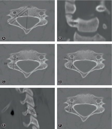
Building upon previous research and employing advanced techniques in pedicle screw and lateral mass screw placement,16-19 we have assessed several critical parameters. These include: D1: The width of the pedicle, representing the distance between the narrowest point of the spinal canal and the vertebral artery. D2: The length of the pedicle, defined as the distance from the anterior edge of the vertebral body to the posterior edge of the lateral mass along the pedicle's longitudinal axis, perpendicular to D1. D3: The height of the pedicle, indicating the height of its narrowest point. D4: The length of the lateral mass, denoting the distance from the posterior midpoint of the lateral mass to the posterior wall of the vertebral artery foramen in the sagittal plane. D5: The width of the lateral mass, measured as the distance between the lateral wall of the spinal canal, perpendicular to the lateral mass's longitudinal axis, and the outer edge of the lateral mass. D6: The longest diameter distance, calculated from the posterior midpoint of the lateral mass to the outer edge of the bone outside the vertebral artery foramen. D7: The height of the side block, referring to the distance between its upper and lower edges. D8: The distance from the posterior midpoint of the lateral mass to the lateral fossa of the vertebral artery foramen. Angle A: The pedicle inclination angle, determined by the angle between D2 and the sagittal plane (the central plane in C4). Angle C: The angle between the directions of D4 and D6. Angle D: The supplementary angle between the long axes of the lateral mass and the vertebral body, roughly representing the angle between the lateral mass's long axis and a horizontal position. Angle E: The angle between D8 and D4 (Figure 1). After obtaining the CT DICOM data, we conducted CT image reconstruction on the PACS workstation (Advantage Workstation, version 4.4, GE HealthCare, United Kingdom). Subsequently, we adjusted the orientation of the images based on the metrics we measured, with C4 as the central focus, and then proceeded with the measurement of the aforementioned parameters. Each indicator is designated by an “L” for left and “R” for right, such as LD1 and RD1. All measurements were conducted by three professionals, with each indicator measured three times to obtain an average value, which was considered the final value.

Statistical Analysis
The statistical analysis utilized the Statistical Package for Social Sciences (SPSS, version 22, IBM, Armonk, NY, USA) and R (version 4.3.1, R Foundation for Statistical Computing, Vienna, Austria). Count, mean, and standard deviation were computed using the aggregate function in R, while the standard error was determined using a specific calculation formula. Confidence intervals were derived employing the tapply function. Following a test for homogeneity of variances, differences in two-sided indicators were assessed using the t-test function from the rstatix package (version 0.7.2, https://cran.r-project.org/web/packages/rstatix/index.html). Linear regression analysis was conducted using the least squares method in SPSS, with regression analysis charts generated using GraphPad v8.0.2 software. Violin plots for each indicator were produced using the R programming language.
Results
Basic Information
The study focused on measuring the C4 pedicle and lateral mass anatomy data of children in southern China. A total of 513 children aged between 0 and 14 years were included, with varying case numbers across different age groups. The highest number of cases (43) was observed in the 13–14 years age group, while the lowest (25) was in the 8–9 years age group. Anatomical measurements were derived from cervical CT images of all 513 children. Statistical analysis encompassed individual and combined indicators for both left and right sides. The parameters measured included the length distances of D1, D2, D3, D4, D5, D6, D7, and D8, along with angles A, C, D, and E. Detailed results of these measurements are provided in Supplementary Data S1.
Indicator Results
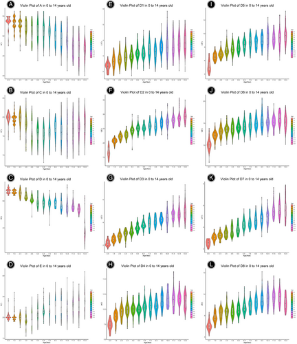
Between the ages of 0 and 14 years, there was a consistent increase in mean values for several parameters. Specifically, the mean D1 increased from 2.31 mm to 3.88 mm, D2 from 16.75 mm to 29.2 mm, D3 from 2.21 mm to 4.92 mm, and D5 from 5.2 mm to 9.71 mm. Notably, mean values for D4, D6, and D8 exhibited fluctuations, with D4 ranging between 7.34 mm and 11.84 mm, D6 between 10.19 mm and 16.16 mm, and D8 between 6.11 mm and 11.45 mm. Overall, there was a general increasing trend with age, except for D4, D6, and D8, which showed a declining trend in the 13–14 years age group, possibly due to anatomical angle changes during growth. Angle A varied between 49.12° and 54.97°, angle C between 15.28° and 19.83°, angle D between 39.91° and 53.7°, and angle E between 18.63° and 28.08° (Figure 2). For detailed results, refer to Supplementary Data S1. While some age groups exhibited individual differences, most bilateral indicators did not demonstrate significant statistical disparities.

Fitting Analysis
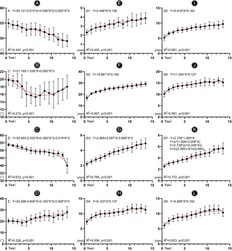
During regression fitting analysis, we adhered to specific principles: prioritizing meaningful p-values, favoring higher R2 values for better fit, and preferring larger F-values when R2 values are consistent. In cases where R2 values, F-values, and p-values were equal, all functions were included. Our findings indicated that D1, D2, D4, D5, D6, and D8 followed power functions, while D3 conformed to a quadratic function. D7, however, adhered to a composite model, growth model, exponential model, and logistic distribution, fitting into all four functions, whether unilaterally or bilaterally. Notably, LD4 exhibited a logarithmic function fit on the left side. Angles A, D, and E were best represented by cubic functions, whereas angle C demonstrated a quadratic function fit (Figure 3). Despite significant p-values, the R2 values obtained for C and E were relatively low, leading us to question their reference value. For detailed results, refer to Supplementary Data S1.

Discussion
Main Findings
In this work, our study comprehensively conducted C4 imaging anatomical measurements on a significant sample of 513 children aged 0–14 in southern China. Overall, as age increased, there was a gradual increase in the size of the vertebral arch and lateral mass. The overall trend of D1-D8 showed a relatively fast growth rate in the early stage, while it remained slow and stable in the later stage. There is no trend of change in the angle indicator. Most indicators show no significant difference on both sides in most age groups.
Indicators Selection
Posterior internal fixation surgeries of the lower cervical spine commonly employ lateral mass screw (LMS) fixation and pedicle screw fixation. Surgical risks include spinal cord injury, vertebral artery damage, and inadequate fixation stability.20, 21 While pedicle screws offer superior pullout resistance compared with LMS screws,22, 23 they also pose a significantly increased risk of spinal cord and vertebral artery injuries.22, 24 Techniques for LMS fixation, such as the Magerl method,18 Roy-Camille method,19 and An method,25 differ primarily in the trajectory of the screw within the lateral mass. The choice of entry point and angle determines this trajectory, influencing the length of the LMS screw path. Optimal selection of the LMS screw path length enhances fixation stability and reduces the risk of nerve root injury.
In previous studies, we conducted radiographic anatomical measurements of the atlas and axis vertebrae using CT images.26, 27 Extending our analysis, this study focuses on the C4 vertebral pedicle and lateral mass using CT imaging data. Given the variable entry angles (between the screw entry direction and the sagittal and axial planes) associated with lateral mass screw techniques, we measured multiple angles for the C4 lateral mass, along with their corresponding lengths. The D6 direction permits the use of longer screws but entails a higher risk of vertebral artery injury. Conversely, the D8 direction presents a lower risk of vertebral artery damage, albeit with shorter screws compared to the D6 direction, resulting in relatively poorer pullout resistance. Additionally, we measured the length of the lateral mass, denoted as D4. Screw placement in the D4 direction is relatively easier due to surrounding bone; however, strict control of screw length is necessary.
Growth Trend
We conducted a regression analysis on the data, revealing a general trend of increasing length indicators with age. However, D4, D6, and D8 showed a decreasing trend at the age of 13–14 years, while angle D exhibited a significant decrease during this period. These changes are hypothesized to be attributed to anatomical variations during growth. According to our criteria, a moderate fitting effect is observed when the R2 value exceeds 0.4, indicating a strong fit when the value surpasses 0.6, and suggesting a weak fitting regression when the value falls below 0.4. Therefore, despite statistically significant p-values obtained from the fitting analysis of angles C and E, their relatively low R2 values lead us to conclude that they lack a well-defined trend. Previous studies have conducted simple trend analyses on various C4 measurement indices.14, 15 However, this is the first time a fitting curve analysis has been performed on these indices. Introducing the concept of growth trend curves provides a better understanding of the growth patterns in children at different stages.
Indicator Size
We were pioneers in employing a substantial sample size to conduct imaging anatomical measurements of the C4 pedicle and lateral mass, specifically focusing on children. Our comprehensive study spanned a broad age range from 0 to 14 years, meticulously segmented into distinct age groups. Previous research has predominantly focused on adult imaging anatomy, often overlooking the pediatric population, resulting in significantly smaller sample sizes in studies.9, 10, 14, 15, 28-34 The dimensions we measured for the C4 pedicle, specifically its length (D1) and width (D2), closely resembled the values reported by Christopher S. Vara14 in his study on American cadavers. However, in comparison to Indian children, the pedicle dimensions (width, length, and height) of the C4 in the southern Chinese children we examined were notably smaller.15 George Al-Shamy conducted a study on 70 patients in Texas, USA, with an average age of 4.93 years (2 months to 16 years), and the results of his study showed that the overall C4 axial width (8.49 ± 1.47 mm) and coronal height (6.29 ± 1.64 mm) were slightly larger than the D4 and D3 index in our study, respectively.35 Interestingly, while our findings on C4 pedicle length align with a study conducted on 60 children aged 4–12 in northern China,34 the pedicle width in our subjects was slightly narrower. Regarding pedicle height, it was comparable to that of northern Chinese children in the 7-year-old group but slightly shorter in the 3–4-year and 10–12-year groups. These variations in vertebral body morphology and structure could potentially be attributed to ethnic or geographical differences. Our findings hold particular relevance in the context of posterior cervical fixation surgery in children, a procedure reported across various age groups, including 2–3-year-olds,9, 32 and 8-year-olds.33 The insights gleaned from our study can serve as valuable morphological and anatomical references for surgeons performing such surgeries on pediatric patients.
Limitations and Strengths
As mentioned before, previous research has predominantly focused on adult imaging anatomy, often overlooking the pediatric population, resulting in significantly smaller sample sizes in studies.9, 10, 14, 15, 28-34 We were pioneers in employing a substantial sample size to conduct imaging anatomical measurements of the C4 pedicle and lateral mass, specifically focusing on children. Our comprehensive study spanned a broad age range from 0 to 14 years, meticulously segmented into distinct age groups. In addition, we still perform growth trend fitting analysis on all indicators. We are the first to conduct a fitting analysis of the C4 pedicle and lateral mass image anatomy in children. Given that our study involves measuring and researching the anatomical structures of the C4 pedicle and lateral mass in a large sample size, our analysis is based solely on CT imaging data. In the future, we will need to conduct extensive cadaveric studies to further validate our findings and perform internal fixation procedures on cadavers based on our measurements. Additionally, our study conducted detailed subgroup analyses for each age group, but we did not analyze differences between male and female subjects. Finally, the cases we included in our study were mainly from southern China, with fewer cases from northern China.
Conclusion
Our study conducted comprehensive imaging anatomical measurements of the C4 vertebra in a substantial sample of 513 children aged 0–14 in southern China. We meticulously measured both the size and orientation of the C4 pedicle and lateral mass. Additionally, we conducted a detailed regression analysis to elucidate their age-related trends. These extensive data provide valuable insights into the anatomical characteristics of the C4 vertebra in children from southern China. These findings have significant potential for clinical applications, surgical planning, and future research endeavors in this field.
Acknowledgments
The present research was supported by the National Natural Science Foundation of China (82360422); Joint Project on Regional High-Incidence Diseases Research of Guangxi Natural Science Foundation (2023JJA140227); Guangxi Young and Middle aged Teacher's Basic Ability Promoting Project (2023KY0115); the “Medical Excellence Award” funded by the Creative Research Development Grant from the First Affiliated Hospital of Guangxi Medical University; Clinical Research Climbing Plan Project of the First Affiliated Hospital of Guangxi Medical University in 2023; Bethune Charity Foundation's “Constant Learning and Improvement-Medical Research” project.
Conflict of Interest Statement
The authors have no conflict of interest to declare that are relevant to the content of this article.
Ethics Statement
Due to the retrospective nature of the study, informed consent was waived. And this study was approved by the Ethics Committee of the First Affiliated Hospital of Guangxi Medical University (2024-E111-01).
Author Contributions
Conceptualization: JRC, TYC, JCZ, SFW, and CL.
Investigation: JRC, CXZ, SFW, CQH, JX, BZ, and SSH.
Design: JRC, SSH, ZXZ, JKL, SXP, and XTX.
Writing–Original Draft: JRC, CQH, and SSH.
Writing–Review & Editing: JRC, XLZ, CL.
Supervision: XLZ and CL.
Open Research
Data Availability Statement
The data of this study were derived from cervical CT data of outpatients and inpatients who underwent CT examinations in the First Affiliated Hospital of Guangxi Medical University from June 2018 to June 2020.


