Integrated functions of membrane-type 1 matrix metalloproteinase in regulating cancer malignancy: Beyond a proteinase
Funding Information
This work was supported by Grants-in-Aid for Scientific Research (S), grant 22220014 and (B), grant 16H04695 from MEXT (to M.S.) and by a Grant-in-Aid for Scientific Research (C), grant 15K06829 from MEXT, a P-CREATE (Project for Cancer Research and Therapeutic Evolution) grant from The Japan Agency for Medical Research and Development, and funding from the Kobayashi Foundation for Cancer Research (to T.S).
Abstract
Membrane-type 1 matrix metalloproteinase (MT1-MMP) is expressed in different types of invasive and proliferative cells, including cancer cells and stromal cells. MT1-MMP cleaves extracellular matrix proteins, membrane proteins and other pericellular proteins, thereby changing the cellular microenvironment and regulating signal activation. Critical roles of protease activity in cancer cell proliferation, invasion and metastasis have been demonstrated by many groups. MT1-MMP also has a non-protease activity in that it inhibits the oxygen-dependent suppression of hypoxia-inducible factors (HIFs) via Munc18-1-interacting protein 3 (Mint3) and thereby enhances the expression of HIF target genes. Elevated HIF activity in MT1-MMP-expressing cancer cells is a fundamental mechanism underlying the Warburg effect, a well-known phenomenon where malignant cancer cells exhibit a higher rate of glucose metabolism. Because specific intervention of HIF activation by MT1-MMP suppresses tumor formation by cancer cells in mice, both the proteolytic and non-proteolytic activities of MT1-MMP are important for tumor malignancy and function in an integrated manner. In this review, we summarize recent findings relating to how MT1-MMP activates HIF and its effects on cancer cells and stromal cells.
The extracellular matrix (ECM) is a fundamental component of tissue architecture. During physiological and pathological tissue remodeling, the ECM is degraded and replaced with a newly formed ECM. Zinc-dependent proteinases, such as matrix metalloproteinases (MMP), play a major role in ECM turnover.1, 2 Although most MMP are secreted, some are membrane-type (MT) MMP. Among them, MT1-MMP (also known as MMP14) was the first membrane-anchored MMP discovered to act as a potent protease that promotes cancer invasion and metastasis.3
The activities of both soluble and MT MMP in cancer contribute to loosening of the ECM. In contrast to soluble MMP, the cell-surface localization of MT-MMP makes them suitable for regulating signals in producer cells, as the cell surface is the place where clusters of signaling molecules are formed. MT1-MMP acts as a processing enzyme for many membrane proteins, including cell-adhesion molecules, receptors, and ligands such as growth factors and cytokines/chemokines, and thereby regulates signals that promote cell motility, proliferation, and differentiation by altering the activities of the processed substrate proteins.1, 2, 4 For example, MT1-MMP cleaves the extracellular ligand-binding domain of Eph receptor A2 (EphA2), a tyrosine kinase type receptor that shows ligand-dependent inhibition activity of ErbB receptor signaling.5, 6 The processing converts EphA2 into a ligand-independent signal transducer that shows opposite activity of the ligand-bound receptor by promoting cell motility.5, 6 MT1-MMP also sheds CD44 and its homolog lymphatic vessel endothelial hyaluronan receptor 1 and enhances cell motility7 and lymphangiogenesis, respectively.8 Likewise, biological feedback due to MT1-MMP expression in producer cells reflects the altered functions of the processed substrate proteins. Therefore, the diverse functions of MT1-MMP in tissues reflect the available substrates in the cellular microenvironment.4
While MT1-MMP is a potent membrane proteinase, it also has activities that are independent of its catalytic activity.9-13 For example, MT1-MMP induces expression of the angiogenic vascular endothelial growth factor (VEGF) and the cytoplasmic tail (CPT), but not the catalytic domain, is responsible for the activity.14, 15 Analysis of macrophages derived from MT1-MMP-knockout (MT1-KO) mice revealed that the levels of glycolytic activity and VEGF production were greatly reduced compared to those in wild-type macrophages.12 These alterations in deficient cells depended on the reduced activities of hypoxia-inducible factors (HIF). Indeed, HIF-1 activity in macrophages was high in wild-type cells and low in MT1-KO cells, even during normoxia.12 HIF are key transcription factors that mediate hypoxia-induced stress responses and regulate the expression of hundreds of genes, thereby affecting many cell functions, such as migration, invasion, proliferation, angiogenesis and cellular metabolism.16, 17 Even though the oxygen-dependent suppression of HIF activity during normoxia and its release upon exposure to hypoxia has been studied extensively,16, 18 it remains largely unclear whether and how HIF are activated during normoxia. Therefore, it is of particular interest to determine how MT1-MMP activates HIF during normoxia and what the biological consequences of such activation might be.
MT1-MMP is a Key Regulator of Mesenchymal Cell Traits
Production of ECM components and their remodeling are largely mediated by fibroblasts in tissue stromata.19 MT1-MMP is usually expressed in activated fibroblasts and cancer cells, and cleaves various ECM molecules such as type-I collagen and laminin.2 The affected substrates are not restricted to the direct target ECM proteins, as MT1-MMP triggers activation cascade of other MMP, such as MMP-2, MMP-9 and MMP-13, by cleaving the propeptide sequence of their latent forms.4 Among the ECM substrates, type I collagen is particularly important as it is the major component forming the connective tissue architecture.20 Other than MT1-MMP, it has been shown that MMP-1, MMP-8 and MMP-13 also have collagenase activity and are responsible for collagenolysis in the connective tissue space.2 In contrast, the collagenase activity of MT1-MMP is rather important for collagenolysis-associated cellular functions, such as invasion and cell proliferation.4, 21, 22 The importance of the collagenolytic activity has been experimentally demonstrated by MT1-MMP-dependent cancer cell invasion and proliferation in 3-dimensional collagen gels.23, 24 During invasion, MT1-MMP localizes at the invasion front of cells where special cell membrane structures known as invadopodia are formed, accompanying intracellular actin reorganization.25 Evidence suggests that particular mechanisms exist to recruit MT1-MMP to invadopodia.26, 27 MT1-KO mice displayed diverse defects related to aberrant collagen turnover such as fibrosis and dwarfism, indicating the importance of the collagenase activity.28, 29 Altered stiffness of the ECM by MT1-MMP also influences cellular conditions such as senescence30 and differentiation via the Hippo signaling pathway.31
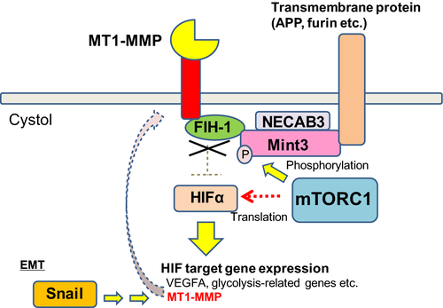
Fully differentiated epithelial cells do not express MT1-MMP, while activated stromal cells such as fibroblasts, stellate cells, endothelial cells and macrophages do.2 Cancerous cells of epithelial origin, particularly invasive ones, also express MT1-MMP.2 Therefore, MT1-MMP expression is likely regulated by signals that induce the epithelial–mesenchymal transition (EMT) or maintain the stromal cell phenotype. One of the key regulators of EMT is a transcriptional suppressor, Snail, that suppresses the expression of genes that maintain epithelial cell traits (such as E-cadherin)32, 33 and that indirectly promotes MT1-MMP expression.34 Hypoxia is also an environmental factor that promotes EMT and enhances the migratory and invasive characteristics of cells.32 HIF activated during hypoxia upregulate MT1-MMP expression in cells.35, 36 Thus, MT1-MMP expression and HIF activation may form a positive feedback loop in mesenchymal cells and cancer cells (Fig. 1). Based on the expression pattern and known functions of MT1-MMP, it is plausibly an important key regulator of activated mesenchymal cell and cancer cell traits.
Oxygen-dependent Suppression of Hypoxia-inducible Factor Transcription Factors
The HIF family of transcription factors is composed of 3 members; namely, HIF-1, HIF-2 and HIF-3, which can bind the hypoxia response elements of target genes after the α and β subunits heterodimerize.18 All HIF use a common β subunit, HIF-1β, but their respective α subunits are encoded by different genes. The transcriptional activity of HIF is mediated by the α subunits that bind the transcriptional co-factor p300/CREB-binding protein (CBP) at the C-terminal activation domain.17 HIF-1α is expressed ubiquitously, whereas HIF-2α and HIF-3α are tissue-specific.18 The hallmark of HIFs is an immediate activation upon exposure of cells to hypoxia.16, 18 Both subunits are transcribed and translated constitutively, but the activities of the α subunits are severely suppressed by oxygen-dependent mechanisms at 2 different levels.17, 37 Quick induction of HIF activity upon the exposure of cells to hypoxia is simply attained by stopping the oxygen-dependent inhibition against the α subunits.17, 18, 38
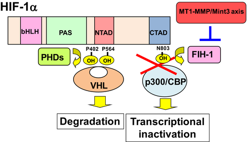
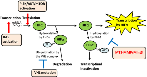
Two types of oxygen-dependent hydroxylases, prolyl hydroxylase domain-containing (PHD1, 2 and 3) and factor-inhibiting HIF-1 (FIH-1), introduce hydroxylation at the proline and asparagine residues of HIFα, respectively (Fig. 2).17, 37 The hydroxylated proline residues of HIFα are recognized by the von Hippel–Lindau tumor-suppressor protein, which successively induces HIFα ubiquitination and proteasomal degradation. Thus, PHD reduce HIFα protein levels greatly during normoxia.17, 37 To ensure suppression of residual HIFα, FIH-1 hydroxylates the asparagine residue of HIFα at the C-terminus transcriptional-activation domain to prevent HIFα from binding p300/CBP.17, 37 Both mechanisms acting at different levels are important in suppressing HIFα activity effectively. For example, the residual level of HIFα that escapes proteasomal degradation is sufficient to activate target genes when FIH-1 is absent.39
MT1-MMP Unlocks the Suppression of Hypoxia-inducible Factors during Normoxia
It is well known that some types of cells exhibit HIF-1 activity during normoxia, even when the expression levels of PHD and FIH-1 are normal.12, 40, 41 Invasive cancer cells often show augmented glycolysis, exhibiting a phenomenon known as the Warburg effect, a century-old observation for cancer cells.42 Data from many studies have shown that elevated HIF-1 activity serves as an underlying driving force of the Warburg effect by upregulating expression of rate-limiting glycolytic enzymes and transporters.38 In addition to HIF-1, several oncogenic signals in cancer cells also affect glycolytic pathways and contribute to the Warburg effect.43 Constitutive HIF-1 upregulation is reminiscent of patients with Von Hippel–Lindau disease, where the VHL tumor suppressor gene is inheritably inactivated by mutation and promotes the development of disease-related tumors.37 However, inactivating PHD, FIH-1 or VHL mutations in tumors do not appear to be common based on recent next-generation cancer-genome sequencing data. These findings suggest that some other mechanisms may activate HIF transcription factors in cancer cells.
Enhanced glycolysis can be observed in some types of normal cells, such as macrophages and activated fibroblasts, during normoxia.44 In particular, macrophages produce most ATP via glycolysis supported by elevated HIF-1 activity, while many other cell types do via oxidative phosphorylation (OXPHOS).12, 40 Interestingly, MT1-KO macrophages exhibit a shortage of cellular ATP and a defect in ATP-dependent cell functions.11, 12, 45 In MT1-KO cells, the protein levels of HIF-1, PHD and FIH-1 were not different from those in wild-type macrophages, even though HIF-1 activity was markedly suppressed.12 Knockdown of genes related to HIF regulation in macrophages demonstrated that FIH-1-dependent HIF-1 inhibition was specifically abrogated by MT1-MMP in the wild-type cells.12 Thus, MT1-MMP looks like an activator of HIF-1 by inactivating FIH-1. Indeed, re-expression of MT1-MMP in MT1-KO macrophages induced FIH-1 inactivation and promoted HIF-1 activation.12 FIH-1 suppressed HIF-2 as well; therefore, MT1-MMP had a similar effect on HIF-2.46, 47
The MT1-MMP-dependent mechanism of HIF-1 activation is not restricted to macrophages. For example, cancer cells exhibit clear correlations between MT1-MMP expression, FIH-1 inactivation, HIF-1 activation and the Warburg effect.47, 48 However, differences in OXPHOS activities between macrophages and other cell types cause different effects of MT1-MMP expression on ATP levels. In macrophages, MT1-MMP depletion reduces ATP levels to less than 40% of normal cells, although this effect was not as high in other cell types.12, 48
MT1-MMP Employs Mint3/APBA3 to Inhibit FIH-1
Because the effect of MT1-MMP on HIF-1 can be observed in different cell types,11, 12, 47, 48 it is of particular interest to understand how MT1-MMP inactivates FIH-1 and whether it is mediated by proteolytic activity against a specific substrate. Domain analysis of MT1-MMP revealed that the CPT, but not the protease domain, is responsible for HIF activation.12 The CPT showed binding activity to FIH-1, but did not affect the enzymatic activity of FIH-1, at least in vitro.12
Munc18-1-interacting protein 3 (Mint3), also known as amyloid beta A4 precursor protein-binding family A member 3 (APBA3), is a member of the APBA family that was identified as an FIH-1-binding factor that cooperates with the CPT of MT1-MMP to inhibit FIH-1 in cells.39 Mint3 shares common C-terminal domain structures composed of a phosphotyrosine binding (PTB) domain and 2 PDZ domains, whereas the N-terminus portion is unique to each member protein. The C-terminal domain of Mint3 is responsible for anchoring it to the cytoplasmic domain of transmembrane proteins, such as amyloid precursor protein and furin.49, 50 Consistent with the localization of these proteins to the Golgi apparatus, Mint3 also localizes there.49, 51 Because furin serves as a proteolytic activator of proMT1-MMP before it is transported to the cell surface, Mint3 that binds to the cytoplasmic portion of furin presumably localizes in close proximity to the CPT of MT1-MMP in the Golgi apparatus.52 The N-terminal portion of Mint3 has a naturally denatured structure and competes with HIFα for binding FIH-1 and thereby inhibits the hydroxylation of HIFα by FIH-1.39
Mint3 is ubiquitously expressed, although some cancer cells express the protein at relatively higher levels.53 However, Mint3 does not inhibit FIH-1 when MT1-MMP is absent. Genetic depletion of Mint3 (Mint3-KO) in mice does not exhibit severe phenotypes, except for macrophages that exhibit an ATP shortage, similarly to MT1-KO cells.45, 54, 55 Even though Mint3 expression is ubiquitous, it requires MT1-MMP to inhibit FIH-1, and during inhibition, these three factors co-localize at the Golgi apparatus. Without MT1-MMP, FIH-1 localizes to the cytoplasm and Mint3 localizes to the Golgi apparatus.12
The CPT of MT1-MMP binds FIH-1 in a sequence-specific manner and helps recruit cytoplasmic FIH-1 to the Golgi apparatus.12 The recruited FIH-1 is successively inhibited by the neighboring Mint3 protein by forming a stable complex. Translocation of FIH-1 from the cytoplasm to the Golgi upon MT1-MMP expression in cells was clearly observed by immunostaining.12 The CPT of MT1-MMP is composed of 20 amino acids and has sequence similarity to other membrane-type MMPs (MT2-MMP, MT3-MMP and MT5-MMP), but the amino acid residues responsible for FIH-1 binding and HIF activation are specific to MT1-MMP.12
Crosstalk of MT1-MMP-mediated Hypoxia-inducible Factor Activation with Oncogenic Signals
Mint3 can be phosphorylated at the N-terminus by the mTOR complex 1 (mTORC1), which increases its interaction with FIH-1 in cells.47 This finding indicates that MT1-MMP-mediated HIF activation is affected by the phosphoinositide 3-kinase (PI3K)/Akt signaling pathway, which is usually activated in cancer cells.56 In addition, N-terminal EF-hand calcium binding protein 3 (NECAB3), which structurally resembles monooxygenase, binds to the PTB domain of Mint3 and promotes Mint3-FIH-1 interactions in cells.53 The monooxygenase activity of NECAB3 is presumably necessary for this function, but the precise mechanism remains unclear. Thus, further studies are needed to clarify the mechanism whereby NECAB3 promotes Mint3-FIH-1 interaction in cooperation with oncogenic signals.
PI3K/Akt is reported to augment HIF-1α translation via mTOR activation. 38 However, it may not contribute to HIF-1α transcription activity so far as FIH-1 remains suppressive. Thus, oncogenic signals that activate MT1-MMP and PI3K/Akt cooperatively use the Mint3/FIH-1 axis and mTOR to activate HIF effectively (Fig. 3).
In addition to HIFα, FIH-1 can hydroxylate the inhibitor of κBα (IκBα), which inhibits nuclear factor κB (NF-κB).57 NF-κB is involved in inflammatory responses and plays an important role in tumor development.58, 59 Interestingly, IκBα is accumulated in Mint3-KO macrophages.55 Thus, the MT1-MMP/Mint3/FIH-1 axis might also control cellular NF-κB signaling via IκBα.
Effect of Hypoxia-inducible Factor Activation on Tumorigenesis
The Warburg effect in cancer cells was reported a century ago42 and this characteristic phenomenon is applied to cancer diagnosis using the glucose derivative 18F-fludeoxyglucose and positron-emission tomography scanning.60 Nevertheless, the impact of the phenomenon on cancer is not necessarily clear because of its complexity. One explanation is that elevated glucose uptake increases the supply of carbon blocks for rapidly dividing cancer cells that need to double their cellular components.43 Another possibility is that reduced oxygen consumption in the normoxic tumor mass area may save available oxygen for the inner cells to mitigate hypoxic stress.16 Among cancer cell lines, invasive cells usually exhibit the Warburg effect, while non-invasive cell lines rarely do so. A pair of human breast carcinoma cell lines was used to analyze the Warburg effect.60 The invasive cell MDA-MB-231 line showed elevated glycolytic activity, whereas the non-invasive MCF-7 cell line did not, and these activities correlated with HIF-1 activity and cellular MT1-MMP expression.48 Gene-expression knockdown experiments demonstrated that HIF-1 activity in MDA-MB-231 cells reflect MT1-MMP and Mint3 dependent FIH-1 inactivation, whereas Mint3 did not inhibit FIH-1 in MCF-7 cells.48 Therefore, the MT1-MMP/Mint3/FIH-1 axis operates in MDA-MB-231 cells to overcome inhibition of HIF activity and activate glycolysis. MT1-MMP was expressed in 10 out of 14 human cancer cell lines tested, where lactate production representing glycolysis and HIF-1 activity was upregulated via Mint3-dependent FIH-1 inactivation.48 However, the remaining 4 cell lines showed lower lactate production and negligible MT1-MMP expression.48 Therefore, a clear correlation was demonstrated between MT1-MMP expression, FIH-1 inactivation and elevated glycolysis in cancer cells. Mint3 knockdown using short hairpin RNA in MDA-MB-231 cells abrogated MT1-MMP-dependent HIF-1 activation and reduced both tumor formation in mice and blood vessel density in tumor tissue.48
Von Hippel–Lindau syndrome is a representative case that indicates the importance of HIF activity in tumor development.37 In this syndrome, the VHL gene is genetically inactivated, which results in constitutively high HIF activity. Patients with the disease develop different types of tumors, such as hemangioblastomas, kidney cancer and pancreatic cancer,37 indicating that HIF activity may increase the rate of tumor development in the disease. In addition, HIF promote malignant conversion of cancer cells by enhancing cell motility and invasion via upregulating multiple genes.18
Because HIF are potent modulators of tumor malignance, they have been regarded as potential targets for cancer therapy. However, the global inhibition of HIF affects their physiological functions, such as hypoxia response.61-63 Because MT1-MMP-dependent activation of HIF is mostly specific to malignant cancer cells and activated stromal cells,12, 48 targeting the MT1-MMP/Mint3/FIH-1 axis is more specific and appropriate for cancer therapy. Application of pathway-specific inhibitors might be suitable for treating patients, in particular for preventing tumor recurrence when residual cancer cells start to grow again.
Intervention of Hypoxia-inducible Factor Activation by MT1-MMP in Stromal Cells
Most cells produce ATP via OXPHOS in the mitochondria, but macrophages mainly utilize glycolysis for ATP production, even during normoxia.40 Among leukocytes, MT1-MMP is expressed in macrophages.11 MT1-MMP-deficient and Mint3-deficient macrophages exhibit decreased expression of HIF-1 target genes, ATP production and ATP-dependent cellular functions, such as motility, invasion and cytokine production.11, 12, 45, 54 In cancer, tumor-associated macrophages (TAM) promote tumor malignancy, such as tumor growth and metastasis.64 Thus, targeting the MT1-MMP/Mint3 axis might also control TAM-mediated tumor malignancy.
Other than macrophages, fibroblasts also show increased expression of HIF-1 target genes due to MT1-MMP activity during normoxia.12 Fibroblasts are one of the major cell types constituting the various types of tumor tissues, and cancer-associated fibroblasts (CAF) are known to regulate tumor growth, metastasis and drug resistance.65, 66 Thus, CAF are also potential target cells of anti-MT1-MMP-mediated HIF-activation therapy.
Conclusion
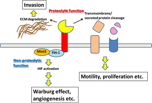
The MT1-MMP proteinase has been characterized as one of the key regulators of cancer cell proliferation, invasion and metastasis. In addition, MT1-MMP activates HIF transcription factors independently of its proteinase activity, as reviewed above. In physiological settings, such as during wound healing, epithelial cells can exhibit EMT and activated stromal cells start to express MT1-MMP, which promotes their proliferation and migration. In invasive cancer cells, MT1-MMP expression is turned on by oncogenic and EMT-inducing signals. MT1-MMP activity promotes cellular invasion and proliferation by processing key substrates and also by activating HIF transcription factors through a CPT-dependent mechanism. Thus, extracellular protease activity and the intracellular non-protease activity are collectively integrated into malignant cancer phenotypes (Fig. 4). Systemic inhibition of the protease activity of MT1-MMP might cause fibrosis, as observed for MT1-KO mice.28, 29 In addition, MT1-MMP proteinase inhibitors cannot suppress the intracellular non-protease activity of MT1-MMP. Thus, targeting both the focused proteolytic function and the non-proteolytic function of MT1-MMP may be preferable for blocking MT1-MMP-mediated malignant cancer phenotypes completely without adverse effects. HIF-1 activation in cancer cells itself is important for tumorigenicity and invasion/metastasis, and inhibition of the HIF-activation pathway by MT1-MMP is an attractive new approach for developing cancer therapeutics. We think that specifically inhibiting HIF-1 activation is better for cancer treatment than global HIF inhibition because a more targeted approach may avoid unnecessary adverse effects. Inhibitors targeting the MT1-MMP/Mint3/FIH-1 axis might be suitable for preventing recurrence in cancer patients following treatment with standard therapy. However, large tumors that contain hypoxic areas inside may not be good targets, as the effect of inhibiting the MT1-MMP-dependent axis is presumably overwhelmed by responses to hypoxia.
Acknowledgments
This work was supported by Grants-in-Aid for Scientific Research (S) and (B) from MEXT (to M.S.) and by a Grant-in-Aid for Scientific Research (C) from MEXT, a P-CREATE (Project for Cancer Research and Therapeutic Evolution) grant from The Japan Agency for Medical Research and Development, and funding from the Kobayashi Foundation for Cancer Research (to T.S).
Disclosure Statement
The authors have no conflict of interest to declare.


