An early Eocene fossil from the British London Clay elucidates the evolutionary history of the enigmatic Archaeotrogonidae (Aves, Strisores)
Abstract
Archaeotrogons have long been known from late Eocene and Oligocene localities in France, where limb bones are abundantly represented. The phylogenetic affinities of these birds, however, have remained elusive. Although archaeotrogons are now considered to be representatives of the Strisores, the clade including ‘caprimulgiform’ and apodiform birds, their exact position in this clade is unresolved. Here, a partial skeleton of a new species of the Archaeotrogonidae is described from the early Eocene London Clay of Walton-on-the-Naze (Essex, UK). In addition, a new specimen of the archaeotrogon Hassiavis from the latest early or earliest middle Eocene of the Messel fossil site in Germany is reported, which is the best preserved skeleton of this species. Archaeodromus anglicus gen. et sp. nov. from the London Clay is the earliest archaeotrogon known to date, and the holotype shows previously unknown skeletal characteristics of archaeotrogons that bear on the phylogenetic affinities of these birds. The quadrate in particular exhibits a distinctive morphology and shows derived morphological characteristics of the Strisores. Although the primary analysis did not conclusively resolve the affinities of archaeotrogons, analyses constrained to a molecular scaffold resulted in a sister group relationship to the Caprimulgidae (nightjars). The evolutionary history of this most species-rich group of nocturnal Strisores is virtually unknown, and recognition of archaeotrogons as archaic stem group representatives of the Caprimulgidae would fill a striking gap in the fossil record.
Strisores is an avian clade, which includes swifts (Apodidae) and hummingbirds (Trochilidae) as well as various crepuscular or nocturnal broad-billed birds (Mayr 2010). Most of these latter groups (the traditional ‘Caprimulgiformes’) are today species-poor and have a restricted geographical distribution, which is true for the Neotropic Steatornithidae (oilbird) and Nyctibiidae (potoos), as well as the Australasian Podargidae (frogmouths) and the Australo-Papuan Aegothelidae (owlet-nightjars).
Analyses of nuclear sequence data yielded well-resolved phylogenies (Hackett et al. 2008; Prum et al. 2015; White & Braun 2019; Kuhl et al. 2021), which do, however, not conform to phylogenies derived from morphological data (Mayr 2002, 2010; Nesbitt et al. 2011; Chen et al. 2019, fig. 4). A major incongruence concerns the position of the Caprimulgidae (nightjars), which in molecular analyses were identified either as the sister taxon of all other Strisores (Prum et al. 2015; White & Braun 2019; Kuhl et al. 2021) or as the sister taxon of the clade formed by Aegothelidae and Apodiformes (Hackett et al. 2008; that study recovered a clade formed by Nyctibiidae and Steatornithidae as the sister taxon of all the remaining Strisores). Morphological data, by contrast, suggest a sister group relationship between the Caprimulgidae and the Nyctibiidae and recover both taxa as nested within Strisores (Mayr 2010). Whereas morphological data suggest that the Steatornithidae are the earliest branching taxon of the Strisores, most current molecular analyses support a sister group relationship between the Steatornithidae and Nyctibiidae (this topology is, however, sensitive to the data type analysed and is not recovered in analyses of non-coding sequences; Braun & Kimball 2021).
The Strisores have a comparatively comprehensive early Palaeogene fossil record, and stem group representatives of the Steatornithidae, Nyctibiidae, Podargidae, Apodidae and Trochilidae occurred in the early Eocene of Europe (Mayr 2009, 2017). Curiously, the evolutionary history of the most species-rich and most widely distributed group of nocturnal aerial insectivores, the Caprimulgidae, remains elusive (Mayr 2009, 2017). Putative Caprimulgidae were reported from the early Eocene of North America (Olson 1999) and the late Eocene of France (Mourer-Chauviré 1988), but these records are based on a few fragmentary bones (a partial carpometacarpus and coracoid, respectively), and an unambiguous identification is not possible without further material. The earliest definitive record of the Caprimulgidae stems from the Pliocene of Africa (Manegold 2010).
However, there is one well-represented extinct taxon, the deceptively named Archaeotrogonidae, for which affinities to the Strisores were assumed, but which has so far defied an unambiguous phylogenetic placement. Archaeotrogons were first reported from the Quercy fissure fillings in France, where isolated bones are abundantly represented and occur in late Eocene and Oligocene sites (1995, 2014). Four species of the taxon Archaeotrogon are currently recognized, and of the best known species, A. venustus, all major limb bones have been identified (Mourer-Chauviré 1980).
Earlier authors already commented on the existence of archaeotrogons from the early Eocene British London Clay in a private collection (Mayr 1998, 2009; Feduccia 1999, table 4.1), but the oldest formally described representative of the Archaeotrogonidae is Hassiavis laticauda from the latest early or earliest middle Eocene (48 Ma) of the Messel fossil site in Germany (Mayr 1998, 2004). This species is known from complete, articulated skeletons, but all of these are compression fossils, which allow the recognition of only a few osteological details.
As indicated by their name, archaeotrogons were originally considered to be representatives of the Trogoniformes (trogons; Milne-Edwards 1892; Mourer-Chauviré 1980). Already in her initial study of the Archaeotrogon fossils, however, Mourer-Chauviré (1980) noted similarities to the Caprimulgidae, and Mourer-Chauviré (1995, 2014) subsequently assigned the Archaeotrogonidae to the ‘Caprimulgiformes’ (the paraphyletic taxon including the nocturnal representatives of the Strisores; Mayr 2002, 2010). A position of archaeotrogons within the Strisores is also supported by the morphology of the skull of Hassiavis, which shows a resemblance to the skull of the Aegothelidae (Mayr 2004). Still, the exact affinities of archaeotrogons within the Strisores have so far remained elusive, although Chen et al. (2019) recently hypothesized that Hassiavis is a stem group representative of the Aegothelidae (Archaeotrogon was not considered in that study).
Here, a partial skeleton of an archaeotrogon from the London Clay of Walton-on-the-Naze (Essex, UK) is described, which stems from the collection of the late Paul Bergdahl (Kirby-le-Soken) and which was recently acquired by Senckenberg Research Institute Frankfurt. This specimen is the oldest record of the Archaeotrogonidae, and the three-dimensionally preserved bones of the fossil show previously unknown osteological features, which add to an improved understanding of the skeletal morphology of these birds. In addition, a new specimen of Hassiavis from the latest early or earliest middle Eocene of the Messel fossil site in Germany is reported, which is the best preserved skeleton of this taxon.
Material and method
The fossils are deposited in the collections of the Natural History Museum London, UK (NHMUK) and Senckenberg Research Institute Frankfurt, Germany (SMF).
A phylogenetic analysis of 86 morphological characters (for character descriptions and matrix, see Mayr 2021) was based on the emended character matrix of Mayr (2010). The scoring of Archaeotrogon is based on A. venustus, which is the type species of the genus and also the best-represented species of Archaeotrogon (data were gathered from the description and illustrations in Mourer-Chauviré 1980). The primary analysis was run with the heuristic search modus of NONA 2.0 (Goloboff 1993) through the WINCLADA 1.00.08 interface (Nixon 2002), using the commands hold 10 000, mult*1000, hold/10, and max*. Bootstrap support values were calculated with 1000 replicates, ten searches holding ten trees per replicate, and tree bisection and reconnection (TBR) branch swapping without max*. Outgroup comparisons were made with the Tinamiformes and Anseriformes. Tree length (L), consistency index (CI), and retention index (RI) were calculated.
In a second analysis, dummy characters were added to constrain the topology of the extant taxa according to the results of comprehensive recent molecular analyses, which congruently recover the Caprimulgidae as the earliest diverging taxon of the Strisores and which support a sister group relationship between the Steatornithidae and Nyctibiidae (Prum et al. 2015; Kuhl et al. 2021). Fifteen dummy characters were scored as 0 for the outgroup taxa and the Caprimulgidae, and as 1 for all other extant Strisores; 15 further characters were scored as 1 for the Nyctibiidae and Steatornithidae and as 0 for all other extant taxa; all fossil taxa were coded as unknown for these additional characters.
To test the influence of the molecular scaffold in a third analysis, the trees were constrained to a sister group relationship between the clade (Steatornithidae + Nyctibiidae) and all remaining Strisores, following the results of the earlier analysis of Hackett et al. (2008). For this analysis, 15 dummy characters were added to the primary dataset, which were scored as 0 for the outgroup taxa, the Steatornithidae, and the Nyctibiidae and as 1 for all other extant Strisores. Fifteen further characters were scored as 1 for the Nyctibiidae and Steatornithidae and as 0 for all other extant taxa; all fossil taxa were coded as unknown for these additional characters.
Institutional abbreviations
NHMUK, Natural History Museum London, UK; SMF, Senckenberg Research Institute Frankfurt, Germany.
Systematic palaeontology
AVES Linnaeus, 1758
STRISORES sensu Mayr (2010)
ARCHAEOTROGONIDAE Mourer-Chauviré, 1980
Emended diagnosis
Humerus stout and with a wide proximal end (ratio proximal width : total length of bone, 0.34–0.37); caput humeri proximally protruding and distal margin abruptly set off from the caudal surface of bone; tuberculum dorsale marked and proximodistally long; tuberculum ventrale prominent; ulna only slightly longer than humerus; extremitas sternalis of coracoid with short processus lateralis with markedly convex lateral margin; tibiotarsus with wide distal end and widely splayed condyles.
Genus ARCHAEODROMUS nov.
LSID
urn:lsid:zoobank.org:act:E0437199-0CB4-44ED-8C90-DCA4973F9ED7
Derivation of name
The genus name is derived from archaios (Greek): ‘old’, and dromos (Greek): ‘racetrack’, a suffix that appears in the names of two distantly related extant aerial avian insectivores (Nyctidromus (Caprimulgidae) and, in a slightly modified form, Aerodramus (Apodidae)). The name refers to the presumed foraging method of the animal and has also been chosen because of its phonetic closeness to Archaeotrogon.
Type species
Archaeodromus anglicus sp. nov.
Diagnosis
Mandible with cotyla lateralis situated far caudally; quadrate with cotyla quadratojugalis cup-shaped and facing dorsally; humerus with transverse ridge in the incisura capitis and with the tuberculum ventrale separated from the crista bicipitalis by a notch; and coracoid with a distinct medial protrusion in the sternal section of the shaft, with the facies articularis clavicularis being bipartite and forming two projections.
Differential diagnosis
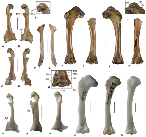
Distinguished from Archaeotrogon Milne-Edwards, 1892, by the medial margin of the proximal end of the humerus not forming a point (Fig. 1C); the coracoid having a proportionally shorter processus acrocoracoideus and a bipartite facies articularis that forms two distinct projections, the more ventral one of which gives the processus acrocoracoideus a hook-like shape; the coracoid shaft being more slender and having a prominent medial projection (Fig. 1G); and the facies articularis sternalis being mediolaterally wider; it further differs from Archaeotrogon venustus, A. zitteli and A. cayluxensis in that the humerus is proportionally longer and has a more slender shaft (ratio humerus : coracoid 1.49 vs 1.31–1.36; based on measurements in Mourer-Chauviré 1980, tables 2, 3).
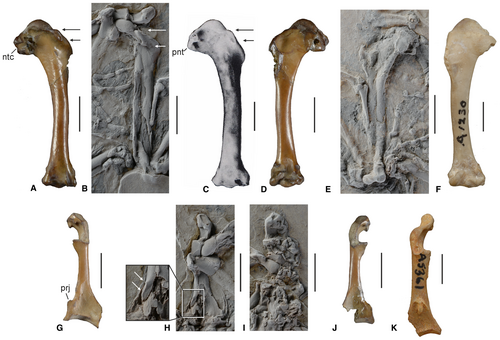
It is distinguished from Hassiavis Mayr, 1998, by the humerus being proportionally longer and having a more slender shaft (ratio humerus : coracoid 1.49 vs c. 1.39 in the holotype of H. laticauda); the tuberculum dorsale being proportionally shorter (Fig. 1A–C); the processus acrocoracoideus of the coracoid less markedly hook-shaped; the scapula having a proportionally narrower shaft; and the phalanx proximalis digiti majoris having a small but distinct processus internus indicis.
Remarks
Most of the previously described birds from Walton-on-the-Naze are clearly distinguished from Archaeodromus, but the humerus of the putatively coliiform taxon Eocolius Dyke & Waterhouse, 2001 shows a superficial similarity. Eocolius differs from Archaeodromus in the coracoid not having a foramen nervi supracoracoidei; the acromion of the scapula being proportionally longer and not forming a narrow, pointed tip; and the humerus with the tuberculum ventrale (proximal end) being proportionally larger, the caput humeri more protruding, and the tuberculum supracondylare ventrale (distal end) being proportionally longer (Dyke & Waterhouse 2001).
The humerus and coracoid of Archaeodromus also resemble the corresponding bones of trogons (Trogoniformes). In both, trogons and archaeotrogons, the humerus is stout and has a wide proximal end, but in archaeotrogons, the tuberculum ventrale is more prominent, the incisura capitis wider, and the sulcus transversus more marked; the coracoid of archaeotrogons has a reduced processus lateralis with a convex lateral margin. Archaeodromus is furthermore clearly distinguished from trogons in the morphology of the quadrate (see description and discussion).
Archaeodromus anglicus sp. nov.
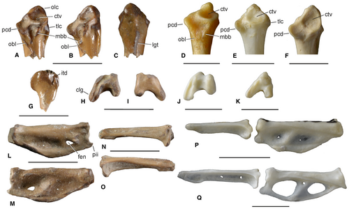
Figures 1A, D, G, J, 2A–F, N–O, 3, 4A–M, 5A–C, G–I, L–O
LSID
urn:lsid:zoobank.org:act:EA5FC86F-EF95-4A0B-9120-536D2735EE5D
Derivation of name
The species epithet refers to the geographic provenance of the new species.
Holotype
SMF Av 654: partial skeleton including the caudal portion of the right mandibular ramus, right quadrate, at least seven vertebrae or fragments thereof, both coracoids, right scapula, right humerus, left humerus lacking proximal end, proximal end of right ulna, fragment of distal end of left ulna, right os carpi ulnare, left phalanx proximalis digiti majoris, right phalanx distalis digiti majoris, and various unidentifiable bone fragments. The fossil was found in 1988 by Paul Bergdahl (original collector’s number BC 8814).
Diagnosis
As for genus. The new species is slightly larger than Hassiavis laticauda (humerus length 24.2 vs 20.3–20.6 mm; Mayr 1998) and its humerus measures only slightly less than that of Archaeotrogon venustus, the smallest Archaeotrogon species, whose humerus has a length of 25.0–29.7 mm (Mourer-Chauviré 1980; table 2). Compared with extant birds, the new species is about the size of the Australian owlet-nightjar (Aegotheles cristatus).
Type locality and horizon
Walton-on-the-Naze, Essex, UK; Walton Member of the London Clay Formation (previously Division A2; Jolley 1996; Aldiss 2012), early Eocene (early Ypresian, 54.6–55 Ma; Collinson et al. 2016).
Measurements
Right coracoid, length, 16.2 mm. Right humerus, length, 24.2 mm; proximal width, 8.9 mm; distal width, 5.0 mm. Phalanx proximalis digiti majoris, length, 8.9 mm. Phalanx distalis digiti majoris, length, 7.7 mm.
Description and comparisons
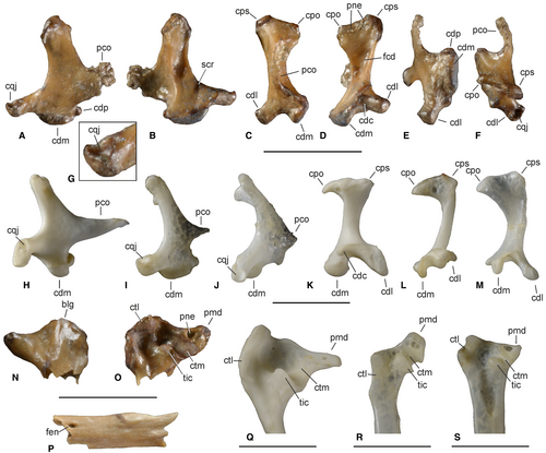
The right quadrate is nearly complete, with only the tip of the processus orbitalis being damaged (Fig. 2A–F). The most unusual feature of the bone is the deeply excavated and dorsally facing, cup-like cotyla quadratojugalis (Fig. 2G). In most birds, including the Steatornithidae, Podargidae and Trochilidae among the Strisores, the cotyla quadratojugalis faces laterally. It is dorsally directed in the Caprimulgidae, Nyctibiidae, Aegothelidae, Apodidae and Hemiprocnidae, but in the latter three taxa it forms a shallow articulation facet rather than a distinct cup-shaped cotyla (in the Caprimulgidae and Nyctibiidae the cotyla is cup-like, even though it is less excavated than in Archaeodromus). The capitulum squamosum is continuous with a small protrusion on the lateral surface of the processus oticus (tuberculum subcapitulare of Elzanowski & Stidham 2010). The capitulum oticum is not strongly medially projecting, unlike in the Caprimulgidae (Fig. 2M), Nyctibiidae, Aegothelidae (Fig. 2L), and Apodiformes; the incisura intercapitularis is very shallow. As in non-apodiform Strisores, but unlike most other neornithine birds, the condylus lateralis faces ventrolaterally. There are small pneumatic foramina along the caudal margin of the tip of the processus oticus, whereas a foramen pneumaticum mediale is absent; with regard to the distribution of pneumatic foramina, the fossil agrees with the quadrate of the Aegothelidae and apodiform birds. The well-developed processus orbitalis is dorsoventrally deep and slightly medially bent; its medial surface is concave. In extant Caprimulgidae (Fig. 2J), Nyctibiidae, Aegothelidae (Fig. 2I), Apodidae and Hemiprocnidae the processus orbitalis is greatly reduced. As in other Strisores but unlike, for example, the Trogoniformes (Fig. 2H) and Coraciiformes, the condylus medialis is proximodistally extensive but only weakly protruding in the ventral direction; its lateral surface forms a distinct bulge, which also occurs in the Podargidae, Nyctibiidae and Aegothelidae. The condylus pterygoideus is well-defined. An unusual feature of the bone is a distinct, scar-like sulcus on the caudomedial surface next to the base of the condylus lateralis (Fig. 2B); if this is a true osteological feature, this sulcus would distinguish the quadrate of Archaeodromus anglicus from that of other neornithine birds.
The dorsoventrally shallow caudal end of the mandible (Fig. 2N, O) has a distinctive shape in that the cotyla lateralis is situated caudally rather than laterally so that, in dorsal or ventral view, the caudal margin has a markedly convex outline and forms a distinct bulge (Fig. 2N). The sulcus intercotylaris is shallow and the tuberculum intercotylare has a crest-like shape. The cotyla medialis forms a deep but narrow trough. The well-developed processus medialis bears a pneumatic foramen. The caudal bulge is also present in the Podargidae and Apodidae, whereas the caudal end of the mandible of the Caprimulgidae (Fig. 2R), Nyctibiidae and Aegothelidae (Fig. 2S) is proportionally smaller and has a straight caudal margin.
In the holotype, a short section of the caudal portion of the right mandibular ramus is preserved (Fig. 2P), which most closely resembles the corresponding section of the mandible of the Aegothelidae. The fragment bears a small fenestra caudalis (absent in the Caprimulgidae and Nyctibiidae) and is not as wide mediolaterally as the caudal section of the mandibular ramus of the Caprimulgidae and Nyctibiidae.
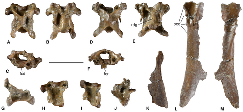
The vertebral series preserved in the holotype includes at least seven vertebrae or fragments thereof (Fig. 3A–J). Two nearly complete cervical vertebrae (Fig. 3A–F) are from the cranial series (4th–8th cervicals). Both bear long and thin processus costales. The processus transversi are moderately long and exhibit ridge-like tori dorsales. In one of the two vertebrae, the ventral surface of the corpus forms a narrow ridge (Fig. 3E); in the other vertebra (Fig. 3A–C), the facies articularis cranialis has a convex surface (rather than being saddle-shaped). Both vertebrae are dorsoventrally compressed as in the Aegothelidae. The foramina transversaria are small, also as in the Aegothelidae and Podargidae (larger in the Caprimulgidae). Two further fragmentary cervicals are from the more caudal series. A fragmentary thoracic vertebra appears to be the 14th (if compared to the Steatornithidae) or 13th (if compared to other Strisores); its corpus bears lateral depressions and there is a pair of wing-like lateral projections at the base of the processus ventralis.
The coracoid (Fig. 4A–D) is distinguished from the corresponding bone of Archaeotrogon in the shape of the processus acrocoracoideus and the more slender shaft. Overall, the bone resembles the coracoid of the Trogonidae (Fig. 4N). With regard to the more slender shaft, the bone also differs from the coracoid of Hassiavis with which it, however, agrees in that the processus acrocoracoideus has a hook-like outline. In Archaeodromus, the facies articularis clavicularis is bipartite and forms two protrusions; the larger, ventrally situated one is for the attachment of the ligamentum acrocoracoclaviculare superficiale and contributes to the hook-like appearance of the processus acrocoracoideus. As in Archaeotrogon, the cotyla scapularis is cup-like but very shallow. The processus procoracoideus appears to have been well developed and of subtriangular shape, even though its tip is broken in both coracoids. A foramen nervi supracoracoidei is absent. The sternal portion of the shaft forms a large medial projection, which is situated next to an indistinct notch. In Archaeotrogon the medial margin of the shaft also exhibits a shallow notch, but there is no pronounced medial projection even though in some specimens a minute protuberance is present (e.g. Mourer-Chauviré 1980, fig. 3c). A raised, elongated scar is situated in the medial portion of the impressio musculi sternocoracoidei. The extremitas sternalis is nearly complete in the right coracoid, in which only a short portion of the midsection of the rim of the processus lateralis is damaged. As in Archaeotrogon, the processus lateralis is short and has a strongly convex outline; the facies articularis sternalis is, however, mediolaterally wider than in Archaeotrogon (the morphology of the sternal end of the coracoid is poorly known for Hassiavis).
The scapula (Fig. 4F, G) has a narrow shaft; the caudal end is only slightly widened and moderately deflected. The acromion is proportionally longer than in Archaeotrogon.
Of the sternum, the left margo costalis with four processus costales as well as various small fragments are preserved (Fig. 3L, M). Another plate-like bone fragment that tapers into a narrow tip (Fig. 3K) defies a straightforward identification and may either be another part of the sternum or, more likely, a skull fragment.
The humerus (Fig. 4H–M) is a comparatively short and stout bone with a wide proximal end and closely resembles the humerus of Archaeotrogon hoffstetteri in its proportions (Fig. 1A, C). As in all Archaeotrogon species and in Hassiavis, the prominent caput humeri protrudes proximally. Also as in Hassiavis and the species of Archaeotrogon, the distal margin of the caput humeri is abruptly set off from the caudal surface of the bone (Fig. 4L). There is a transverse ridge in the incisura capitis (Fig. 4L), which is also found in Archaeotrogon hoffstetteri (Mourer-Chauviré 1980, fig. 10), but which is absent in other Archaeotrogon species. The sulcus transversus is long and distinct. The well-developed and caudally protruding tuberculum ventrale is separated from the crista bicipitalis by a notch (Fig. 1A); the incisura capitis is wide. Unlike in Archaeotrogon (Fig. 1C), the medial margin of the proximal humerus does not form a distinct point. The base of the fossa pneumotricipitalis is damaged so that it cannot be discerned whether there were pneumatic openings. The crus dorsale fossae is broken. The tuberculum dorsale is large and proximodistally elongated; in extant Strisores a pronounced tuberculum dorsale occurs only in the Caprimulgidae and in some apodiform birds. The crista deltopectoralis is damaged but appears to have had a convex margin. The shaft of the bone is narrower than the humerus shaft of all Archaeotrogon species except for A. hoffstetteri, which has a proportionally more slender shaft (Fig. 1C, F; Mourer-Chauviré 1980); on its caudal surface there is an elongate scar for the insertion of the musculus latissimus dorsi pars caudalis. The sulcus scapulotricipitalis, on the distal end of the bone, is very marked; its distolateral edge forms a distally projected prominence. The fossa musculi brachialis is shallow. A tuberculum supracondylare dorsale is absent, but there is a longitudinal insertion scar for the attachment of the musculus extensor carpi radialis (Fig. 4M). The scar for the attachment of the tendon of musculus pronator superficialis, on the ventral surface of the distal humerus, is a raised tubercle. The distoventral portion of the bone (processus flexorius and epicondylus ventralis) forms a short but well-defined projection. The large tuberculum supracondylare ventrale reaches as far proximally as the condylus dorsalis. The condyli are of similar shape to those of Archaeotrogon and are separated by a distinct incisura intercondylaris.
The proximal end of the right ulna (Fig. 5A–C) has a distinctive morphology. It resembles the proximal ulna of the Caprimulgidae (Fig. 5D) and Nyctibiidae, except for the short olecranon, which has a broadly rounded tip in Archaeodromus and is more similar to the olecranon of the Podargidae and Aegothelidae (Fig. 5E), whereas the olecranon of the Caprimulgidae and Nyctibiidae is narrower. The tuberculum ligamenti collaterale ventrale is small. A marked, oblique ridge extends distally past the processus cotylaris dorsalis. The cotyla ventralis is large. On the cranial surface of the bone, distal of the cotyla ventralis, there are two tubercles for the attachment of the tendon of musculus biceps brachii; these tubercles are a derived characteristic of the ulna of the Caprimulgidae (Fig. 5A, D). Also as in the Caprimulgidae, the impressio scapulotricipitalis forms a deeply marked pit. A midline ridge runs along the caudal surface of the bone.
The holotype includes a fragment of the caudal portion of the distal end of the left ulna (Fig. 5G). The condylus dorsalis does not exhibit the derived shape of the Caprimulgidae, in which it is caudally splayed, and the incisura tendinosa is very distinct.
In its shape, the os carpi ulnare (Fig. 5H, I) resembles the corresponding bone of the Caprimulgidae (5J). As in all other Strisores (Mayr 2010), the crus longum is pronounced.
The phalanx proximalis digiti majoris (Fig. 5L, M) is similar to that of the Aegothelidae (Fig. 5P) in its shape and morphology. It bears a well-defined, albeit very small, processus internus indicis (Stegmann 1963). There is a small fenestra in the distal portion of the bone, which represents a true osteological feature rather than a breakage owing to damage of the bone; the proximal section of the phalanx exhibits only two minute perforations. The phalanx distalis digiti majoris (Fig. 5N, O) is proportionally longer than in the Aegothelidae, being as long as the phalanx proximalis digiti majoris.
Genus HASSIAVIS Mayr, 1998
Hassiavis cf. laticauda Mayr, 1998
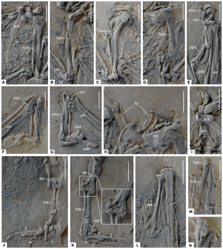
Figures 1B, E, H, I, 6, 7

Referred specimen
SMF-ME 11702A + B (postcranial skeleton on two slabs; Fig. 6).
Locality and horizon
Messel near Darmstadt, Germany; latest early or earliest middle Eocene (48 Ma; Lenz et al. 2015).
Measurements
Given as: left/right, maximum length [measurements of the holotype of H. laticauda from Mayr 1998] (all in mm). Humerus, 22.1/c. 22 [c. 20.5/20.5]. Ulna, 24.0/c. 24.6 [c. 24.1/c. 23.9]. Carpometacarpus, 13.8/13.6 [c. 12.7/13.1]. Femur, c. 14.5/c. 14.5. Tibiotarsus, c. 23.0/c. 23.0 [25.0/24.6]. Tarsometatarsus, c. 13.5/12.4 [12.0/12.2]. Pedal phalanges: I1, c. 4.8 [4.4]; I2, 2.5 [2.5]; II1, c. 4.2 [3.9]; II2, c. 4.0 [3.9]; II3, 2.2 [2.8]; III1, 4.1 [3.2]; III2, 3.1 [3.8]; III3, >3.6 [4.1]; III4, 2.4 [2.9]; IV1, 3.6 [2.8]; IV2, 2.4 [2.5]; IV3, 2.1 [2.5]; IV4, 2.8 [2.5]; IV5, 2.2 [c. 2.3].
Description and comparisons
The new fossil is the best-preserved skeleton of Hassiavis found so far and the only one in which the postcranial bones are not substantially flattened and crushed. Whereas the overall shape of the major limb bones could be assessed in previous fossils, the new specimen reveals several osteological details that were unknown for Hassiavis and that are described in the following. The new specimen closely resembles previously reported fossils of H. laticauda, but has a proportionally slightly longer humerus and carpometacarpus, and a slightly shorter tibiotarsus. Whether these differences indicate distinctness at the species level or are due to the taphonomic distortion of the fossils is difficult to determine, so that the new fossil is classified as Hassiavis cf. laticauda.
In the new specimen, the hook-like shape of the processus acrocoracoideus of the coracoid is less apparent than it is in previously described specimens (Mayr 1998, 2004), which is due to the fact that the processus acrocoracoideus lacks the ventral portion in the right coracoid and is distorted in the left one (Figs 1H, I, 7I). However, the processus acrocoracoideus of the right coracoid shows that the facies articularis clavicularis was bipartite, as it is in Archaeodromus. The shaft of the bone appears to be somewhat stouter than that of Archaeodromus, although it is not as stout as in Archaeotrogon. The processus procoracoideus is not fully preserved in the fossil, but seems to have been broad and short. The medial margin of the sternal end is damaged, but the right coracoid allows the recognition of the base of a broken medial projection (Fig. 1H); whether this projection was as pronounced as in Archaeodromus remains, however, unknown. The processus lateralis is likewise damaged in both coracoids and although it appears to have been short, its exact shape cannot be discerned.
The humerus (Fig. 7A–D) closely resembles that of Archaeodromus. As in the latter, the tuberculum dorsale is distinct and proximodistally elongated and the sulcus transversus is deeply marked. The tuberculum dorsale is proportionally longer than in Archaeodromus (Fig. 1A–C; the humerus length is 24.3 vs 22.1 mm for Archaeodromus and Hassiavis, respectively, whereas the length of the tuberculum dorsale is 1.7 vs 2.0 mm, respectively). As far as comparisons are possible, the distal end of the bone resembles the distal humerus of Archaeodromus. The distolateral edge of the sulcus scapulotricipitalis forms a distally projected prominence. The scar for the attachment of the tendon of musculus pronator superficialis forms a raised tubercle.
The proximal end of the ulna agrees with Archaeodromus in that there is a deeply marked impressio scapulotricipitalis. The visible part of the distal end of the bone is also similar to the fragment of the distal ulna preserved in the Archaeodromus anglicus holotype, and exhibits a distinct incisura tendinosa.
The comparatively short carpometacarpus (Fig. 7E–G) has similar proportions to that of Archaeotrogon. Unlike in the latter, however, the long and cranially prominent processus extensorius does not form a spur-like, pointed tip.
The os carpi ulnare (Fig. 7F) resembles that of Archaeodromus in its shape and has a long crus longum. The os carpi radiale is similar to the corresponding bone of the Caprimulgidae, Nyctibiidae and Aegothelidae, whereas the bone has a distinctive derived morphology in the Trogoniformes and in coraciiform birds (compare Fig. 7E with Mayr 2014, figs 3, 6). Unlike in Archaeodromus, the phalanx proximalis digiti majoris is not fenestrated.
As in Archaeotrogon, the tibiotarsus has a very wide distal end, with widely splayed condyli (Fig. 7K). The trochlea cartilaginis tibialis is proximodistally extensive and has a distinctly concave articular surface.
The tarsometatarsus (Fig. 7J–M) likewise has similar proportions to that of Archaeotrogon, and in concordance with the wide distal end of the tibiotarsus, it has a wide proximal end. The tuberositas musculi tibialis cranialis is situated near the medial margin of the bone. The hypotarsus forms a broad platform (Fig. 7N), which is distally continuous with a short crista medianoplantaris; deep hypotarsal sulci or canals for the flexor tendons of the toes appear to be absent. The trochleae metatarsorum resemble those of Archaeotrogon in their proportions. The foramen vasculare distale is not clearly visible. The proximal phalanges of the fourth toe are not abbreviated.
The tail feathers are well preserved in the fossil (Fig. 6C, D), with the longest one measuring c. 62 mm. Although the tail is therefore as long as that of other Hassiavis specimens with preserved tail feathers, the new fossil shows no banding pattern, which is pronounced in previously reported fossils (Mayr 1998, 2004).
Phylogenetic analysis
Analysis of the primary character matrix resulted in six most parsimonious trees (L = 161; CI = 0.55; RI = 0.64), the strict consensus tree of which is shown in Figure 8A. The analysis supported a clade including Archaeodromus, Hassiavis and Archaeotrogon, but the position of this clade was controversially resolved. In two trees, archaeotrogons were obtained as the sister taxon of the Caprimulgidae, in two further trees they were obtained as the sister taxon of the clade (Caprimulgidae + Nyctibiidae), in one they resulted as the sister taxon of the Apodiformes, and in another one they resulted as the sister taxon of the clade (Aegothelidae + Apodiformes).
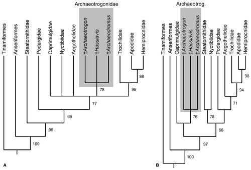
As in other analyses of morphological data, the primary analysis did not support a sister group relationship between the Caprimulgidae and all other taxa of the Strisores, whereas this is congruently recovered in current analyses of nuclear sequence data (Prum et al. 2015; Kuhl et al. 2021). A second analysis was therefore constrained to the tree topologies obtained by the latter studies. This analysis resulted in two most parsimonious trees (L = 209; CI = 0.56; RI = 0.63), which supported a sister group relationship between the Archaeotrogonidae and the Caprimulgidae (Fig. 8B).
A third analysis was finally run that was constrained to a sister group relationship between the clade (Steatornithidae + Nyctibiidae) and all remaining Strisores, following the results of an earlier study (Hackett et al. 2008). This analysis likewise resulted in two most parsimonious trees (L = 210; CI = 0.59; RI = 0.67) and supported a sister group relationship between the Archaeotrogonidae and the Caprimulgidae (not shown).
Discussion
Although the humerus and coracoid of Archaeodromus anglicus gen. et sp. nov. show a resemblance to the corresponding bones of trogons (Trogoniformes), an assignment of the fossil to the Strisores is clearly evident from the morphology of the quadrate. One of the most distinctive features of this bone is the dorsally directed, cup-like cotyla quadratojugalis. In extant birds, a dorsally (or laterodorsally) directed cotyla quadratojugalis is found only in taxa of the Strisores, that is, in the Caprimulgidae, Nyctibiidae, Aegothelidae, Apodidae and Hemiprocnidae. In all other birds (including the Steatornithidae and Podargidae), the cotyla quadratojugalis faces laterally, and its dorsal orientation in broad-billed representatives of the Strisores with large nostrils may be due to the fact that in these avian insectivores the jugal is strained differently than in other neornithine birds. In addition to the highly characteristic morphology of the cotyla quadratojugalis, the quadrate of Archaeodromus corresponds to that of the Caprimulgidae, Nyctibiidae, Aegothelidae and apodiform birds in that the condylus caudalis is reduced. Archaeodromus anglicus furthermore exhibits an os carpi ulnare with a long crus longum, which was identified as an apomorphy of the Strisores by Mayr (2010), as well as a fenestrated phalanx proximalis digiti majoris (which is absent in trogons and coraciiform birds).
Archaeodromus anglicus shares a characteristic humerus morphology with Hassiavis and Archaeotrogon. Derived traits include a wide proximal end (ratio proximal width : total length of bone, 0.34–0.37) and a proximally protruding caput humeri, the distal margin of which is abruptly set off from the caudal surface of the bone (compare Fig. 4J, L with Mourer-Chauviré 1980, fig. 3a), as well as a marked and proximodistally long tuberculum dorsale. The combination of these traits does not occur in any other taxon of the Strisores. The coracoid of Archaeodromus and Archaeotrogon is characterized by a short processus lateralis with a convexly curved lateral margin (the shape of the processus lateralis is unknown for Hassiavis).
Unpublished further fossils of Archaeodromus anglicus from Walton-on-the-Naze in a private collection (see introduction) corroborate a classification of the new species into the Strisores as well as an assignment to the Archaeotrogonidae. These specimens exhibit a caprimulgid-like mandible with a sigmoidally curved ramus, a sternum with a short, bifurcate spina externa (as in Hassiavis), a carpometacarpus with a spur-like processus extensorius (as in Archaeotrogon), and a short tarsometatarsus of similar proportions to that of Archaeotrogon, but with a broader hypotarsus that does not form distinct sulci or canals. Archaeodromus anglicus is the oldest fossil record of the Archaeotrogonidae, pre-dating Hassiavis from the 48-myr-old Messel oilshale by 6–7 myr and the oldest Archaeotrogon fossils from the Quercy fissure fillings (40 myr; 2006) by 15 myr.
Judging from the similarities in quadrate morphology (Archaeodromus; the present study) and bill shape (Hassiavis; Mayr 2004), archaeotrogons probably had a foraging strategy similar to that of the Aegothelidae, which capture insects by sallying flights from perches (Holyoak 1999). The marked spur formed by the processus extensorius of Archaeotrogon (Mourer-Chauviré 1980) and Archaeodromus (pers. obs. based on unpublished specimens; see Mayr 2017) suggests that archaeotrogons used their wings as weapons in intraspecific combat, a behaviour not found in extant representatives of the Strisores.
As far as comparisons between the fossils are possible, Archaeodromus closely resembles Hassiavis in the morphology of most major limb bones. As noted in the introduction, Chen et al. (2019) proposed that Hassiavis is a stem group representative of the Aegothelidae. Although this hypothesis is not supported by the present analyses, one of the trees resulting from the unconstrained analysis likewise placed archaeotrogons within the clade (Aegothelidae + Apodiformes), as the sister taxon of the Apodiformes. However, a position of archaeotrogons within the clade including the Aegothelidae and Apodiformes conflicts with the fact that the coracoid of the Archaeotrogonidae lacks a foramen nervi supracoracoidei, which is one of the key apomorphies of the (Aegothelidae + Apodiformes) clade. In its overall morphology, the coracoid of archaeotrogons is also very different from the coracoid of the Aegothelidae and Apodiformes, and more closely resembles the coracoid of the Caprimulgidae (Fig. 4O, P). The hook-shaped processus acrocoracoideus, which was the main supporting synapomorphy listed by Chen et al. (2019), is furthermore differently formed in the Archaeotrogonidae (Archaeodromus and Hassiavis) and Aegothelidae, and a hook-shaped processus acrocoracoideus is altogether absent in Archaeotrogon (Fig. 1K) and the apodiform Trochilidae. Other similarities between the archaeotrogon taxa Archaeodromus and Hassiavis and the Aegothelidae may well be plesiomorphic for Strisores or a subclade thereof (e.g. the similar shapes of the beak and distal wing phalanges).
Although archaeotrogons are therefore unlikely to be within the clade (Aegothelidae + Apodiformes), Archaeodromus shares one potential synapomorphy with the latter clade, that is, the presence of pneumatic foramina in the caudal surface of the processus oticus of the quadrate. This derived characteristic might support a sister group relationship between archaeotrogons and the clade (Aegothelidae + Apodiformes), which was obtained in one tree resulting from the primary analysis.
However, most trees resulting from the primary analysis supported closer affinities between archaeotrogons and the Caprimulgidae, and two of the former trees as well as those from the constrained analyses recovered a sister group relationship of both taxa. Derived characteristics shared by Archaeodromus and the Caprimulgidae include a humerus with (1) a large tuberculum dorsale; (2) two distinct tubercles for the attachment of the tendon of musculus biceps brachii on the proximal end of the ulna (George & Berger 1966: p. 330); and (3) an impressio scapulotricipitalis (ulna) forming a deeply marked pit. The presence of a transverse ridge in the incisura capitis of the humerus was already listed as a caprimulgid feature of archaeotrogons by Mourer-Chauviré (1980). This ridge is present in Archaeotrogon hoffstetteri but is absent in other Archaeotrogon species, which have a stouter humerus than A. hoffstetteri. The ridge is also present in Archaeodromus and, if plesiomorphic for the Archaeotrogonidae, may be another derived characteristic shared by these birds and the Caprimulgidae (the humeri of Hassiavis do not allow the recognition of morphological details in the corresponding area of the bone).
Here it is concluded that current evidence conforms best to a sister group relationship between the Archaeotrogonidae and the Caprimulgidae, which are the most speciose group of nocturnal Strisores. Certainly, however, more data on the osteology of archaeotrogons are needed for a robust and unambiguous placement of these birds, and it is noted that the Caprimulgidae distinctly differ from archaeotrogons in some osteological characteristics. For example, the intertarsal joint (distal tibiotarsus and proximal tarsometatarsus) is exceptionally wide in archaeotrogons, but unusually narrow in the Caprimulgidae. These differences may possibly be associated with the highly derived foot morphology of the Caprimulgidae, in which one phalanx of the fourth toe is lost, so that there are only four instead of five phalanges. Functionally, this derived foot morphology of nightjars may be related to the terrestrial breeding ecology of these birds, which are the sole ground-breeding taxon of the Strisores. With regard to the derived morphology of some skull elements (e.g. the quadrate and proximal end of the mandible), the Caprimulgidae are likewise distinguished from archaeotrogons and closely resemble the Nyctibiidae (Mayr 2010). In morphology-based analyses (Mayr 2010), these similarities are optimized as synapomorphies and would conflict with a sister group relationship between the Caprimulgidae and the Archaeotrogonidae, which lack these derived morphologies. According to molecular and combined morphological and molecular analyses, however, the Caprimulgidae and Nyctibiidae are more distantly related (Hackett et al. 2008; Prum et al. 2015; Chen et al. 2019; Kuhl et al. 2021), which suggests that the derived similarities are of convergent origin.
Chen et al. (2019) identified a reduced lacrimal as a synapomorphy of non-caprimulgid Strisores. Provided that the Caprimulgidae are indeed the sister taxon of other taxa of the Strisores and if archaeotrogons are the sister taxon of caprimulgids, it is to be expected that archaeotrogons had a well-developed lacrimal. A better knowledge of the skull morphology of archaeotrogons may therefore provide further evidence to confirm or reject the proposed sister group relationship to caprimulgids.
Nightjars are the only nocturnal taxon of the Strisores that occurs today in Europe. The absence of stem group nightjars in the early Eocene of Europe contrasts with the fact that early Eocene European avifaunas included stem group representatives of the Steatornithidae, Nyctibiidae and Podargidae that were already quite similar to their extant relatives (Mayr 2017). Recognition of archaeotrogons as stem group representatives of the Caprimulgidae would not only fill a major gap in the fossil record of nocturnal Strisores, but may also indicate that the radiation of modern-type Caprimulgidae took place well after the Eocene.
Conclusion
A well-preserved partial skeleton of an archaeotrogon from the early Eocene London Clay of Walton-on-the-Naze (Essex, UK) is the earliest fossil record of the Archaeotrogonidae and is described as Archaeodromus anglicus.
The holotype of the new species and a newly identified specimen of Hassiavis from the latest early or earliest middle Eocene of the Messel fossil site in Germany inform the osteology of the Archaeotrogonidae.
The quadrate of Archaeodromus shows derived morphological characteristics of the Strisores, and new osteological data suggest that archaeotrogons are stem group representatives of the Caprimulgidae (nightjars and allies).
If confirmed by future studies, this phylogenetic placement would shed light on the early evolutionary history of the most speciose extant taxon of nocturnal Strisores, whose evolutionary history has so far been very poorly known.
Acknowledgements
I thank Jason Bergdahl for facilitating the acquisition of the collection of his father, the late Paul Bergdahl. Anika Vogel (SMF) is thanked for access to the Messel fossil and Bruno Behr (SMF) for its excellent preparation. The photos were taken by Sven Tränkner (SMF). Michael Daniels is acknowledged for access to his private collection of London Clay birds during earlier visits. Cécile Mourer-Chauviré is thanked for access to Archaeotrogon fossils and for permission to reproduce a photo of the humerus of Archaeotrogon hoffstetteri. Comments from Ursula Göhlich, two anonymous reviewers, and the Technical Editor Sally Thomas improved the manuscript. Open Access funding enabled and organized by Projekt DEAL.
Open Research
Data archiving statement
This published work and the nomenclatural acts it contains, have been registered in ZooBank (http://zoobank.org/References/168B3C6E-8BA9-4235-93C7-47EFA1C2D8D7). The matrix and character list are available in MorphoBank: http://morphobank.org/permalink/?P3914


