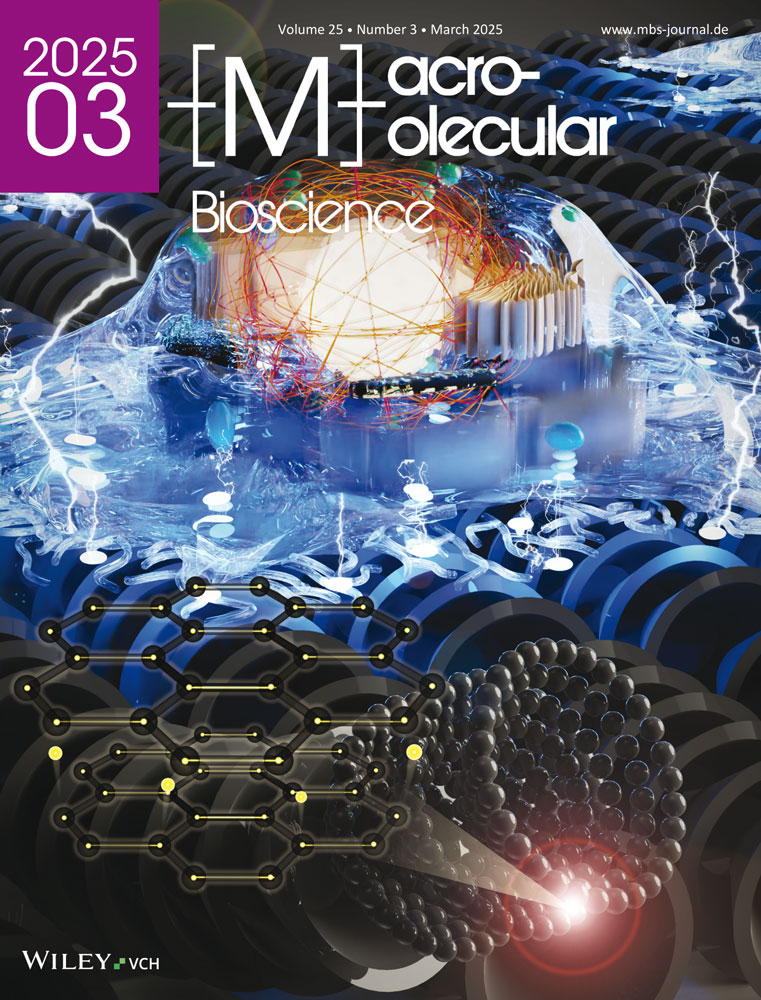
Cellular Behaviors of Human Dermal Fibroblasts on Pyrolytically Stripped Carbon Nanofiber's Surface
Abstract
There has been limited exploration of carbon nanofiber as a scaffold for cellular attachment and proliferation. In this work, commercially available, pyrolytically stripped carbon nanofiber (cCNF) is deposited over electrospun nanofiber mats, polycaprolactone (PCL) and poly(D-lactide) (PDLA), to immobilize them and investigate whether the 3D cCNF layer's surface augments cell proliferation of human dermal fibroblasts (nHDF). Spectral characterizations, such as XRD and Raman, show that cCNF exhibited crystalline structure with a high graphitization degree. cCNF layers are modified to have an irregular or planar surface by simple agitation (s-cCNF) or probe sonication (p-cCNF) of the solution. The in vitro cell line studies revealed that p-cCNF is better than s-cCNF in providing a platform that supports a homogenous spread of the fibroblasts all over the nanofiber's surface. The p-cCNF-deposited PCL mat (p-cCNF@PCL) demonstrated cellular growth, similar to that of the neat PCL mat. However, the p-cCNF@PCL mat exhibited remarkable antibacterial properties by reducing the E. coli numbers, ≈16 times greater than the PCL mat. It is concluded that the immobilized, pyrolytically stripped carbon nanofiber's surface has the potential to accommodate cellular growth and inhibit bacterial colonies, suggesting the biomaterial scaffold is promising for in vivo and clinical applications of skin tissue regeneration.
Conflict of Interest
The authors declare no conflict of interest.
Open Research
Data Availability Statement
The data that support the findings of this study are available from the corresponding author upon reasonable request.


