Volume 11, Issue 12 e201800187
EDITOR'S CHOICE

Thermal effect on dispersive infrared spectroscopic imaging of prostate cancer tissue

Cai L. Song

Cai L. Song

Department of Chemical Engineering, Imperial College London, London, UK

Search for more papers by this author
Meguya Ryu

Meguya Ryu

Department of Materials Science and Engineering, Tokyo Institute of Technology, Tokyo, Japan

Search for more papers by this author
Junko Morikawa

Corresponding Author

Junko Morikawa

Department of Materials Science and Engineering, Tokyo Institute of Technology, Tokyo, Japan

Correspondence

Junko Morikawa, Department of Materials Science and Engineering, Tokyo Institute of Technology, Ookayama, Meguro, Tokyo 152-8552, Japan.

Email: [email protected]

Sergei G. Kazarian, Department of Chemical Engineering, Imperial College London, South Kensington Campus, London SW7 2AZ, UK.

Email: [email protected]

Search for more papers by this author
Archana Kothari

Archana Kothari

Department of Pathology, Kingston Hospital, Surrey, UK

Search for more papers by this author
Sergei G. Kazarian

Corresponding Author

Sergei G. Kazarian

Department of Chemical Engineering, Imperial College London, London, UK

Correspondence

Junko Morikawa, Department of Materials Science and Engineering, Tokyo Institute of Technology, Ookayama, Meguro, Tokyo 152-8552, Japan.

Email: [email protected]

Sergei G. Kazarian, Department of Chemical Engineering, Imperial College London, South Kensington Campus, London SW7 2AZ, UK.

Email: [email protected]

Search for more papers by this author
First published: 13 July 2018
Citations: 4
Funding information Tokyo Institute of Technology; Imperial College London

Abstract

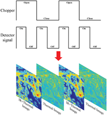
A system that combines dispersive infrared micro-spectroscopic imaging and thermography has been developed to study the effect of thermal radiation on the infrared absorption spectra of prostate biopsy samples. The system allows the distribution of thermal signal intensity as a function of emissivity to be interpreted from the integrated absorbance obtained by spectroscopic imaging. Biochemical differences between cancer and benign areas within the specimens are identified in the spectra. Side-by-side comparison of H&E stained adjacent tissue sections with infrared images constructed before and after the removal of thermal effect showed that the latter strongly support differentiation of regions within tissues. The use of spectral bands at discrete wavelengths significantly reduced spectral acquisition time, making this technique promising as a future clinical diagnostic tool. A systemic methodology was implemented to process the data, first by k-means clustering on the second derivative spectra without a priori knowledge, followed by principal component analysis (PCA). Four distinct regions within the tissue samples were successfully classified based on the antisymmetric stretching mode of the methylene functional group. Separation between data in clusters occurs when projecting spectra on a PCA score plot on a plane made by first 2 principal components. The significance of the disparity was verified with statistical test. Regulation of signal to chopper and detector enables simultaneous acquisition of infrared and thermal images of the prostate biopsy tissues.

image

The full text of this article hosted at iucr.org is unavailable due to technical difficulties.