Surface enhanced Raman spectroscopy-detection of the uptake of mannose-modified nanoparticles by macrophages in vitro: A model for detection of vulnerable atherosclerotic plaques
Vera Dugandžić
Institute of Physical Chemistry, Friedrich-Schiller University Jena, Jena, Germany
Leibniz Institute of Photonic Technology, Jena, Germany
Abbe Center of Photonics, Friedrich Schiller University Jena, Jena, Germany
Search for more papers by this authorDenis Drikermann
Institute for Organic Chemistry and Macromolecular Chemistry, Friedrich Schiller University Jena, Jena, Germany
Search for more papers by this authorOleg Ryabchykov
Institute of Physical Chemistry, Friedrich-Schiller University Jena, Jena, Germany
Leibniz Institute of Photonic Technology, Jena, Germany
Abbe Center of Photonics, Friedrich Schiller University Jena, Jena, Germany
Search for more papers by this authorAndreas Undisz
Otto Schott Institute of Materials Research, Friedrich Schiller University Jena, Jena, Germany
Search for more papers by this authorIvan Vilotijević
Institute for Organic Chemistry and Macromolecular Chemistry, Friedrich Schiller University Jena, Jena, Germany
Search for more papers by this authorStefan Lorkowski
Abbe Center of Photonics, Friedrich Schiller University Jena, Jena, Germany
Institute of Nutrition, Friedrich Schiller University Jena, Jena, Germany
Competence Cluster for Nutrition and Cardiovascular Health (nutriCARD), Halle-Jena-Leipzig, Germany
Jena Centre for Soft Matter, (JCSM), Friedrich Schiller University Jena, Jena, Germany
Search for more papers by this authorThomas W. Bocklitz
Institute of Physical Chemistry, Friedrich-Schiller University Jena, Jena, Germany
Leibniz Institute of Photonic Technology, Jena, Germany
Abbe Center of Photonics, Friedrich Schiller University Jena, Jena, Germany
Search for more papers by this authorChristian Matthäus
Institute of Physical Chemistry, Friedrich-Schiller University Jena, Jena, Germany
Leibniz Institute of Photonic Technology, Jena, Germany
Abbe Center of Photonics, Friedrich Schiller University Jena, Jena, Germany
Search for more papers by this authorKarina Weber
Institute of Physical Chemistry, Friedrich-Schiller University Jena, Jena, Germany
Leibniz Institute of Photonic Technology, Jena, Germany
Jena Centre for Soft Matter, (JCSM), Friedrich Schiller University Jena, Jena, Germany
Search for more papers by this authorCorresponding Author
Dana Cialla-May
Institute of Physical Chemistry, Friedrich-Schiller University Jena, Jena, Germany
Leibniz Institute of Photonic Technology, Jena, Germany
Abbe Center of Photonics, Friedrich Schiller University Jena, Jena, Germany
Correspondence
Dana Cialla-May, Institute of Physical Chemistry, Friedrich-Schiller University Jena, Helmholtzweg 4, 07743 Jena, Germany.
Email: [email protected]
Search for more papers by this authorJürgen Popp
Institute of Physical Chemistry, Friedrich-Schiller University Jena, Jena, Germany
Leibniz Institute of Photonic Technology, Jena, Germany
Abbe Center of Photonics, Friedrich Schiller University Jena, Jena, Germany
Jena Centre for Soft Matter, (JCSM), Friedrich Schiller University Jena, Jena, Germany
Search for more papers by this authorVera Dugandžić
Institute of Physical Chemistry, Friedrich-Schiller University Jena, Jena, Germany
Leibniz Institute of Photonic Technology, Jena, Germany
Abbe Center of Photonics, Friedrich Schiller University Jena, Jena, Germany
Search for more papers by this authorDenis Drikermann
Institute for Organic Chemistry and Macromolecular Chemistry, Friedrich Schiller University Jena, Jena, Germany
Search for more papers by this authorOleg Ryabchykov
Institute of Physical Chemistry, Friedrich-Schiller University Jena, Jena, Germany
Leibniz Institute of Photonic Technology, Jena, Germany
Abbe Center of Photonics, Friedrich Schiller University Jena, Jena, Germany
Search for more papers by this authorAndreas Undisz
Otto Schott Institute of Materials Research, Friedrich Schiller University Jena, Jena, Germany
Search for more papers by this authorIvan Vilotijević
Institute for Organic Chemistry and Macromolecular Chemistry, Friedrich Schiller University Jena, Jena, Germany
Search for more papers by this authorStefan Lorkowski
Abbe Center of Photonics, Friedrich Schiller University Jena, Jena, Germany
Institute of Nutrition, Friedrich Schiller University Jena, Jena, Germany
Competence Cluster for Nutrition and Cardiovascular Health (nutriCARD), Halle-Jena-Leipzig, Germany
Jena Centre for Soft Matter, (JCSM), Friedrich Schiller University Jena, Jena, Germany
Search for more papers by this authorThomas W. Bocklitz
Institute of Physical Chemistry, Friedrich-Schiller University Jena, Jena, Germany
Leibniz Institute of Photonic Technology, Jena, Germany
Abbe Center of Photonics, Friedrich Schiller University Jena, Jena, Germany
Search for more papers by this authorChristian Matthäus
Institute of Physical Chemistry, Friedrich-Schiller University Jena, Jena, Germany
Leibniz Institute of Photonic Technology, Jena, Germany
Abbe Center of Photonics, Friedrich Schiller University Jena, Jena, Germany
Search for more papers by this authorKarina Weber
Institute of Physical Chemistry, Friedrich-Schiller University Jena, Jena, Germany
Leibniz Institute of Photonic Technology, Jena, Germany
Jena Centre for Soft Matter, (JCSM), Friedrich Schiller University Jena, Jena, Germany
Search for more papers by this authorCorresponding Author
Dana Cialla-May
Institute of Physical Chemistry, Friedrich-Schiller University Jena, Jena, Germany
Leibniz Institute of Photonic Technology, Jena, Germany
Abbe Center of Photonics, Friedrich Schiller University Jena, Jena, Germany
Correspondence
Dana Cialla-May, Institute of Physical Chemistry, Friedrich-Schiller University Jena, Helmholtzweg 4, 07743 Jena, Germany.
Email: [email protected]
Search for more papers by this authorJürgen Popp
Institute of Physical Chemistry, Friedrich-Schiller University Jena, Jena, Germany
Leibniz Institute of Photonic Technology, Jena, Germany
Abbe Center of Photonics, Friedrich Schiller University Jena, Jena, Germany
Jena Centre for Soft Matter, (JCSM), Friedrich Schiller University Jena, Jena, Germany
Search for more papers by this authorAbstract
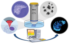
Atherosclerosis is a process of thickening and stiffening of the arterial walls through the accumulation of lipids and fibrotic material, as a consequence of aging and unhealthy life style. However, not all arterial plaques lead to complications, which can lead to life-threatening events such as stroke and myocardial infarction. Diagnosis of the disease in early stages and identification of unstable atherosclerotic plaques are still challenging. It has been shown that the development of atherosclerotic plaques is an inflammatory process, where the accumulation of macrophages in the arterial walls is immanent in the early as well as late stages of the disease. We present a novel surface enhanced Raman spectroscopy (SERS)-based strategy for the detection of early stage atherosclerosis, based on the uptake of tagged gold nanoparticles by macrophages and subsequent detection by means of SERS. The results presented here provide a basis for future in vivo studies in animal models.The workflow of tracing the SERS-active nanoparticle uptake by macrophages employing confocal Raman imaging.
Supporting Information
| Filename | Description |
|---|---|
| jbio201800013-sup-0001-AppendixS1.docxWord 2007 document , 1.5 MB |
File S1. Supporting Information Figure S1 Raman images of human THP-1 macrophages incubated with AuNP@PDI for 2 hours. (A-D) Raman images of macrophages together with corresponding point spectra, in which the signal of the Raman reporter was found (marker mode labeled with red). White circles indicate the location of red points in images for improved visibility; (E-K) Raman images of human THP-1 macrophages, in which the signal of the Raman reporter was not found. The corresponding spectra present a Raman spectrum in a single pixel of a microphage image. The scale bar represents 10 μm Figure S2 Raman images of human THP-1 macrophages incubated with AuNP@PDI@Silica for 2 hours. (A-I) Raman images of macrophages together with corresponding point spectra, in which the signal of the Raman reporter was found (marker mode labeled with red). White circles indicate the location of red points in images for improved visibility; (J) Raman image of a human THP-1 macrophage, in which the signal of the Raman reporter was not found. The corresponding spectra present a Raman spectrum in a single pixel of a microphage image. The scale bar represents 10 μm Figure S3 Raman images of human THP-1 macrophages incubated with AuNP@PDI@Silica-man for 2 hours together with corresponding point spectra extracted from each red point in the Raman image. The specific signal of the Raman reporter was found in all inspected cells (marker mode labeled with red). White circles indicate the location of red points in images for improved visibility. The scale bar represents 10 μm. *Only 10 randomly chosen spectra were plotted in the cases of the cells where number of red points exceeded 10. |
Please note: The publisher is not responsible for the content or functionality of any supporting information supplied by the authors. Any queries (other than missing content) should be directed to the corresponding author for the article.
REFERENCES
- 1World Health Organization, www.who.int/mediacentre/factsheets/fs317/en/ (accessed: September, 2017).
- 2M. Naghavi, A. A. Abajobir, C. Abbafati, et al., Lancet, 2017, 390, 1151.
- 3M. C. Fishbein, Cardiovasc. Pathol. 2010, 19, 6.
- 4M. J. Davies, Circulation, 1996, 94, 2013.
- 5A. C. van der Wal, A. E. Becker, C. M. van der Loos, P. K. Das, Circulation 1994, 89, 36.
- 6K. J. Moore, F. J. Sheedy, E. A. Fisher, Nat. Rev. Immunol. 2013, 13, 709.
- 7P. Greenland, J. S. Alpert, G. A. Beller, E. J. Benjamin, M. J. Budoff, Z. A. Fayad, E. Foster, M. A. Hlatky, J. M. Hodgson, F. G. Kushner, M. S. Lauer, L. J. Shaw, S. C. Smith Jr., A. J. Taylor, W. S. Weintraub, N. K. Wenger, J. Am. Coll. Cardiol., 2010, 56, e50.
- 8A. L. Catapano, A. Pirillo, G. D. Norata, Br. J. Pharmacol. 2017, 174(22), 3973.
- 9E. Corrado, M. Rizzo, G. Coppola, K. Fattouch, G. Novo, I. Marturana, F. Ferrara, S. Novo, J. Atheroscler. Thromb. 2010, 17, 1.
- 10F. Wiesmann, M. Szimtenings, A. Frydrychowicz, R. Illinger, A. Hunecke, E. Rommel, S. Neubauer, A. Haase, Magn. Reson. Med. 2003, 50, 69.
- 11J. A. Rumberger, D. B. Simons, L. A. Fitzpatrick, P. F. Sheedy, R. S. Schwartz, Circulation, 1995, 92, 2157.
- 12S. Schroeder, A. F. Kopp, A. Baumbach, C. Meisner, A. Kuettner, C. Georg, B. Ohnesorge, C. Herdeg, C. D. Claussen, K. R. Karsch, J. Am. Coll. Cardiol. 2001, 37, 1430.
- 13X. Li, J. Li, J. Jing, T. Ma, S. Liang, J. Zhang, D. Mohar, A. Raney, S. Mahon, M. Brenner, P. Patel, K. K. Shung, Q. Zhou, Z. Chen, IEEE J. Sel. Top. Q. Electron. 2014, 20, 196.
- 14D. H. O'Leary, J. F. Polak, S. K. Wolfson, M. G. Bond, W. Bommer, S. Sheth, B. M. Psaty, A. R. Sharrett, T. A. Manolio, Stroke 1991, 22, 1155.
- 15M. Rogosnitzky, S. Branch, Biometals 2016, 29, 365.
- 16A. Alame, E. S. Brilakis, Catheter. Cardiovasc. Interv. 2016, 87, 241.
- 17S. Lee, M. W. Lee, H. S. Cho, J. W. Song, H. S. Nam, D. J. Oh, K. Park, W.-Y. Oh, H. Yoo, J. W. Kim, Circ. Cardiovasc. Interv. 2014, 7, 560.
- 18L. Marcu, J. A. Jo, Q. Fang, T. Papaioannou, T. Reil, J.-H. Qiao, J. D. Baker, J. A. Freischlag, M. C. Fishbein, Atherosclerosis 2009, 204, 156.
- 19J. Bec, D. M. Ma, D. R. Yankelevich, J. Liu, W. T. Ferrier, J. Southard, L. Marcu, J. Biophotonics 2014, 7, 281.
- 20A. S. Peters, J. Backhaus, A. Pfützner, M. Raster, G. Burgard, S. Demirel, D. Böckler, M. Hakimi, Vib. Spectrosc. 2017, 92, 20.
- 21S. K. Zacharias, R. D. Safian, R. D. Madder, I. D. Hanson, M. C. Pica, J. L. Smith, J. A. Goldstein, A. E. Abbas, Vasc. Med. 2016, 21, 337.
- 22I. James, F. Brennan, Y. Wang, R. R. Dasari, M. S. Feld, Appl. Spectrosc. 1997, 51, 201.
- 23A. S. Haka, J. R. Kramer, R. R. Dasari, M. Fitzmaurice, J Biomed Opt. 2011, 16, 011011.
- 24O. R. Šćepanović, M. Fitzmaurice, A. Miller, C.-R. Kong, Z. Volynskaya, R. R. Dasari, J. R. Kramer, M. S. Feld, J Biomed Opt. 2011, 16, 011009.
- 25S. W. E. van de Poll, D. J. M. Delsing, J. W. Jukema, H. M. G. Princen, L. M. Havekes, G. J. Puppels, A. van der Laarse, Atherosclerosis 2002, 164, 65.
- 26K. M. Marzec, T. P. Wrobel, A. Rygula, E. Maslak, A. Jasztal, A. Fedorowicz, S. Chlopicki, M. Baranska, J. Biophotonics 2014, 7, 744.
- 27A. Lattermann, C. Matthäus, N. Bergner, C. Beleites, B. F. Romeike, C. Krafft, B. R. Brehm, J. Popp, J. Biophotonics 2013, 6, 110.
- 28S. W. E. van de Poll, K. Kastelijn, T. C. Bakker Schut, C. Strijder, G. Pasterkamp, G. J. Puppels, A. van der Laarse, Heart 2003, 89, 1078.
- 29J. T. Motz, M. Fitzmaurice, A. Miller, S. J. Gandhi, A. S. Haka, L. H. Galindo, R. R. Dasari, J. R. Kramer, M. S. Feld, J Biomed Opt. 2006, 11, 021003.
- 30C. Matthäus, S. Dochow, G. Bergner, A. Lattermann, B. F. M. Romeike, E. T. Marple, C. Krafft, B. Dietzek, B. R. Brehm, J. Popp, Anal. Chem. 2012, 84, 7845.
- 31C. Stiebing, L. Schmölz, M. Wallert, C. Matthäus, S. Lorkowski, J. Popp, J. Lipid Res. 2017, 58, 876.
- 32C. Stiebing, T. Meyer, I. Rimke, C. Matthäus, M. Schmitt, S. Lorkowski, J. Popp, J. Biophotonics 2017, 10, 1217.
- 33C. Stiebing, C. Matthäus, C. Krafft, A.-A. Keller, K. Weber, S. Lorkowski, J. Popp, Anal. Bioanal. Chem. 2014, 406, 7037.
- 34A. Oseledchyk, C. Andreou, M. A. Wall, M. F. Kircher, ACS Nano 2017, 11, 1488.
- 35R. Ankri, D. Leshem-Lev, D. Fixler, R. Popovtzer, M. Motiei, R. Kornowski, E. Hochhauser, E. I. Lev, Nano Lett. 2014, 14, 2681.
- 36M. Schnoor, I. Buers, A. Sietmann, M. F. Brodde, O. Hofnagel, H. Robenek, S. Lorkowski, J. Immunol. Methods 2009, 344, 109.
- 37M. Schnoor, P. Cullen, J. Lorkowski, K. Stolle, H. Robenek, D. Troyer, J. Rauterberg, S. Lorkowski, J. Immunol. 2008, 180, 5707.
- 38G. H. Jeong, Y. W. Lee, M. Kim, S. W. Han, J. Colloid Interface Sci. 2009, 329, 97.
- 39W. Stöber, A. Fink, E. Bohn, J. Colloid Interface Sci. 1968, 26, 62.
- 40M. K. Patel, B. Vijayakrishnan, J. R. Koeppe, J. M. Chalker, K. J. Doores, B. G. Davis, Chem. Commun. 2010, 46, 9119.
- 41S. Kralj, M. Drofenik, D. Makovec, J. Nanopart. Res. 2011, 13, 2829.
- 42 R Development Core Team, R: A Language and Environment for Statistical Computing. Vienna, Austria, the R Foundation for Statistical Computing. 2015.
- 43S. Barthelme. 2017, imager: Image Processing Library Based on 'CImg' R package, version 0.41.1
- 44M. Morhac 2012, Peaks, R package, version 0.2
- 45S. Urbanek. 2013, Package “tiff”, R package, version 0.1-5.
- 46C. G. Ryan, E. Clayton, W. L. Griffin, S. H. Sie, D. R. Cousens, Nucl. Instrum. Methods Phys. Res., Sect. B 1988, 34, 396.
- 47O. Ryabchykov, T. Bocklitz, A. Ramoji, U. Neugebauer, M. Foerster, C. Kroegel, M. Bauer, M. Kiehntopf, J. Popp, Chemom. Intell. Lab. Syst. 2016, 155, 1.
- 48S. P. Mulvaney, M. D. Musick, C. D. Keating, M. J. Natan, Langmuir 2003, 19, 4784.
- 49S. M. Kang, B. S. Lee, S.-g. Lee, I. S. Choi, Colloids Surf. A Physicochem. Eng. Asp. 2008, 313–314, 150.
- 50L. M. Liz-Marzán, M. Giersig, P. Mulvaney, Langmuir 1996, 12, 4329.
- 51K. Nakamoto, Applications in Coordination Chemistry, John Wiley & Sons, Inc., Hoboken, New Jersey, 2008, p. 1.
- 52S. Zhu, M. Niu, H. O'Mary, Z. Cui, Mol. Pharm. 2013, 10, 3525.
- 53 J. D. Ernst, Infect. Immun. 1998, 66, 1277.
- 54Z. Cui, C.-H. Hsu, R. J. Mumper, Drug Dev. Ind. Pharm. 2003, 29, 689.
Citing Literature