Repression of MicroRNA-30e by Hepatitis C Virus Enhances Fatty Acid Synthesis
Abstract
Chronic hepatitis C virus (HCV) infection often leads to end-stage liver disease, including hepatocellular carcinoma (HCC). We have previously observed reduced expression of microRNA-30e (miR-30e) in the liver tissues and sera of patients with HCV-associated HCC, although biological functions remain unknown. In this study, we demonstrated that HCV infection of hepatocytes transcriptionally reduces miR-30e expression by modulating CCAAT/enhancer binding protein β. In silico prediction suggests that autophagy-related gene 5 (ATG5) is a direct target of miR-30e. ATG5 is involved in autophagy biogenesis, and HCV infection in hepatocytes induces autophagy. We showed the presence of ATG5 in the miR-30e–Argonaute 2 RNA-induced silencing complex. Overexpression of miR-30e in HCV-infected hepatocytes inhibits autophagy activation. Subsequent studies suggested that ATG5 knockdown in Huh7.5 cells results in the remarkable inhibition of sterol regulatory element binding protein (SREBP)-1c and fatty acid synthase (FASN) level. We also showed that overexpression of miR-30e decreased lipid synthesis-related protein SREBP-1c and FASN in hepatocytes. Conclusion: We show new mechanistic insights into the interactions between autophagy and lipid synthesis through inhibition of miR-30e in HCV-infected hepatocytes.
Abbreviations
-
- Ago
-
- Argonaute
-
- ATG5
-
- autophagy related gene 5
-
- C/EBP-β
-
- CCAAT/enhancer binding protein β
-
- DAPI
-
- 4´,6-diamidino-2-phenylindole
-
- FASN
-
- fatty acid synthase
-
- GFP
-
- green fluorescent protein
-
- HCC
-
- hepatocellular carcinoma
-
- HCV
-
- hepatitis C virus
-
- Ig
-
- immunoglobulin
-
- LC3B
-
- microtubule-associated protein 1 light chain 3 beta
-
- LD
-
- lipid droplet
-
- miR
-
- microRNA
-
- mRNA
-
- messenger RNA
-
- mTOR
-
- mammalian target of rapamycin
-
- p-mTOR
-
- phosphorylated-mammalian target of rapamycin
-
- RT-qPCR
-
- reverse-transcription real-time polymerase chain reaction
-
- siRNA
-
- small interfering RNA
-
- SREBP-1c
-
- sterol regulatory element binding protein 1c
-
- UPR
-
- unfolded protein response
-
- UTR
-
- untranslated region
Chronic hepatitis C virus (HCV) infection is one of the etiologic causes of developing cirrhosis and eventually hepatocellular carcinoma (HCC).1 Approximately 55% of patients infected with HCV develop steatosis because HCV uses host lipid metabolism for its life cycle.2 Abnormal lipid metabolism influences metabolic disorders, cirrhosis, and liver carcinogenesis.3-5 Histologically, HCV infection induces lipid droplet (LD) formation in hepatocytes.6 We and others have shown that HCV infection induces autophagy7, 8 and modulates signaling pathways involved in lipogenesis, resulting in the accumulation of LDs,9, 10 although the precise mechanism is not fully understood.
MicroRNAs (miRNAs) are small noncoding RNAs that modulate the translation and stability of messenger RNAs (mRNAs) through a mechanism involving specific binding to mRNA based on base pair complementarity, controlling protein production. Most human miRNAs are processed into mature 20-26 nucleotide duplexes through endonucleolytic cleavage by ribonuclease (RNase) III enzymes and Argonaute (Ago) proteins. Dicer and Ago cooperate to form the RNA-induced silencing complex (RISC), consisting of Ago-bound single-strand miRNAs, which target host RNAs and induce a combination of translational repression.11 miRNAs have a widespread impact on the etiologies of disease in viral infections12 either directly or indirectly through regulation of virus-associated host pathways.11 Moreover, several miRNAs interact directly with host lipid metabolism.13, 14
In the present study, we observed that miR-30e expression is inhibited at the transcriptional level following HCV infection by down-regulating the transcription factor CCAAT/enhancer binding protein β (C/EBP-β). miR-30e targets autophagy-related gene 5 (ATG5), a key molecule of autophagy signaling, and inhibits de novo lipogenesis-related protein, implicating its role in HCV-mediated pathogenesis.
Materials and Methods
Cell Culture, HCV Infection, and Transfection
Huh7.5 cells and Huh7.5 cells harboring the HCV genotype 2a genome-length replicon (Rep2a cells; kindly provided by Hengli Tang, Florida State University, FL, USA) were used. Cells were maintained in Dulbecco's modified Eagle's medium supplemented with 10 % fetal bovine serum and penicillin–streptomycin at 37°C in a 5% CO2 atmosphere.
HCV genotype 2a (clone JFH1) was grown in Huh7.5 cells as described.15 Virus released into cell culture supernatant was filtered through a 0.45-μm pore cellulose acetate membrane and quantitated in standard IU/mL. For infection, cells were incubated with HCV genotype 2a (clone JFH1) (multiplicity of infection, 0.1) in a minimum volume of medium as described.16 The cellular RNA was extracted 3 days postinfection.
Transfection of ATG5 small interfering RNA (siRNA) into Huh7.5 cells was performed using lipofectamine RNAiMAX (Invitrogen, Carlsbad, CA). Briefly, Huh7.5 cells were plated at a density of approximately 1 × 10⁵ cells/well in a 12-well plate and transfected with 50 nM ATG5 siRNA (sc-41445; Santa Cruz, Dallas, TX) or control siRNA and lysed for western blot analysis at 48 hours after transfection. Transfection of an miR-30e-5p mimic (002223; ThermoFisher Scientific, Waltham, MA) into Huh7.5 cells was performed using lipofectamine (Invitrogen). Briefly, Huh7.5 cells were plated at a density of approximately 1 × 10⁵ cells/well in a 12-well plate and transfected with 20 nM of miR-30e-5p mimic or control miR. Cells were lysed for western blot analysis 48 hours after transfection. Total RNA was prepared in a separate transfection.
RNA Quantitation and Reverse-Transcription Real-Time Polymerase Chain Reaction
Total RNA was isolated by using a TRIzol reagent (Invitrogen). RNA was quantified by using a NanoDrop ND-1000 spectrophotometer (Thermo Fisher Scientific). Complementary DNA was synthesized using miR-30e- or a U6-specific primer (Thermo Fisher Scientific) with a TaqMan miRNA reverse transcription (RT) kit or random hexamers and a Superscript III RT kit (Invitrogen). Real-time polymerase chain reaction (qPCR) was performed with a 7500 real-time PCR system (Applied Biosystems, Foster City, CA). TaqMan universal PCR master mix and a 6-carboxyfluorescein (FAM)-minor groove binder (MGB) probe for ATG5 (Hs00169468_m1; Thermo Fisher Scientific) and 18S ribosomal RNA (rRNA; Hs03928985_g1; Thermo Fisher Scientific) were used. Relative expression level was calculated by normalizing with U6 or 18S rRNA, using the 2−ΔΔCT formula (ΔΔCT = ΔCT of the sample − ΔCT of the untreated control).
Western Blot Analysis
Cells were lysed by using a sodium dodecyl sulfate–polyacrylamide gel electrophoresis (SDS-PAGE) sample loading buffer. The lysates were subjected to PAGE and transferred onto a nitrocellulose membrane. The membranes were blocked with 5% nonfat dried milk and probed with the following specific primary antibodies: C/EBP-β, sterol regulatory element binding protein (SREBP)-1 (2A4), and fatty acid synthase (FASN) (A-5; Santa Cruz); ATG5 (Proteintech, IL); and mammalian target of rapamycin (mTOR; 7C10) and phosphorylated-mTOR (p-mTOR; D9C2) (Cell Signaling Technology, Danvers, MA). After washing, the blots were incubated with secondary antibody for 1 hour. Proteins were detected by using an enhanced chemiluminescence western blot substrate (Pierce; ThermoFisher Scientific). Membranes were reprobed with antibody to actin (Santa Cruz) as an internal control for normalization of the protein load. ImageJ software (National Institutes of Health [NIH]) was used for densitometric scanning of western blot images.
Luciferase Reporter Assays
The miR-30e promoter (nucleotides −1813 to +1; P0) or promoter fragment of the deletion mutant of miR-30e (P1) were PCR amplified from Huh7.5 genomic DNA by using specific primers (Table 1). The fragments were digested with MluI and HindIII and cloned into the pGL3-Basic luciferase vector (Promega, Madison, WI). Huh7.5 cells and Rep2a cells were transfected with a luciferase reporter plasmid containing the miR-30e promoter (1 µg/mL) by using Lipofectamine 3000 (Invitrogen). Luciferase activity was determined as described.17
| Primers | Sequence |
|---|---|
| miR-30e-full promoter_For | 5′-ATTAGCCTTCTGACGCGTGTGTTCTCTTAC-3′ |
| miR-30e-F1 promoter_For | 5′-ATTCAATACGCGTGAACCAAAGG-3′ |
| miR-30e promoter_Rev | 5′-TGACAGAAGCTTGCTAATCC-3′ |
Huh7.5 cells were plated at a density of 0.8 × 10⁵ cells/well in a 24-well plate and cotransfected with 25 nM C/EBP-β siRNA (Santa Cruz) or control siRNA and the promoter construct of miR-30e using Endofectin (GeneCopoeia, Rockville, MD). A luciferase reporter assay was performed 48 hours after transfection.
Ago2-RNA Co-Immunoprecipitation
The hepatocyte RNA–miR-30e complex was precipitated as described.18 Briefly, miR-30e transfected Huh7.5 cells were lysed with lysis buffer (150 mM KCl, 25 mM Tris-HCl [pH 7.4], 5 mM ethylene diamine tetraacetic acid, 1% Triton X-100, 5 mM dithiothreitol, protease inhibitor mixture, and 100 U/mL RNase-OUT [Invitrogen]). Lysates were clarified, incubated with an anti-Ago2 monoclonal antibody (11A9; Sigma-Aldrich, St. Louis, MO) or isotype control immunoglobulin (Ig)G2a at 4°C for 7 hours, and mixed with protein G-Sepharose (GE Healthcare, Piscataway, NJ) overnight. The beads were washed 4 times, and RNA was isolated by using TRIzol reagent (Invitrogen). Target RNA was quantitated by RT-qPCR using specific primers.
Immunofluorescence
Huh7.5 cells were transfected with miR-30e mimic and infected with green fluorescent protein (GFP)-tagged HCV. Cells were fixed for 30 minutes with 4% paraformaldehyde after 72 hours of infection and then treated with 0.2% Triton X-100 for 5 minutes. After blocking with 3% bovine serum albumin, cells were incubated with primary antibody rabbit polyclonal microtubule-associated protein light chain 3B (LC3B; Sigma-Aldrich) and then conjugated to Alexa Fluor 594 (Molecular Probes, Eugene, OR) with anti-rabbit Ig. 4´,6-Diamidino-2-phenylindole (DAPI) was incubated for nuclear staining. Three-channel optical images (green for HCV, red for LC3, and blue for nucleus) were collected using the sequential scanning mode of the Olympus FV1000 confocal system (Olympus, Center Valley, PA). Images were superimposed digitally for fine comparisons.
Huh7.5 cells were transfected with ATG5 siRNA (sc-41445; Santa Cruz) or control siRNA for 48 hours. Cells were stained by boron-dipyrromethene (BODIPY) 493/503 (Invitrogen) for 1 hour, and DAPI was incubated for nuclear staining. Lipid accumulation was observed by using the Olympus FV1000 confocal system (Olympus) in cells with DAPI-stained nuclei. Images were superimposed digitally for fine comparisons. ImageJ software (NIH) was used for densitometric scanning of immunofluorescence images.
Statistical Analysis
Results are presented as mean ± SD. Data were analyzed by the Student t test with a two-tailed distribution. P < 0.05 was considered statistically significant.
Results
HCV Infection Down-Regulates miR-30e Expression
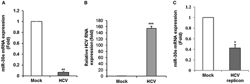
We have previously shown that miR-30e expression was significantly lower (P < 0.003) in the sera of patients with HCV-infected HCC compared with healthy volunteers.19 We also observed down-regulation of miR-30e in liver samples from patients with HCV-associated HCC, although the mechanism of miR-30e down-regulation remains unknown. We examined the expression level of miR-30e in HCV-infected human hepatocytes. The level of miR-30e was significantly reduced following HCV infection in Huh7.5 cells (Fig. 1, panel A). HCV RNA was measured from HCV-infected Huh7.5 cells by RT-qPCR (Fig. 1, panel B). A similar result was obtained when we examined the level of miR-30e in Huh7.5 cells harboring the genome-length HCV replicon (Rep2a cells) compared to parental Huh7.5 cells (Fig. 1, panel C). Together, our results indicate that HCV infection suppresses miR-30e expression in hepatocytes.
HCV Infection Down-Regulates C/EBP-β-Mediated miR-30e Promoter Activation
We next examined how HCV modulates miR-30e expression. For this, an miR-30e promoter sequence (nucleotides −1,813 to +1; P0) was cloned into pGL3 luciferase reporter plasmid DNA, using specific primers (Table 1). Huh7.5 cells and Huh7.5 cells harboring the HCV genome-length replicon cells were transfected with the P0 construct to examine promoter activity. miR-30e promoter activity was significantly down-regulated in HCV replicon-expressing cells compared to control parental cells (Fig. 2, panel A). To determine the cis-regulatory elements present in the miR-30e promoter region (nucleotides −1,813 to +1), we searched PROMO, using version 8.3 of TRANSFAC. Several regulatory elements were found in the designated consensus sequence of the miR-30e promoter, including binding sites for C/EBP-β. C/EBP-β, which belongs to the leucine zipper family, is an important transcriptional regulator.20 Moreover, we have previously shown that HCV down-regulates promoter activity of complement C3 and miR-181c by C/EBP-β.21, 22
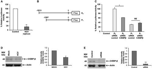
We next identified the specific region on the miR-30e promoter responsible for HCV-mediated repression. We generated deletion mutants of the miR-30e promoter constructs (nucleotides −1567 to +1 [P1]) and cloned these into pGL3 luciferase reporter plasmid DNA (Fig. 2, panel B). We knocked down C/EBP-β using a specific siRNA and examined the activity of the P0 or P1 construct of the miR-30e promoter in Huh7.5 cells to analyze the role of C/EBP-β in the regulation of HCV-mediated miR-30e expression. Knockdown of C/EBP-β inhibited the activity of the miR-30e P0 promoter, but the P1 fragment did not exert an effect (Fig. 2, panel C), suggesting that the trans-suppressor element may be located between regions –1,813 to –1,565. We further confirmed that HCV infection in Huh7.5 cells reduces C/EBP-β expression (Fig. 2, panel D), in agreement with previous reports.21, 22 Transfection of siRNA to C/EBP-β in Huh7.5 cells reduced protein expression (Fig. 2, panel E). Together, these results suggest that HCV-mediated C/EBP-β suppression is an important mechanism for miR-30e down-regulation.
ATG5 IS a Direct Target of miR-30e
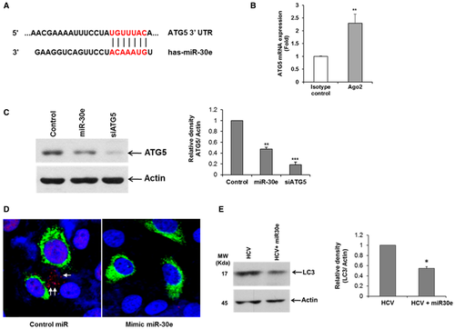
In silico prediction suggests that ATG5 is a direct target of miR-30e (Fig. 3, panel A). A recent report showed that miR-30e targets the 3′-untranslated region (UTR) of ATG5 and inhibits its translation in gastric cancer cells.23 We examined the interaction between miR-30e and ATG5 in hepatocytes. For this, miR-30e-transfected Huh7.5 cell lysates were immunoprecipitated with an Ago2-specific monoclonal antibody or an unrelated isotype control. RNA was isolated from the immunoprecipitated lysates and analyzed by RT-qPCR. Higher ATG5 level was noted in the miR-30e–Ago2 complex compared to immunoprecipitated control, suggesting its presence in their complex (Fig. 3, panel B). Overexpression of miR-30e inhibits ATG5 expression (Fig. 3, panel C). siRNA to ATG5 was used as a positive control. As reported,23 we did not observe the inhibition of ATG5 mRNA by miR-30e in our experimental system. We and others have shown that HCV induces autophagy.17, 24 ATG5 is an important molecule in the autophagy signaling pathway. We therefore predicted that overexpression of miR-30e in HCV-infected hepatocytes will reduce ATG5 expression and may impair the induction of autophagy. For this, we overexpressed miR-30e in GFP-tagged HCV-infected cells and stained these for LC3, a marker for autophagy. Our results demonstrated that overexpression of miR-30e inhibited HCV-induced LC3 lipidation compared to control miR-treated cells (Fig. 3, panel D). We also observed that overexpression of miR-30e inhibited HCV-induced LC3 lipidation by western blot analysis (Fig. 3, panel E).
Depletion of ATG5 Inhibits Lipid Synthesis in Hepatocytes
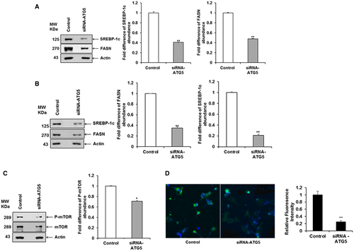
LDs have been implicated as an essential link for efficient autophagy in the yeast system,25 and autophagy is inhibited following depletion of free fatty acids. In HCV infection, both autophagy and LDs are induced in hepatocytes. Because ATG5 is a direct target of miR-30e, we wanted to understand the interplay between ATG5 and lipid synthesis. ATG5 and ATG7 are essential molecules for the induction of autophagy; ATG7 conjugates ATG5 to ATG12 for autophagosome formation.26 ATG7-deficient mice suppress LD formation,27 and SREBP-1c forms LDs through de novo lipogenesis.28 To investigate the relationship between ATG5 and SREBP-1c expression, we knocked down ATG5 using a specific siRNA and performed western blot analysis using specific antibodies. A significant down-regulation of SREBP-1c and FASN expression level was observed compared to that of the control cell after normalization with actin (Fig. 4, panel A). A similar result was obtained when we examined the expression of SREBP-1c and FASN in Huh7.5 cells harboring the genome-length HCV replicon (Rep2a cells) compared to parental cells (Fig. 4, panel B). We also observed a significant down-regulation of p-mTOR, an intermediate between ATG5 and SREBP-1c expression level, compared to that of the control cell (Fig. 4, panel C). We further showed an inhibition of LDs in ATG5-depleted cells (Fig. 4, panel D). Together, these data suggest that depletion of ATG5 inhibits SREBP-1c and FASN expression.
Overexpression of miR-30e Inhibits Lipid Synthesis in Hepatocytes
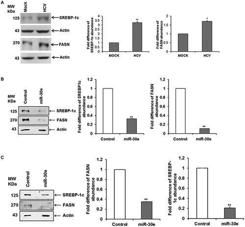
SREBP-1 and FASN are important players in lipid metabolism, and SREBPs are key transcriptional factors in lipogenesis and lipid uptake.29, 30 We observed that HCV infection in Huh7.5 cells increased expression of SREBP-1c and its downstream target FASN (Fig. 5, panel A). We also observed ATG5 depletion inhibits SREBP-1c and FASN expression. We therefore examined whether miR-30e overexpression modulates lipid synthesis in hepatocytes. For this, we performed western blot analysis in Huh7 cells transfected with an miR-30e mimic or control miR. A significant down-regulation of SREBP-1c and FASN expression level compared to that of the control parental cell was observed after normalization with the housekeeping protein actin (Fig. 5, panel B). A similar result was obtained when we examined the expression of SREBP-1c and FASN in Huh7.5 cells harboring the genome-length HCV replicon (Rep2a cells) compared to parental cells (Fig. 5, panel C). Together, our data suggest that mir-30e inhibits ATG5 and eventually SREBP-1c and FASN in hepatocytes.
Discussion
In the present study, we demonstrated that (i) HCV infection transcriptionally down-regulates miR-30e, (ii) down-regulation of miR-30e targets ATG5 and modulates induction of autophagy, and (iii) overexpression of miR-30e or ATG5 knockdown inhibits de novo lipogenesis.
We previously showed that HCV infection increased lipid accumulation through metabolic gene regulation.9 Increased accumulation of LDs frequently causes liver steatosis in patients chronically infected with HCV.2, 31 In this report, we observed that HCV infection modulates lipogenesis processes primarily by regulating miR-30e transcription. Overexpression of miR-30e inhibits the expression of SREBP-1c and FASN, which are known to be involved in de novo lipogenesis and an increased expression of LDs, promoting lipid accumulation. Together, these results suggest that HCV increases de novo lipogenesis in hepatocytes by inhibiting miR-30e expression.
miRNAs impact the regulation of many cellular processes, and viruses modulate the host miRNA expression either directly or indirectly to accelerate pathogenesis.12, 32 In reaction to viral infection, signaling pathways in hepatocytes direct a variety of intracellular events that generate de novo lipogenesis in the infected cells.13, 14 In earlier reports, miR-30e was implicated as a subtype-specific prognostic marker in breast cancer and ovarian cancer33, 34 and is inhibited in chronic myeloid leukemia, lung cancer, and patients with HCC.19, 35, 36 miR-30 has been implicated as a tumor suppressor and inhibits epithelial-to-mesenchymal transition in HCC through metastasis-associated protein 1 modulation.37, 38 In cancer development, fatty acids are understood to be the most relevant because lipid metabolism participates in the regulation of many cellular processes, such as cell growth, proliferation, differentiation, and survival.39 Moreover, miR-30e has been implicated in the regulation of fatty acid metabolism in goat mammary epithelial cells.40
We observed inhibition of miR-30e expression by HCV infection or HCV replicon in human hepatocytes, suggesting direct evidence for an association with HCV infection. C/EBP-β is an important transcriptional factor belonging to the leucine zipper family and is involved in several cellular functions, such as cell proliferation and differentiation.20 We previously showed that C/EBP-β is down-regulated following HCV infection.21 In our experiments, we demonstrated that HCV inhibits miR-30e promoter activity by down-regulating C/EBP-β expression. Deletion of the C/EBP-β binding site on an miR-30e promoter (P1 fragment) or knockdown of C/EBP-β showed suppression of miR-30e promoter activity.
We demonstrated an interaction between miR-30e and ATG5 in our complex by RISC assay; this concurs with a report that ATG5 is a direct target of miR-30e.23 ATG5 and ATG7 are autophagy-specific genes involved in the initiation of autophagosome formation and completion through a ubiquitin-like conjugation system.41 Interestingly, ATG7-deficient mice suppressed LD formation,27 and liver-specific ATG5-knockout mice displayed impaired LD biogenesis.42 HCV infection induces autophagy. We postulate that HCV-mediated inhibition of miR-30e facilitates ATG5 for autophagy induction following HCV infection. We showed previously that autophagy acts as an upstream positive regulator of mTOR complex 1 (mTORC1),17 which regulates SREBP-1c at the transcriptional level43; however, further study is needed to dissect this mechanism. SREBP-1c forms LDs through de novo lipogenesis.28 Furthermore, unfolded protein response (UPR) activates autophagy in HCV-infected hepatocytes,44 and UPR activates SREBP maturation in cultured cells.45, 46 Moreover, the activation of multiple UPR responses and the SREBP pathway significantly overlapped.47 Processing of SREBP-1c has been reported to depend on mTORC1.48 In our study, SREBP-1c and FASN were inhibited by knockdown of ATG5 in hepatocytes. These results demonstrated that de novo lipogenesis is regulated through autophagy in hepatocytes following HCV infection. HCV causes steatosis, with the reported mean prevalence of HCV-related NAFLD being 55% and nonalcoholic steatohepatitis being 4%-10% of cases.49 We previously showed that HCV-infected hepatocytes increased expression of SREBP-1c and its downstream target FASN, both of which are involved in lipogenesis.9 We showed here HCV-mediated down-regulation of miR-30e is responsible, in part, for up-regulating SREBP-1c and FASN expression.
We observed that expression levels of de novo lipogenesis-related protein SREBP-1c and FASN are significantly inhibited in miR-30e-overexpressed hepatocytes. miR-30e also directly targets the 3′ UTR of ATG5. Moreover, knockdown of ATG5 expression negatively regulates SREBP-1c and FASN protein expression level. In summary, we have demonstrated that HCV infection transcriptionally down-regulates miR-30e expression by targeting C/EBP-β, which in turn enhances the expression of de novo lipogenesis-related protein SREBP-1c and FASN through autophagy. Our observations contribute to the understanding of the mechanistic role of miR-30e in HCV–host interactions toward pathogenesis.


