Feasibility and Safety of a Novel Cable-Transmitted Magnetically Controlled Capsule Endoscopy System for Upper Gastrointestinal Examination
ABSTRACT
Background
To evaluate the feasibility and safety of a novel cable-transmitted magnetically controlled capsule endoscopy (CT-MCE) system for upper gastrointestinal examination.
Methods
Twenty-six participants (19 healthy volunteers and seven patients with gastrointestinal symptoms) willing to undergo upper gastrointestinal endoscopy were recruited. Each participant underwent CT-MCE followed by conventional gastroscopy within 24 h. Maneuverability and visibility of the CT-MCE capsule in the upper gastrointestinal tract, adverse events, and discomfort during the procedure were evaluated. The sensitivity and specificity of CT-MCE for diagnosing upper gastrointestinal lesions were evaluated using conventional gastroscopy findings as the standard.
Results
Maneuverability was graded as “good” for all segments of the esophagus. The percentage of participants in which maneuverability was good according to gastric region was as follows: cardia (100.00%), pylorus (96.15%), angulus (92.31%), antrum (88.46%), fundus (84.62%), and body (73.08%). In the duodenal bulb and descending duodenum, it was good in only 20.83% and 16.67% of participants, respectively. Visibility was graded as “excellent” or “good” in the esophagus, Z line, and duodenal bulb in all participants; excellent/good visibility was achieved in the stomach and descending duodenum in 96.15% and 79.17% of participants, respectively. Forty-one lesions were detected overall. The sensitivity and specificity of CT-MCE in diagnosing upper gastrointestinal lesions were 85.00% and 98.15%, respectively. The CT-MCE capsule was successfully removed through the mouth in all participants. No serious adverse events or capsule retention occurred.
Conclusions
CT-MCE showed good feasibility and safety for upper gastrointestinal examination. The system was effective in examining the esophagus and stomach with no risk of capsule retention.
1 Introduction
Capsule endoscopy was first approved for clinical examination of the small bowel in 2000. Since then, it has been widely used owing to its noninvasiveness and high patient tolerability [1, 2]. The first wireless capsule endoscopes were propelled by peristalsis and passively passed through the gastrointestinal tract, without an ability to control travel speed or camera orientation. This precluded repeated inspection of suspicious lesions and areas of interest, and made capsule endoscopy unsuitable for detailed examinations of the esophagus or stomach. However, esophageal capsule endoscopy is effective for screening and evaluating reflux esophagitis, Barrett's esophagus, and esophageal varices [3-5]. Nonetheless, the esophageal examination can be limited owing to the high travel speed [6].
The concept of magnetically controlled capsule endoscopy (MCE), which involves guiding a capsule endoscope using an external magnetic field control device, was first proposed by Carpi et al. in 2006 [7]. MCE for gastric examination is safe and feasible [8-11] and as accurate as conventional gastroscopy [11, 12]. To enhance capsule control during MCE in the esophagus, a capsule controlled by a detachable thin string was designed, which slows passage through the esophagus to enable a more thorough examination [13]. The string can be detached from the capsule after the esophageal examination to allow investigation of the stomach and small intestine. Nevertheless, owing to the presence of a built-in battery, MCE systems in clinical use are large and limited by a low frame rate and low image resolution. These factors render it difficult for some patients to ingest and impede the ability to detect small lesions in the mucosa of the digestive system. Moreover, the risk of capsule retention prevents their use in patients with gastrointestinal stenosis or obstruction.
To address the limitations of current MCE systems in the examination of the upper gastrointestinal tract, we have developed a novel cable-transmitted MCE (CT-MCE) system. It employs a flexible cable for power and signal transmission, is relatively small, obtains high-resolution images at a high frame rate, and is easily removed. This pilot study investigated its feasibility and safety for upper gastrointestinal examination.
2 Materials and Methods
2.1 CT-MCE System
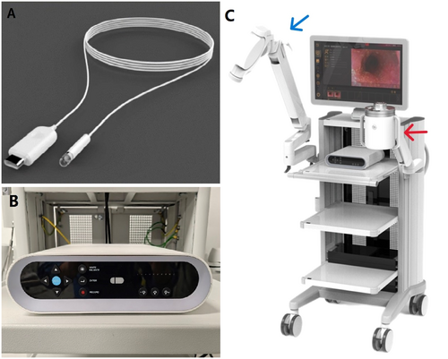
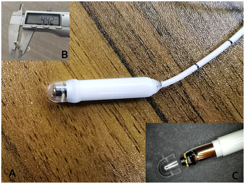
The AICAM-S-1 system (ShanXing Medical Technologies Co. Ltd, Beijing, China) consists of an MCE connected to a cable, an image processor, a mechanical arm for magnetic field control, and a cable control device (Figures 1 and 2). The 4.5 × 25.0 mm capsule includes a built-in permanent magnet and a complementary metal oxide semiconductor sensor. The sensor has a resolution of 1280 × 720 pixels, 140° field of view, and a frame rate of 30/second (Table S1). The flexible cable is 1.1 mm in diameter and 1450 mm long. Manipulation of the magnetic field using a mechanical arm is combined with cable traction to control the movement of the capsule endoscope in the digestive tract. The cable is connected to a cable control device to provide electric power and real-time signal transmission from the capsule endoscope. The capsule endoscopy images are transmitted to an image processor and displayed in real-time on a monitor. The image processor can be used to freeze and save individual frames and videos.
2.2 Study Design
This prospective, single-center, blinded, self-controlled pilot study was approved by the institutional review board of the Chinese PLA General Hospital (approval no. S2022-188-01) and was registered at chictr.org.cn (ChiCTR2200060597). Written informed consent was provided by all participants.
2.3 Study Patients
Patients with gastrointestinal symptoms or healthy volunteers aged 18–75 years willing to undergo upper gastrointestinal endoscopy were recruited at the Chinese PLA General Hospital from July 2022 to December 2022. The exclusion criteria were as follows: (1) the presence of electronic devices containing magnetic metal in the body (e.g., pacemaker, electronic cochlear implant, drug infusion pump, and nerve stimulator); (2) inability to undergo endoscopy because of psychosomatic factors or poor general physical condition (American Society of Anesthesiologists Class III/IV); (3) pregnancy or suspected pregnancy; and (4) symptoms of dysphagia.
2.4 Study Intervention
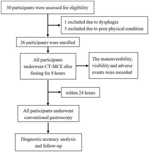
The participants underwent CT-MCE followed by conventional gastroscopy (GIF-H260; Olympus, Tokyo, Japan) within 24 h. CT-MCE was performed by an endoscopist who was well-trained and skilled in operating the CT-MCE system. Participants received intravenous anesthesia and underwent gastroscopy by another experienced endoscopist; biopsies were performed if necessary. The second endoscopist and participants were blinded to the results of the CT-MCE. Participants were evaluated 24 h after CT-MCE and gastroscopy: any discomfort or adverse events were recorded (Figure 3).
2.5 CT-MCE Examination
The upper gastrointestinal tract was prepared as described in a previous study [12]. Participants fasted for at least 8 h before the examination. To eliminate air bubbles and mucus within the stomach and to improve the visibility of the gastric mucosa, simethicone (Menarini Group, Florence, Italy) and Pronase granules (Beijing Tide Pharmaceutical Co. Ltd., Beijing, China) were administered 30 min before the examination. Immediately before the procedure, participants drank 500 mL of water to fill the gastric cavity; an additional 200–500 mL of water was consumed as needed during the examination based on the filling volume of the gastric cavity. The maximum volume of consumed water was limited to 1000 mL.
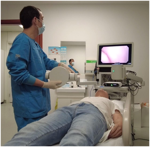
Each participant was seated and provided with a small amount of water to aid capsule ingestion. The entire esophagus and esophageal Z line were slowly examined under the combined action of esophageal peristalsis and cable traction. Once the CT-MCE capsule entered the stomach, the participant was turned to the supine position. The mechanical arm was used to manipulate the magnetic field and thus control the travel speed and camera orientation of the capsule endoscope. A systematic gastric examination was performed in the following order: cardia, fundus, body, angulus, antrum, and pylorus. During the examination, the participant was asked to change position as needed to optimize the visualization of the target area. Additional water intake was considered if gastric filling was inadequate. The duodenum examination followed once the stomach had been fully evaluated. The specific time spent on each segment was recorded. Detailed information was collected regarding the number, type, location, and size of any lesions; the maneuverability of the CT-MCE capsule, and the visibility of the digestive mucosa (Figure 4). The capsule was removed through the mouth using the cable after the examination was complete. The esophagus was observed a final time as the capsule ascended through it.
2.6 Study Outcomes
The primary study endpoints were the maneuverability of the CT-MCE capsule and the visibility of the upper gastrointestinal mucosa. The secondary endpoints included examination time, diagnostic accuracy, and safety.
Following the methodology outlined in a previous study [14], maneuverability was evaluated in the upper, middle, and lower esophagus and at the Z line; in the gastric cardia, fundus, body, angulus, antrum, and pylorus; and the duodenal bulb and descending duodenum. Maneuverability was classified as follows: good, the capsule endoscope could be precisely controlled to reach the target site for a detailed examination; moderate, the capsule endoscope could be controlled to reach the target site but without achieving a detailed examination; and poor, the capsule endoscope could not be controlled to reach the target site for observation.
The visibility of the mucosa was evaluated at the same points maneuverability was and was classified into four grades [14]: excellent, high visibility and 100% of the mucosa visible; good, moderate visibility and > 75% of the mucosa visible; fair, low visibility and 50%–75% of the mucosa visible; and poor, very low visibility and < 50% of the mucosa visible.
The sensitivity and specificity of CT-MCE for detecting esophageal, focal gastric, and duodenal lesions were evaluated. Conventional gastroscopy findings were used as the standard. Focal gastric lesions included polyps, erosions, ulcers, neoplasms, xanthomas, and diverticula. Diffuse lesions caused by gastritis and gastric mucosal atrophy were defined as negative because they are easily diagnosed using capsule endoscopy.
2.7 Safety and Adverse Events
Discomfort during CT-MCE was evaluated using a questionnaire. The level of discomfort was graded as mild, moderate, or severe. Adverse events within 24 h of CT-MCE completion were recorded.
2.8 Statistical Analysis
Continuous data are presented as means ± standard deviation or medians with range, as appropriate. Categorical data are presented as frequencies with percentage. Statistical analyses were performed using SPSS software version 25.0 (IBM Corp, Armonk, NY, USA).
3 Results
A total of 26 participants were enrolled in this study, including 19 asymptomatic volunteers and 7 patients with gastrointestinal symptoms (e.g., abdominal pain, abdominal distension, acid reflux, heartburn, and belching) (Figure 3). The cohort consisted of 12 males and 14 females, with a mean age of 46.69 ± 18.29 years (range: 25–74 years). All participants successfully ingested the CT-MCE and completed the study protocol. The mean duration of the CT-MCE examination was 3.77 ± 1.34 min for the esophagus, 23.27 ± 3.38 min for the stomach, and 4.58 ± 2.48 min for the duodenum. CT-MCE failed to pass through the pylorus and access the duodenum within the expected timeframe in two participants.
3.1 Maneuverability
As shown in Table S2, the maneuverability of CT-MCE was good in all segments of the esophagus (upper, middle, lower, and Z line). The maneuverability varied in different parts of the stomach, and the percentage of participants with good maneuverability was in the following order: cardia (100%), pylorus (96.15%), angulus (92.31%), antrum (88.46%), fundus (84.62%), and body (73.08%). The maneuverability in the duodenum was low, with 20.83% and 16.67% of participants showing good (75.00% and 83.33% showing moderate) controllability in the duodenal bulb and descending duodenum, respectively.
3.2 Visibility
The percentages of participants with excellent visibility were 88.46%, 73.08%, 76.92%, and 96.15% in the upper, middle, and lower esophagus, and Z line, respectively. The visibility varied considerably in different parts of the stomach, and the percentages of participants with excellent visibility were in the following order: pylorus (100.00%), antrum (96.15%), cardia (92.31%), angulus (92.31%), body (50.00%), and fundus (42.31%). Additionally, 87.50% of participants showed excellent visibility in the duodenal bulb, and 62.50% showed excellent visibility in the descending duodenum (Figure 5).
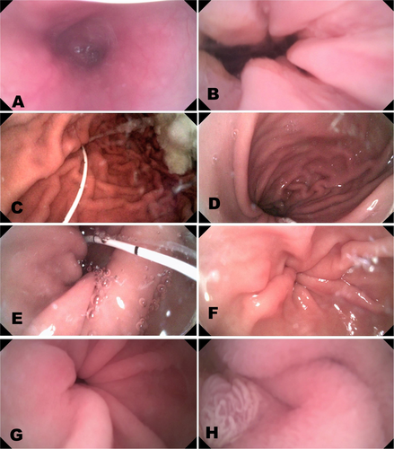
3.3 Diagnostic Accuracy
As shown in Table S3, 41 lesions were detected in the upper gastrointestinal examinations using CT-MCE and conventional gastroscopy (Figure 6). Among these, 34 lesions were detected by both CT-MCE and conventional gastroscopy, six lesions were detected exclusively by conventional gastroscopy, and one lesion was detected exclusively by MCE. Using the findings of conventional gastroscopy as the standard, the sensitivity and specificity of CT-MCE for diagnosing upper gastrointestinal lesions were 85.00% (34/40) and 98.15% (53/54), respectively (Table S4). In the examination of esophageal lesions, CT-MCE failed to detect an ectopic gastric mucosa at the entrance of the esophagus, and a participant was misdiagnosed with Candida esophagitis in which the esophagus presented no abnormality upon using conventional gastroscopy. The sensitivity and specificity of CT-MCE for the diagnosis of esophageal lesions were 83.33% (5/6) and 95.24% (20/21), respectively. In the examination of focal gastric lesions, CT-MCE missed three polyps and did not detect additional lesions in contrast to conventional gastroscopy. CT-MCE showed 88.00% (22/25) sensitivity and 100.00% (13/13) specificity for diagnosis of focal gastric lesions. In the examination of duodenal lesions, CT-MCE missed one polyp and one erosive lesion and did not detect additional lesions in contrast to conventional gastroscopy. CT-MCE showed 77.78% (7/9) sensitivity and 100.00% (20/20) specificity for diagnosis of duodenal lesions.
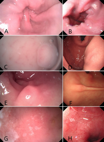
3.4 Safety
All participants successfully underwent both CT-MCE examination and conventional electronic gastroscopy. A total of 15 (57.7%) participants reported discomfort during the CT-MCE examination, including sensation of a foreign body in the pharynx (11 cases), nausea (6 cases), abdominal distension (1 case), sore throat (1 case), and heartburn (1 case). The majority of discomfort symptoms were mild, and all symptoms rapidly subsided following exam completion (Table S5). No serious adverse events were reported throughout the study. The capsules were successfully removed from the mouth after examination.
4 Discussion
The innovative CT-MCE system offers several advantages, including small size (4.5 × 25.0 mm), high resolution (1280 × 720 pixels), high frame rate (30/second), and easy removal. In our study, high maneuverability and visibility were achieved during esophageal and gastric examinations. Using the findings of conventional gastroscopy as the standard, the sensitivity and specificity of CT-MCE in diagnosing upper gastrointestinal lesions reached 85.00% and 98.15%, respectively. All participants successfully ingested the capsule and underwent esophageal and gastric examinations. Upon completion of the procedure, the capsule was removed using a cable through the mouth. CT-MCE showed high safety—there were no capsule detachments or other serious adverse events. Fifteen participants experienced discomfort during CT-MCE and all instances resolved spontaneously. In contrast to the wireless MCE capsule currently in clinical use, the CT-MCE capsule can be removed from the mouth after examination. Therefore, CT-MCE is associated with a low risk of capsule retention and can be safely used for patients with gastrointestinal stenosis or obstruction.
Owing to the influences of both gravitational force and esophageal peristalsis, conventional wireless capsules pass through the esophagus at a high speed, which prevents a thorough examination of the esophageal mucosa. To address this challenge and improve the detection rate of esophageal lesions, Eliakim et al. [15] introduced the dual-camera esophageal capsule endoscope “PillCam ESO” in 2004, which increased visualization of the esophageal mucosa. After other advancements, the third-generation “PillCam ESO 3,” with a frame rate of 35/second, was validated for esophageal examination in 2016 [16]. Although these esophageal capsule endoscopes have increased the accuracy of esophageal examination by increasing the frame rate, their movement cannot be manipulated within the esophagus for detailed examination of suspicious lesions. Chen et al. modified the NaviCam MCE system (Ankon Technologies, Shanghai, China) by incorporating a detachable string for repeated observations of the esophagus [13]. However, owing to constraints imposed by the built-in battery, it has a low frame rate (2/second) and low image resolution (480 × 480 pixels), which decrease its ability to detect esophageal lesions.
The CT-MCE system used in this study is powered by a cable, overcoming the limitations posed by the battery. It achieved a high frame rate (30/second) and high resolution (1280 × 720 pixels) and was controlled via the cable as it traveled through the esophagus. Furthermore, the capsule can be suspended in the esophagus to repeatedly observe any region of interest, and good maneuverability and visibility were achieved in various parts of the esophagus.
The CT-MCE system missed ectopic gastric mucosa at the entrance of the esophagus. This was attributed to the contraction of the upper esophageal sphincter during capsule passage through the cervical esophagus and the inability of the system to inflate and expand the esophageal mucosa. Similarly, owing to the presence of a white, cheese-like substance on the esophageal mucosa, one participant was misdiagnosed with Candida esophagitis. Subsequent conventional gastroscopy revealed that the substance was food residue, and the appearance of the esophageal mucosa returned to normal after rising. This misdiagnosis was attributed to the lack of mucosa rinsing during the examination.
Overall, CT-MCE showed high sensitivity and specificity for the diagnosis of esophageal lesions. The system appears to be a valuable tool for examining esophageal lesions and monitoring esophageal varices, esophageal reflux, and Barrett's esophagus. In addition, it has the potential to serve as a viable alternative for patients averse to or unable to undergo conventional gastroscopy. Further study is warranted.
MCE is effective for diagnosing focal gastric lesions [12, 17, 18]. In our study, CT-MCE showed good maneuverability and visibility in the stomach. Similar to previous reports [14, 19], visibility was significantly better in the pylorus, antrum, cardia, and angulus than in the fundus and body. This divergence can be explained by the suboptimal expansion of the gastric body and the accumulation of gastric mucus within the fundus and body. The visibility of these areas can be improved by changing the position of the patient, having the patient drink additional water during the examination, and administering mucolytic agents. Notably, the cable stimulated the pharynx and increased saliva secretion. The influx of saliva into the upper gastrointestinal tract could further impede stomach visibility. Based on our experience, patients can be instructed to spit rather than ingest the saliva secreted during the examination, effectively improving gastric visibility.
CT-MCE detected 22 of 25 focal gastric lesions, yielding a sensitivity of 88.00% and a specificity of 100.00%. However, it missed one polyp in the gastric body and two in the gastric fundus, which may be attributed to the poor visibility of CT-MCE in these regions. Overall, the sensitivity and specificity of CT-MCE for diagnosis of focal gastric lesions were similar to those reported in large-sample, multicenter clinical studies [12]. These results preliminarily demonstrate the feasibility and effectiveness of CT-MCE for the diagnosis of gastric lesions.
The CT-MCE capsule failed to pass through the pylorus into the duodenum within the expected timeframe of 30 min from the start of the procedure in two patients. The pylorus passage rate was 92.31%, which is higher than those reported in previous studies of MCE [17, 19]. This may be attributed to the smaller diameter of the CT-MCE capsule (4.5 mm). Jiang et al. [20] showed that a small MCE capsule (9.5 × 24.5 mm) traveled more rapidly through the pylorus into the duodenum than a standard-sized one (11.8 × 27 mm); the average time for a small MCE to reach and pass through the pylorus was 38.8 min. Consequently, we speculate that extending the examination time beyond the 30 min we allowed may lead to an increase in the pylorus passage rate.
Within the duodenum, the controllability of the CT-MCE was suboptimal. Controllability was good in the duodenal bulb and the descending duodenum in only 20.83% and 16.67% of cases, respectively. This can be attributed to the constrained space in the duodenal bulb and the rapid peristalsis of the descending duodenum. Furthermore, the extended cable length in the upper gastrointestinal tract could contribute to the low maneuverability in the duodenum.
This study has several limitations. First, semiquantitative grading systems were used to evaluate maneuverability and visibility, which may have introduced subjectivity into the evaluation processes. Second, stimulation of the pharynx by the cable during examination led to increased saliva secretion and ingestion, reducing visibility. Addressing this concern warrants refinement of the cable material and optimization of the upper gastrointestinal tract preparation protocol. Third, the sample size was small, and some participants were asymptomatic healthy volunteers, leading to a limited number of lesions detected in the upper gastrointestinal tract. Controlled clinical studies involving larger sample sizes are needed to validate the effectiveness of CT-MCE in the diagnosis of upper gastrointestinal lesions.
In summary, this study was the first self-controlled clinical investigation of a novel CT-MCE method. The CT-MCE system showed remarkable feasibility and safety for upper gastrointestinal examination, as well as high patient tolerance and no risk of capsule retention. This study provides preliminary evidence for the effectiveness of CT-MCE in examining the upper gastrointestinal tract, especially the esophagus and stomach. We will further validate the accuracy of CT-MCE in the diagnosis of upper gastrointestinal lesions in larger controlled clinical studies.
5 Conclusion
In conclusion, this study was the first self-controlled clinical investigation of a novel CT-MCE method. The CT-MCE system showed remarkable feasibility and safety for upper gastrointestinal examination, as well as high patient tolerance and no risk of capsule retention. This study provides preliminary evidence for the effectiveness of CT-MCE in examining the upper gastrointestinal tract, especially the esophagus and stomach. We will further validate the accuracy of CT-MCE in the diagnosis of upper gastrointestinal lesions in larger controlled clinical studies.
Author Contributions
Ke Meng: data curation (equal), investigation (equal), methodology (equal), writing – original draft (equal). Yan Li: data curation (equal), formal analysis (equal), methodology (equal), software (equal). Bin Yan: data curation (equal), formal analysis (equal), methodology (equal), software (equal). Fei Pan: conceptualization (equal), data curation (equal), formal analysis (equal), resources (equal), software (equal), validation (equal). Jingshuang Yan: conceptualization (equal), data curation (equal), investigation (equal), resources (equal), software (equal). Guanzhou Zhou: conceptualization (equal), data curation (equal), resources (equal), software (equal), supervision (equal). Haixu Chen: conceptualization (equal), funding acquisition (lead), project administration (equal), supervision (lead), writing – review and editing (lead). Xiaomei Zhang: conceptualization (lead), investigation (lead), investigation (equal), project administration (lead), supervision (lead), writing – review and editing (equal).
Acknowledgments
This work was supported by the National Key Research and Development Program (2019YFC0120303) and was sponsored by the Beijing Nova Program (20240484678).
Ethics Statement
The study protocol was approved by the institutional review board of Chinese PLA General Hospital (S2022-188-01) and was registered at chictr.org.cn (ChiCTR2200060597).
Consent
Written informed consent was provided by all participants.
Conflicts of Interest
The authors declare no conflicts of interest.
Open Research
Data Availability Statement
A peer review of empirical data will be conducted to confirm that the data reproduce the analytic results reported in the paper. The data that support the findings of this study are available on request from the corresponding author. The data are not publicly available due to privacy or ethical restrictions.


