Early-onset scoliosis in children aged 4–7 years in Nanjing, China: A cross-sectional study
Abstract
Background
This study aimed to investigate the potential variance in the prevalence of early-onset scoliosis among children aged 4–7 years and analyze the influencing factors. The goal was to establish a crucial reference point for monitoring and evaluating spinal curvature development in preschoolers, ultimately to reduce the occurrence of adverse health outcomes.
Methods
Children aged 4–7 years within the main urban area of Nanjing were selected using a stratified random sampling method. A team of four senior therapists conducted screenings for spinal curvature among children using visual inspection, the Adams forward bending test, and an electronic scoliometer to measure the angle of trunk rotation (ATR) and identify children displaying signs of scoliosis. Children with suspected scoliosis in the initial screening underwent X-ray Cobb angle assessment for confirmation. The prevalence of early-onset scoliosis was then determined from the screening results. R version 4.2.0 software was used to analyze the factors associated with scoliosis among children using partial least squares structural equation modeling.
Results
A total of 2281 children were included in this study, consisting of 1211 boys and 1070 girls, with a mean age of 5.44 ± 0.81 years (ranging from 4 to 7 years). Among them, 7.58% exhibited positive signs of scoliosis, 5.87% had early-onset scoliosis, and the positive predictive value was 77.5%. Significant differences in ATR were observed among children in different age groups (Kruskal–Wallis = 15, p = 0.0104) and by sex (t = 3.17, p = 0.00153). Significant variations in ATR were noted in children with scoliosis (t = −22.7, p < 0.001), with a cutoff at ATR = 4.5°, and auxiliary values of 0.947 and 0.990. Children diagnosed with early-onset scoliosis generally exhibited lower body mass index values, with a statistically significant difference (t = 2.99, p = 0.003).
Conclusions
Using visual inspection, the Adams test, and an electronic scoliometer to measure the ATR, the present triad method is more sensitive for early scoliosis screening in children with abnormal posture aged 4–7 years. A full spine X-ray is advised in children with an ATR over 4.5° and poor posture.
Abbreviations
-
- Adj R2
-
- adjusted R2 values
-
- ATR
-
- angle of trunk rotation
-
- AUC
-
- area under the ROC curve
-
- BMI
-
- body mass index
-
- LM
-
- linear model
-
- MAE
-
- mean absolute error
-
- PLS
-
- partial least squares
-
- PLS-SEM
-
- partial least squares structural equation modeling
-
- R2
-
- coefficient of determination
-
- RMSE
-
- root mean square error
-
- ROC
-
- receiver operating characteristic
-
- SRS
-
- Scoliosis Research Society
1 INTRODUCTION
Scoliosis is the most common three-dimensional deformity of the spine, characterized by a sideways curvature of one or more spinal segments from the body's midline in the coronal plane. This results in a spinal deformity with curvature, typically accompanied by rotational deformity and changes in the anterior or posterior protrusion within the sagittal plane [1]. The Scoliosis Research Society (SRS) defines scoliosis as a condition where the curvature of the spine, measured using the Cobb method on standing anteroposterior X-rays, is 10° or greater [2].
Scoliosis can be categorized into various types based on etiology: neuromuscular, idiopathic, congenital, and other causes [3]. Early-onset scoliosis, which occurs before the age of 10 years, encompasses idiopathic, congenital, neuromuscular, and other spinal deformities associated with scoliosis [4]. Without timely treatment, children may develop postural issues such as uneven shoulder height, limb length discrepancy, and trunk deformity [5]. In severe cases, affected individuals may experience pain, impaired cardiopulmonary function, and spinal cord compression [6]. Prolonged abnormal posture can also lead to psychological issues, reducing social functioning and quality of life in children [7].
In China, the incidence of scoliosis has been on the rise, with an average detection rate of 4.40% and a diagnostic rate of 1.23% among primary and secondary school students in the mainland [8]. According to China's Clinical Practice Guidelines and Pathway Guidelines for Adolescent Scoliosis Screening, screening for scoliosis is more feasible in infants and adolescents than in adults, where the condition is more complex and challenging to manage [9]. Timely detection can effectively prevent disease progression. However, current scoliosis screening primarily targets adolescents aged 10–18 years in primary and secondary school [10-12], with less emphasis on monitoring the early spinal growth and development of preschool children aged 3–6 years.
Although studies indicate that over 80% of cases of early-onset scoliosis tend to resolve spontaneously with growth and development, significant progression of the disease is still experienced in the remaining 20% of cases, necessitating long-term and complex treatment [13, 14]. Therefore, early screening of early-onset scoliosis is particularly important.
In this study, we carried out screening of early-onset scoliosis in preschool children using visual inspection, the Adams forward flexion test, and quantitative measurement of ATR with an electronic scoliometer. We aimed to fill gaps in the research on scoliosis among preschool children in China. Our study will provide an important reference index for monitoring and evaluation of the development of spinal curvature in preschool children. We explored potential differences in the incidence of early-onset scoliosis among children aged 4–7 years and analyzed influencing factors. This will help in the early detection and early correction of scoliosis, enhancing awareness among adolescents and parents regarding the disease, ensuring timely and appropriate treatment, and ultimately reducing the occurrence of adverse health outcomes.
2 MATERIALS AND METHODS
2.1 Study design and sampling
This cross-sectional study, conducted in Nanjing from May 2021 to June 2022, included a survey of the prevalence of early-onset scoliosis in children and a preliminary exploration of the factors affecting scoliosis. Stratified random sampling was used in this study. The study was approved by the Ethics Committee of Nanjing Women and Children's Healthcare Hospital in 2021, with ethics approval number [2021]YL-013.
2.2 Participants
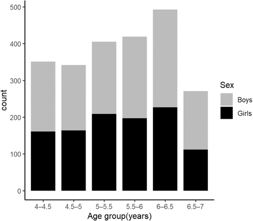
Three kindergartens were randomly selected from four main urban districts: Xuanwu District, Qinhuai District, Jianye District, and Gulou District. A comprehensive list of children aged 4–7 years was compiled from each participating kindergarten. Participating children were categorized into six age groups, as follows: (4.0–4.5), (4.5–5.0), (5.0–5.5), (5.5–6.0), (6.0–6.5), and (6.5–7.0) years. Stratified sampling was used as follows: 4 (districts) × 3 (kindergartens) × 6 (age groups) × 2 (sexes) × n (minimum sample); n children were assessed in each kindergarten and each age group. Based on the prevalence rates of scoliosis in children from international literature, which varied between 2% and 5% [15], we set our error level at 0.05 and an allowable error (Z) of 0.2p (prevalence). Using these parameters, we estimated the required sample size and determined a minimum of approximately 865 participants.
The inclusion criteria were healthy children with normal exercise capacity, whose guardians provided their informed consent. Children were excluded if they had neurological or motor system diseases or serious social and cognitive communication disorders. Exclusion was determined by reviewing each child's prior physical examination and health records.
2.3 Screening protocol
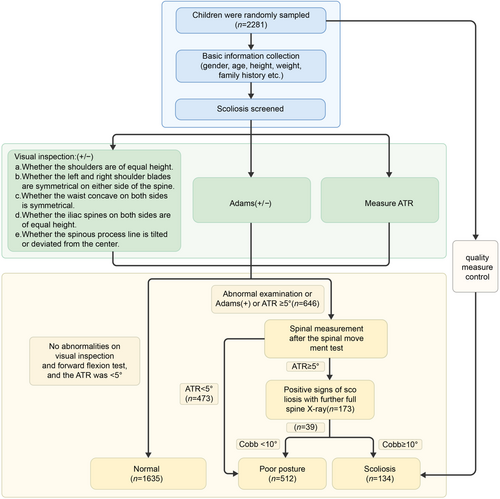
Physical examination methods for scoliosis, including visual inspection and the forward flexion test, are among the most accessible screening modalities in clinical practice, requiring no specialized tools [16]. To enhance the accuracy of the screening process, we used visual measurement, the Adams forward flexion test, and quantitative assessment of the children's trunk inclination angle.
Visual inspection is performed by having the child stand naturally with a bare back, feet and shoulders at equal width, eyes level, and upper limbs in a neutral position. Any trunk asymmetry, identified as one or more abnormalities, is noted [17].
The Adams forward flexion test involves the examinee standing with knees extended and palms together, then bending forward. This position allows the examiner to assess the upper thoracic, thoracic, thoracolumbar, and lumbar segments from the posterior midline for any height discrepancies and asymmetry. The presence of these signs is indicative of a positive result for scoliosis screening [18].
- a)
Mode selection: On the “Change mode” interface, the Start/End button is briefly pressed to navigate to the Trunk Angle screen.
- b)
The examinee's posture is as follows:
-
Stand with feet positioned 5 to 8 cm apart.
-
Extend arms forward, palms facing each other.
-
Lean the torso forward by 90°, keeping the head down, knees straight, and arms relaxed with palms folded.
-
Ensure shoulders are at or below hip level.
-
Gently guide the patient's folded palms downward to achieve full flexion.
-
- c)
The measurement procedure is as follows:
-
Position the examiner behind the examinee.
-
Place the SpineScan electronic scoliometer on the examinee's shoulders, aligning the semicircular notches with the spinous processes of the spine, ensuring both notches contact the patient's back.
-
Without applying pressure, rest the SpineScan on the skin and the examiner slide it along the examinee's spinal process line, from the C7 vertebra to the L4 vertebra.
-
Upon completion, the SpineScan will display the maximum ATR value detected.
-
If the initial examination is abnormal, the forward flexion test is positive, or the ATR is ≥5°, a spinal movement test should be performed. This involves the child performing two cycles each of the following: spinal forward flexion, back extension, lateral bending to the left and right, and axial rotation to the left and right, followed by resuming a natural standing position. Subsequently, the examiner conducts a repeat assessment using a trunk rotation measuring instrument [9]. When the ATR value is ≥5°, a standing radiograph of the entire spine is obtained. The Cobb angle is measured, and scoliosis is diagnosed if the Cobb angle is ≥10° [20].
2.4 Statistical methods
Data management and entry were conducted by designated personnel, with sensitive information encoded for confidentiality purposes. Data were entered into a database using EpiData software. Subsequent analyses were performed using R software version 4.2.0 (The R Project for Statistical Computinga), encompassing general statistical descriptions. Continuous variables exhibiting a normal distribution are reported as mean ± standard deviation, whereas non-normally distributed continuous variables are reported as median and interquartile range. Categorical variables are expressed in terms of frequency and percentage. Group differences for continuous variables were evaluated using independent samples t-tests for parametric data and corresponding nonparametric tests for nonparametric data. Categorical data were compared using Pearson's chi-squared tests or Fisher's exact tests, as appropriate. Statistical significance was defined as a p value < 0.05.
Partial least squares structural equation modeling (PLS-SEM) was used to investigate the determinants of scoliosis in children. The one-way arrows within the model denote directional relationships. These unidirectional arrows, when underpinned by robust theoretical foundations, can be interpreted as indicative of causality. These signify the predictive influence of a measured variable on a latent variable or between latent variables. The analysis was conducted with a two-sided significance level set at α = 0.05 [21].
2.5 Quality control
- a)
Equipment management: Designated personnel oversaw all equipment maintenance and storage, with a dedicated room provided by the department.
- b)
Software management: The project team was in charge of software maintenance, ensuring daily inspection data were backed up on a designated hard disk.
- c)
Inspector training: The project leader provided standardized training before usage. By ‘Inspector,’ we refer to the qualified individuals responsible for conducting the assessments and evaluations as part of our study's methodology. These inspectors are trained professionals who perform the physical examinations and collect data according to the established protocols. Regarding “usage,” we are specifically referring to the utilization of the screening tools and techniques within the context of the scoliosis examination process. This includes the application of visual inspection methods, the Adams forward flexion test, and the electronic scoliometer for measuring the angle of trunk rotation.
- d)
Operational protocol: A standardized inspection process was established, with all inspectors adhering to it under the supervision of the deputy chief physician.
- e)
Data management: Project team members handled the data collection and organization. Data entry and quality control were managed by team members with a background in medical statistics, followed by statistical analysis.
3 RESULTS
3.1 Summary of scoliosis screening
In this study, 2281 children were surveyed, comprising 1211 boys and 1070 girls, with a mean age of 5.44 ± 0.81 years (range: 4–7 years old). During screening, 173 children (7.58%) exhibited positive signs of scoliosis. Additionally, 591 children with poor posture were screened. Following full spinal X-ray reassessment, 134 of these children were diagnosed with scoliosis, including 73 boys and 61 girls (Figure 2).
3.2 Prevalence of early-onset scoliosis
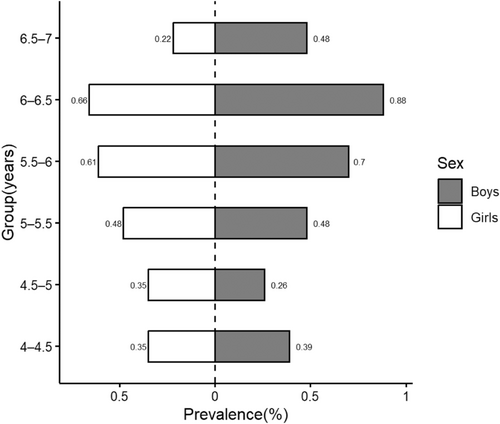
The overall prevalence of early-onset scoliosis was 5.87%, with a rate of 3.20% among boys and 2.67% among girls (Figure 3). There was no significant difference in prevalence between sex (χ2 = 0.0587, p = 0.808). Analysis of scoliosis detection rates across different age groups and sexes revealed that children in the age group 6–6.5 years exhibited a higher prevalence than those in other age groups, with a significant difference (χ2 = 34.3, p = 0.00311). Detailed data are shown in Tables 1 and 2.
| Normal | Scoliosis | ||
|---|---|---|---|
| N = 2147 (%) | N = 134 (%) | p Value | |
| Sex | χ2 = 0.0587, p = 0.808 | ||
| Boys | 1138 (53.0) | 73 (54.5) | |
| Girls | 1009 (47.0) | 61 (45.5) |
| Normal | Scoliosis | ||||
|---|---|---|---|---|---|
| Boys | Girls | Boys | Girls | ||
| N = 1138 (%) | N = 1009 (%) | N = 73 (%) | N = 61 (%) | p Value | |
| Age (years) | χ2 = 13.9, p = 0.535 | ||||
| 4–4.5 | 181 (15.9) | 153 (15.2) | 9 (12.3) | 8 (13.1) | |
| 4.5–5 | 172 (15.1) | 156 (15.5) | 6 (8.2) | 8 (13.1) | |
| 5–5.5 | 185 (16.3) | 198 (19.6) | 11 (15.1) | 11 (18.0) | |
| 5.5–6 | 206 (18.1) | 183 (18.1) | 16 (21.9) | 14 (23.0) | |
| 6–6.5 | 246 (21.6) | 212 (21.0) | 20 (27.4) | 15 (24.6) | |
| 6.5–7 | 148 (13.0) | 107 (10.6) | 11 (15.1) | 5 (8.2) | |
3.3 Distribution of ATR, BMI, age, and Cobb angle in children with early-onset scoliosis
Compared to their normal counterparts, children with scoliosis had significantly higher ATR, older age, and larger Cobb angle measurements. In contrast, body mass index (BMI) was generally lower in children with scoliosis, with all observed differences being statistically significant (p < 0.05). These findings are detailed in Table 3.
| Normal | Scoliosis | ||
|---|---|---|---|
| (N = 2147) | (N = 134) | p Value | |
| ATR | t = −38.4, p < 0.001 | ||
| Mean (SD) | 2.45 (1.11) | 5.39 (0.840) | |
| BMI | t = 2.99, p = 0.00324 | ||
| Mean (SD) | 16.5 (1.91) | 15.9 (2.22) | |
| Age | t = −2.02, p = 0.0452 | ||
| Mean (SD) | 5.44 (0.812) | 5.58 (0.780) | |
| Cobb | t = −17, p < 0.001 | ||
| Mean (SD) | 5.34 (2.13) | 11.5 (1.35) |
- Abbreviations: ATR, angle of trunk rotation; BMI, body mass index.
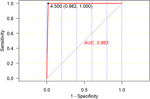
A statistically significant difference in ATR was observed between normal children and those with scoliosis (t = 22.7, p < 0.001). Receiver operating characteristic (ROC) curve analysis was conducted. With an ATR of 4.5°, the model yielded a cutoff value of 0.982 and an area under the ROC curve (AUC) of 0.993 (Figure 4).
3.4 Distribution of ATR among groups of children by age
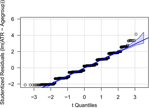
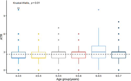
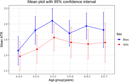
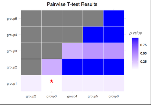
Analysis of the measured ATR showed that the ATR was normally distributed among children of different ages and the difference was statistically significant (Kruskal–Wallis H = 15, p = 0.0104) (Figures 5 and 6). The ATR increased with age from 4 to 5.5 years, then decreased after 5.5 years, and appeared to stabilize by 6 years of age (Figure 7). Based on this, there was a statistically significant difference between groups 1 and 3 (p < 0.05); however, there was no significant difference in ATR between the other groups (p > 0.05) (Figure 8).
3.5 Angle distribution of ATR by age group and sex
A statistically significant difference in ATR was observed between boys and girls aged 4–7 years (t = 3.17, p = 0.00153), as detailed in Table 4. However, although a statistically significant difference in ATR was found among boys of different ages (p = 0.014), no such significance was detected among girls (p = 0.58) (Figure 9).
| Boys | Girls | ||
|---|---|---|---|
| (N = 1211) | (N = 1070) | p Value | |
| ATR | t = 3.17, p = 0.00153 | ||
| Mean (SD) | 2.70 (1.29) | 2.53 (1.31) | |
| Median [Min, Max] | 3.00 [0, 10.0] | 2.00 [0, 10.0] |
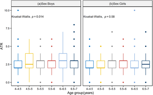
3.6 Partial least squares structural equation modeling
The results showed significant pathways: from ATR to scoliosis (β = 0.507, p < 0.001), from Cobb angle to scoliosis (β = 0.383, p < 0.001), and a mediated pathway from BMI to ATR, to poor posture, and finally to scoliosis (β = −0.069 for BMI → ATR, β = 0.098 for ATR→poor posture, β = 0.06 for poor posture→scoliosis, all p < 0.05) (Figure 10). Our results showed that the endogenous construct had an R² value of [0.507], which is considered to be moderately explanatory (Table 5).
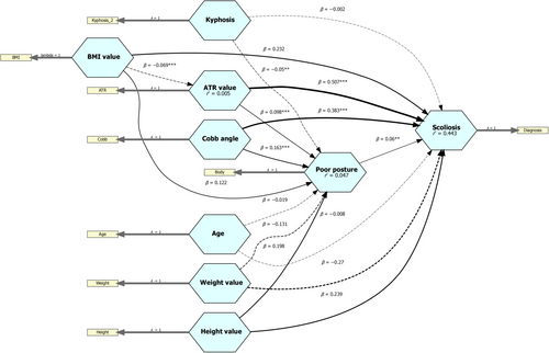
| Poor posture | Scoliosis | ATR value | |
|---|---|---|---|
| R2 | 0.047 | 0.443 | 0.005 |
| Adj R2 | 0.044 | 0.442 | 0.004 |
| Age | −0.019 | −0.008 | |
| Kyphosis | −0.050 | −0.002 | |
| ATR value | 0.098 | 0.507 | |
| Cobb angle | 0.163 | 0.383 | |
| BMI value | 0.122 | 0.232 | −0.069 |
| Height value | 0.198 | 0.239 | |
| Weight value | −0.131 | −0.270 | |
| Poor posture | 0.060 |
- Abbreviations: Adj R2, adjusted R2 values; ATR, angle of trunk rotation; BMI, body mass index; R2, the coefficient of determination.
The R2 (denoted as r2 in Figure 10 for abbreviation) value indicates the degree to which an endogenous structure (node indicated by arrow) interprets the variance of the input data. β indicates how much the data held by the node changes by one unit of data that refers to the endogenous structure, which is equivalent to the regression coefficient. The asterisk of the connecting line refers to the significance of the coefficient obtained using the bootstrap test, where the lower limit of the confidence interval of 5% confidence interval is >0, which means that the coefficient is significant at the 5% level.
4 DISCUSSION
The overall prevalence of early-onset scoliosis in this study was 5.87%, with rates of 3.20% and 2.67% observed in boys and girls, respectively. The positive predictive value for the present triad screening method was 77.5%. Both boys and girls aged 6–6.5 years were found to have a higher prevalence of scoliosis than other age groups. This finding suggests that age 6–6.5 years may be a critical period for monitoring children's spinal health.
In scoliosis screening, the ATR is a critical indicator, with values exceeding 5° being potentially indicative of the condition, as defined by the SRS [22]. However, traditional ATR measurement techniques are prone to error. Consequently, digital-based ATR measurement methods using three-dimensional reconstruction have been developed to enhance measurement precision [23]. The ROC curve is a pivotal tool for assessing classification model performance, with the false positive rate plotted on the horizontal axis and the true positive rate on the vertical axis [24]. A larger AUC signifies superior model performance. In this study, we used an electronic measuring instrument (scoliometer) and achieved an AUC of 0.993 at an ATR threshold of 4.5° [25] (Figure 4). Hence, for early scoliosis screening in preschool children, the ATR serves as a significant predictor, and measurements above 4.5° should be further evaluated alongside clinical assessment and imaging studies to ascertain the presence of scoliosis.
A PLS path model was selected for its effectiveness in handling complex relationships and robustness with small to moderate sample sizes, which are fitting for our study's aims. The model comprises two primary components: the structural model, also known as the inner model within PLS-SEM, which interconnects the constructs and illustrates the relationships (paths) between them; and the measurement models, or outer models, which depict the relationships between the constructs and their corresponding indicator variables [26].
In this study, PLS-SEM was used to analyze the effect of the ATR, Cobb angle, BMI, age, height, and weight on preschool children, thereby distinguishing scoliosis from poor posture [27]. Additionally, we observed that a lower BMI is associated with a larger ATR, thereby potentially increasing the risk of scoliosis [28].
To address concerns regarding the model's fit indices, we acknowledge that PLS-SEM does not rely on the same fit indices as covariance-based structural equation modeling [29]. Instead, we focused on the model's explanatory and predictive power. R² for our endogenous construct was 0.506, indicating moderate explanatory power (Table 5). We also conducted a comparison of our PLS-SEM model with alternative statistical methods, such as linear regression models, to ensure that the most appropriate analytical approach was used [30].
Our comparison indicated that the PLS-SEM model demonstrated lower prediction error across all indicators, suggesting superior predictive performance. For instance, the mean absolute error values for the PLS-SEM model were consistently lower than those for the linear regression models, indicating higher predictive accuracy (Table 6) [30]. This comparison, along with the guidelines provided by Shmueli et al. [29], affirms the suitability of the PLS-SEM model for our research and its strong predictive capabilities.
| Items | RMSE | MAE |
|---|---|---|
| LM_in_sample.Body | 0.441 | 0.363 |
| LM_in_sample.Diagnosis | 0.175 | 0.115 |
| LM_in_sample.ATR | 1.295 | 1.042 |
| LM_out_of_sample.Body | 0.443 | 0.365 |
| LM_out_of_sample.Diagnosis | 0.177 | 0.116 |
| LM_out_of_sample.ATR | 1.297 | 1.044 |
| PLS_in_sample.Body | 0.440 | 0.360 |
| PLS_in_sample.Diagnosis | 0.175 | 0.115 |
| PLS_in_sample.ATR | 1.059 | 0.874 |
| PLS_out_of_sample.Body | 0.442 | 0.362 |
| PLS_out_of_sample.Diagnosis | 0.177 | 0.116 |
| PLS_out_of_sample.ATR | 1.063 | 0.877 |
- Abbreviations: LM, linear model; MAE, mean absolute error; PLS, partial least squares; RMSE, root mean square error.
One limitation of this study is the inability to perform radiological confirmation on all participants to prevent unnecessary exposure to radiation. Secondly, the prevalence of scoliosis was assessed over a single year (2023), and the annual prevalence may fluctuate. Consequently, the reported prevalence reflects the accuracy of screening rather than the actual incidence. To address this issue, the study will be followed by a questionnaire survey to analyze the factors that affect scoliosis, including sex, age, BMI, children's posture management, and parents’ perceptions of children's spinal health.
5 CONCLUSIONS
The overall prevalence of early-onset scoliosis among preschool children in Nanjing, China, was 5.87%, with a rate of 3.20% and 2.67% for boys and girls, respectively. Analysis using the PLS-SEM model revealed that children's BMI and height were independently correlated with the prevalence of the disease, indicating that children with lower BMI are at a higher risk of developing scoliosis. The triad of visual inspection, the Adams forward flexion test, and quantitative assessment of the child's trunk offer a high degree of accuracy and can be readily applied in a short time to assess the risk of scoliosis. When the ATR reaches 4.5° in children, regular follow-up of their spinal growth and development is recommended.
Early screening enables the timely detection of scoliosis, allowing for scientific management and treatment to enhance spinal deformity care. Our study serves as a crucial reference for monitoring spinal curvature development in preschool children, revealing potential differences in early-onset scoliosis incidence among 4- to 7-year-olds, as well as contributing factors. This approach can aid in the early detection and correction of scoliosis, as well as raise awareness among adolescents and parents to ensure prompt and appropriate treatment and minimize adverse health outcomes.
AUTHOR CONTRIBUTIONS
Jun Song: Conceptualization (lead); writing—original draft (lead); formal analysis (lead); writing—review and editing (lead). Hong-xin Rui, Ya-chun Xie, Yan Wang, Ting Li: Data curation; validation. Xia Chi, Mei-lin Tong: Conceptualization; project administration. Feng Lin: Supervised the entire study, contributed to the research idea and data analysis, designed the protocol, and revised the final manuscript. All authors have read and agreed to the published version of the manuscript.
ACKNOWLEDGMENTS
The authors acknowledge all the families for their participation in this study and the medical staff of the Nanjing Women and Children's Healthcare Hospital.
CONFLICT OF INTEREST STATEMENT
The authors declare no conflict of interest.
ETHICS STATEMENT
The research screening project was approved by the Ethics Committee of Nanjing Women and Children's Healthcare Hospital in 2021, with the ethics approval number [2021]YL-013.
INFORMED CONSENT
Written informed consent was obtained from the parents or legal guardians of all minors included in the study.
Open Research
DATA AVAILABILITY STATEMENT
The data that support the findings of this study are available from the corresponding author upon reasonable request.


