COVER IMAGE
Free Access
Cover Image, Volume 73, Issue 1
First published: 20 December 2024
Graphical Abstract
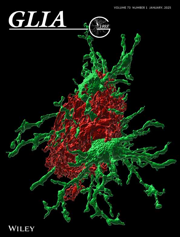
Cover Illustration: 3D IMARIS render of Airyscan confocal z stack image showing the GFP microglia (green) processes diving into the diffuse amyloid plaque shell stained with anti-Aβ antibody D54D2 (red). Activated microglia interacts here with the plaque in two ways: with soma going deep into the plaque, and with thin processes which are intertwined in looser amyloid layers. (See Gotkiewicz, M., et al, https://doi.org/10.1002/glia.24628)