Robust genital gag-specific CD8+ T-cell responses in mice upon intramuscular immunization with simian adenoviral vectors expressing HIV-1-gag
Abstract
Most studies on E1-deleted adenovirus (Ad) vectors as vaccine carriers for antigens of HIV-1 have focused on induction of central immune responses, although stimulation of mucosal immunity at the genital tract (GT), the primary port of entry of HIV-1, would also be highly desirable. In this study, different immunization protocols using chimpanzee-derived adenoviral (AdC) vectors expressing Gag of HIV-1 clade B given in heterologous prime-boost regimens were tested for induction of systemic and genital immune responses. Although i.n. immunization stimulated CD8+ T-cell responses that could be detected in the GT, this route induced only marginal cellular responses in systemic tissues and furthermore numbers of Gag-specific CD8+ T cells contracted sharply within a few weeks. On the contrary, i.m. immunization induced higher and more sustained frequencies of vaccine-induced cells which could be detected in the GT as well as systemic compartments. Antigen-specific CD8+ T cells could be detected 1 year after immunization in all compartments analyzed. Genital memory cells secreted IFN-γ, expressed high levels of CD103 and their phenotypes were consistent with a state of activation. Taken together, the results presented here show that i.m. vaccination with chimpanzee-derived (simian) adenovirus vectors is a suitable strategy to induce a long-lived genital CD8+ T-cell response.
Introduction
The efficacy of most vaccines is linked to their ability to induce neutralizing Ab (NAb). This approach has thus far been elusive for vaccines to HIV-1 as the envelope proteins of this virus are heavily glycosylated 1, variable between isolates 2, and undergo structural changes during binding to their receptors and coreceptors 3. Many HIV-1 vaccines currently in clinical or preclinical testing thus attempt to induce HIV-1-specific T cells to more conserved viral antigens 4.
HIV-1 is most commonly transmitted through sexual contact 5. Women are infected at a higher rate than men and can pass the virus to their offspring. One would assume that vaccine-induced CD8+ T cells at the port of entry, i.e. the genital tract (GT) would be crucial for early clearance of infected cells before HIV-1 spreads to LN and then to the intestinal tract, which provides a rich source of HIV-1 target cells 6. Although our knowledge on T cells within the GALT is rapidly expanding, pertinent characteristics of T cells that home to the female GT remain understudied.
The HIV-1 vaccine efforts of our group have focused on chimpanzee-derived (simian) adenovirus (AdC) vectors that induce potent transgene product-specific B- and CD8+ T-cell responses in mice 7-9 and nonhuman primates 10. NAb to AdC are rare in humans and do not cross-react with human serotypes of Ad. AdC-induced responses are sustained as the vector persists at low levels 11 and can be increased by heterologous prime-boost regimens 10, 12. As we reported recently, i.n. administration of AdC elicits high frequencies of CD8+ T cells that home to the GT of female mice 13.
Here, we extended these studies and our results show that CD8+ T cells that home to the GT can be induced at high frequencies by both mucosal and i.m. immunizations. Briefly, i.m. immunization elicits stronger systemic and mucosal responses than i.n. or intravaginal (i.vag.) immunization. Genital CD8+ T cells express phenotypic markers indicative for activation, and most importantly, are fully functional; they proliferate and secrete cytokines upon reencounter of their cognate antigen.
Results
Frequencies of vaccine-induced CD8+ T cells
Responses were analyzed upon a single immunization of female BALB/c mice with AdC6gag given i.m., i.n. or i.vag. Frequencies of gag-specific CD8+ T cells were determined by tetramer staining using a gag peptide- (AMQMLKETI) and H-2Kd-specific tetramer of cells isolated from spleens, blood, iliac LN (ILN), GT and nasal-associated lymphoid tissue (NALT) at different times after administration. For most experiments, samples from the GT-included cells from the vagina, cervix, uterus, uterine horns and ovaries. For some of the phenotypic analyses, cells from the vagina were isolated separately from the remaining GT, referred to as OUC (ovaries, uterus, uterine horns and cervix). Cells isolated from the same compartments of naïve mice were used as controls.
As reported previously 10 and shown in Fig. 1A, i.m. immunization induced a robust and sustained gag-specific CD8+ T-cell response in systemic compartments. Surprisingly, i.m. immunization induced high frequencies of gag-specific CD8+ T cells within the GT that by week 2 after vaccinations were close to 40% and by 10 wk were still above 10% of all CD8+ T cells. I.n. vaccination induced readily measurable responses within the GT and NALT, but only marginal responses in blood or spleens. I.vag. immunization was ineffective and only induced a low and transient response in all tissues analyzed. Importantly, i.n. or i.vag. immunization induced lower frequencies of Gag-specific CD8+ T cells within the GT than i.m. immunization. Differences in frequencies achieved by i.m. in comparison to i.n. or i.vag. immunization were statistically significant (p<0.05) in spleens, blood, ILN and GT at all post-vaccination time points tested.

Frequencies of Gag-specific CD8+ T cells after administration of AdC vectors expressing HIV-1 gag. (A) Mice were immunized i.n. (open bars), i.m. (black bars) or i.vag. (striped bars) with AdC6gag. Cells from different tissues were analyzed at different time points thereafter, as indicated. Cells were stained with Ab to CD8α, CD44 and specific tetramer and analyzed by flow cytometry. Graphs show frequencies of CD8+, tetramer+ cells over total CD8+ cells. The wk 0 data show the average frequencies in naïve mice obtained for the different time points. (B) Mice were immunized i.vag., i.n. or i.m. with the AdC6gag vector, and boosted 6 wk later with the AdC68gag vector. Briefly, i.n.-primed mice were boosted i.n. (open bars) or i.vag. (angle-striped bars); i.vag.-primed mice were boosted i.vag. (horizontally striped bars) or i.n. (vertically striped bars); i.m.-primed mice were boosted i.vag. (gray bars) or i.m. (black bars). Cells from different tissues were analyzed in (B), 2 wk before the boost (−2 wk), 2 and 4 wk post-boost, and for the i.m./i.m. regimen 1 year after the boost. Samples from ILN, GT and NALT were pooled from 5 to 20 mice, samples from spleen and blood were analyzed from five individual mice. Statistical analysis was performed using unpaired two-sample Student's t-test with p<0.01 (**) and p<0.05 (*). Columns show mean and +/− standard deviations upon comparison of three experiments conducted independently with groups of 20 mice for analysis of the GT and groups of five mice for the remaining tissues; m.t: non tested.
In the next set of experiments, prime-boost regimens were tested to establish whether systemic and mucosal CD8+ T-cell responses could be enhanced by a second immunization with a heterologous AdC vector expressing the same transgene product. For these experiments, mice were primed either i.n., i.m. or i.vag. with AdC6gag. Six weeks later, they were boosted i.n., i.vag. or i.m. (i.m. for the i.m.-primed group only) with AdC68gag. Frequencies of Gag-specific CD8+ T cells were analyzed 2 wk before and 2 and 4 wk after the boost (Fig. 1B). GT and NALT were assessed after immunization with regimens inducing the highest responses against HIV-Gag in systemic compartments. Briefly, i.m.-primed/i.m.-boosted mice were also analyzed for frequencies of tet+CD8+ T cells at 1 year after booster immunization to determine the longevity of the response.
Vaginal booster immunization failed to increase frequencies of Gag-specific CD8+ T cells in systemic compartments of i.m.-primed mice. However, i.vag. boost of i.n.-primed mice elicited an increase of frequencies in spleen and blood, although less pronounced than the i.m./i.m. regimen (p<0.05). Frequencies were higher in spleen, blood, ILN and GT for the group receiving two doses through systemic routes in comparison to groups receiving at least one mucosal administration (p<0.05). Within the GT, frequencies of Gag-specific CD8+ T cells increased after i.n./i.vag. or i.m./i.m. regimens, being more pronounced in the group receiving the vectors systemically (p<0.01). At 2 and 4 wk after the i.m/.im. prime-boost immunization, frequencies at the GT exceeded those from blood (p<0.01). At 1 year after the i.m./i.m. regimen, Gag-specific CD8+ T cells could still be detected in the GT although frequencies were not statistically different from those in blood (p<0.05) and had decreased compared with those detected at 4 wk after boost (p<0.05). At that time, frequencies in spleens and ILN remained stable and those in blood decreased, presumably reflecting a loss of the more activated effector/effector memory cells (p<0.05).
Cytokine secretion by Gag-specific CD8+ T cells
To gain insight into functional properties of Gag-specific T cells, we conducted ELISpot assays for IFN-γ and IL-2. Figure 2A shows IFN-γ secretion by splenocytes isolated from mice that received AdC6gag i.m. Concomitantly with the ELISpot assays, cells were tested by flow cytometry to determine the frequencies of CD8+ T cells and results were normalized to reflect spots per 106 CD8+ T cells. In the ELISpot assay, cells were stimulated with either the AMQMLKETI peptide, which carries an immunodominat MHC class I epitope of gag for H-2kd mice or with a pool of peptides representing the entire Gag sequence. We observed no difference in the response and therefore conclude that, as observed previously, our AdC-based vaccine induces mainly CD8+ T cells rather than CD4+ T cells 14 to the transgene product, and that Gag-specific CD8+ T cells in BALB/c mice are directed mainly to the AMQMLKETI peptide. Consequently, the use of this one peptide for stimulation of specific cells would be expected to detect the majority of Gag-specific CD8+ T cells in this mouse strain.

IFN-γ production by vaccine-induced Gag-specific CD8+ T cells. IFN-γ ELISpot assay was performed with splenocytes from (A) mice immunized i.m. with AdC6gag 2 wk earlier, and stimulated with the total pool of consensus Gag clade B in parallel to the AMQMLKETI peptide or (B) mice immunized i.n. or i.m. with the AdC6gag vector and tested 2 wk later. Each sample was tested in triplicate using the AMQMLKETI peptide as well as media controls for in vitro stimulation. Cells were also tested by flow cytometry to determine the frequencies of CD8+ T cells and results were normalized to reflect spots/106 CD8+ cells. Data obtained with the control, which yielded <12 SFU for any of the samples, were subtracted from data obtained with the peptide. Statistical analysis was performed using unpaired two-sample Student's t-test with p<0.01 (**) and p<0.05 (*) and columns show mean and +/− standard deviations upon comparison of results from three experiments conducted independently with groups of ten mice for analysis of the GT and groups of five mice for the remaining tissues.
Independent of the route and number of immunizations, T cells isolated from different tissues preferentially produced IFN-γ; significant numbers of IL-2-producing cells could not be detected (>55 spot-forming units (SFU)/106 lymphocytes). Examples for the results are shown in Fig. 2B, which presents data from mice immunized 2 wk earlier i.n. or i.m. with AdC6gag. Similar results were obtained at later time points or after prime-boost regimens (data not shown). Numbers of IFN-γ-secreting cells were higher in spleen, blood, ILN and the GT upon i.m. immunization (p<0.05). Although samples from the GT showed secretion of IFN-γ in response to the antigen, we had expected higher SFU numbers from this compartment based on the SFU numbers obtained by tetramer staining (higher in GT than in blood or spleen (p<0.05) for both i.n. and i.m administration). However, ELISpot assays showed significantly higher secretion of IFN-γ in blood than in GT for the i.n. group (p<0.05) and comparable numbers for the i.m.-primed mice. It is feasible that cells from the GT or NALT secrete cytokines other than IFN-γ or IL-2 and therefore escaped detection by the ELISpot assays. Although this was not ruled out, we favor the explanation that vaccine-induced T cells from the GT and NALT are comparatively frail and thus more readily detected by staining procedures that do not require lengthy incubations. In order to further address this issue, mice were immunized with AdC6gag i.m. and tetramer frequencies were evaluated from cells isolated from the GT either directly without further culture, or after an overnight culture at 37°C with or without the specific peptide. Cells were stained with an Ab to CD8α, the specific tetramer, a live cell dye and analyzed by flow cytometry. We observed pronounced cell death after overnight incubation of cells especially upon stimulation with the specific peptide; accordingly numbers of tet+CD8+ T cells declined ∼25- or 150-fold upon overnight in vitro culture in medium or the Gag peptide, respectively (data not shown).
Phenotypes of vaccine-induced CD8+ T cells
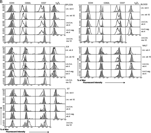
To elucidate potential differences between T cells isolated from distinct compartments, expression levels of CD44, CD27 (two lymphocyte activation markers), CD62L, an LN homing marker differentially expressed by effector and central memory cells, and α4β7, an integrin that favors migration to the gut mucosa, were determined on tet+CD8+ T cells induced by AdC6gag. Figure 3A shows data for naïve CD8+ lymphocytes compared with tet+CD8+ T cells 4 and 10 wk after a single i.n. immunization and 4 wk after a vaginal boost of i.n.-primed mice. Figure 3B shows data for CD8+ T cells tested 4 and 10 wk after i.m. priming, at 4 wk after a booster immunization of i.m.-primed mice given i.vag. or i.m., and at 1 year after an i.m/i.m. prime-boost regimen. In all experiments, tet−CD8+ T cells from immune mice were also analyzed and their phenotypes mirrored those of naïve mice (data not shown).

Phenotype analysis of Gag-specific CD8+ T cells upon mucosal or systemic administration of AdC vectors. Lymphocytes from different tissues were harvested, stained with Ab to CD8α, CD44, CD62L, CD27 and α4β7, and analyzed by flow cytometry. (A) Markers expression in mice that were vaccinated 4 or 10 wk earlier with AdC6gag given i.n., or that were primed i.n. with AdC6gag and boosted 4 wk before the analysis with AdC68gag given i.vag. (B) Markers expression in mice that were vaccinated 4 or 10 wk earlier with AdC6gag given i.m., or that were primed i.m. with AdC6gag and then boosted with AdC68gag given i.m. or i.vag. 4 wk before the analysis. For i.m./i.m.-immunized mice, analyses were also performed 1 year after the booster immunization. Both graphs show the expression of the markers on tet+CD8+ from vaccinated mice (black lines) and tet−CD8+ cells from naïve mice (gray areas). The results are representative of three experiments conducted independently with groups of 20 mice for analysis of the GT and groups of five mice for the remaining tissues.

Continued.
Four weeks after i.n. immunization with AdC6gag, CD44 was upregulated on Gag-specific CD8+ T cells from spleens, blood, ILN and NALT (Fig. 3A). This increase was less pronounced on tet+CD8+ cells from the GT, presumably reflecting that most T cells from the GT were already antigen-experienced. Most of the tet+CD8+ T cells from the GT expressed comparable levels of CD62L although a small population was CD62Lhi. It should be pointed out that expression of CD62L was also low on most of the genital CD8+ T cells from naïve mice. Expression of α4β7 was low on most cells except for a small population of tet+CD8+ T cells present in spleen and blood. The booster immunization did not have a pronounced effect on the expression of CD44, CD62L or CD27. α4β7 expression was again increased on some of the tet+CD8+ T cells from spleens and ILN.
At 4 wk after i.m. immunization, CD44 expression was upregulated on tet+CD8+ T cells from spleens, ILN and GT (Fig. 3B). We detected a downregulation of CD62L expression on tet+CD8+ T cells from spleens, blood and the GT but not on those from ILN. CD27 expression was decreased on a subpopulation of tet+CD8+ T cells from blood, spleens and GT. At 4 wk after i.n. or i.vag. boost, expression levels of CD44, CD62L, CD27 and α4β7 mirrored those seen at 10 wk after priming, and there were no striking differences among groups that received an AdC6gag i.m. prime followed by a heterologous boost through the i.m. or i.vag. routes. At 1 year after the i.m. prime-boost vaccine regimen, expression of CD44 on tet+CD8+ T cells isolated from the different compartments (NALT was not tested in this experiment) overlapped with those seen on part of CD8+ T cells of age-matched naïve mice. This may reflect an increase of CD44 expression on the control CD8+ T cells due to immunosenescence 15. Gag-specific CD8+ T cells isolated from the ILN and GT showed an increase in CD62L expression, which was unexpected for the latter compartment. In blood and spleen, expression of CD62L and CD27 was similar or only slightly increased above those seen on unprimed CD8+ T cells, suggesting that the Gag-specific CD8+ T cells had differentiated into resting memory cells.
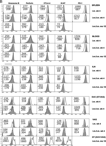
Additional markers were analyzed on Gag-specific CD8+ T cells isolated from different compartments after an i.m./i.m. heterologous prime-boost regimen (Fig. 4). For the two early time points, i.e. 4 wk after priming or boosting, cells isolated from the vaginal mucosa were treated and analyzed separately from OUC. CD44, CD62L and CD27 were tested and found to mirror those shown in Fig. 3. Figure 4, 4 shows the expression levels of CD69, an early activation marker enriched on mucosal cells derived from the intestinal tract, CD127, the IL-7 receptor that allows for homeostatic proliferation of memory CD8+ T cells through IL-7, CD103, also known as integrin αEβ7, which has been linked to infiltration of cells into epithelial tissues 16, 17 and NK group 2D (NKG2D), a transmembrane protein that acts as a primary activation receptor on NK cells and serves as a costimulator for CD8+ T cells 18.

Phenotype of Gag-specific CD8+ T cells induced by i.m. administration of AdC vectors. Lymphocytes from different tissues were harvested and analyzed 4 wk after i.m. prime with AdC6gag vector, and 4 wk and 1 year after boost with the heterologous AdC68gag vector. Lymphocytes were stained with Ab to CD8α, CD44, the tetramer and the markers indicated in the graphs, and analyzed by flow cytometry. (A) Expression of CD127, CD103, NKG2D and CD69. (B) Expression of lytic enzymes, CTLA-4, Ki-67 and PD-1. For the analysis of Gag-specific responses, cells were gated on CD8+, tetramer+ cells (black lines) and compared with tet−CD8+ cells from naïve mice (gray areas). Upper values show median fluorescence intensity for tetramer−CD8+ cells from naïve mice, lower values show median fluorescence intensity for tetramer+CD8+ cells from vaccinated animals. The results are representative of three experiments conducted independently with groups of 20 mice for analysis of the GT and groups of five mice for the remaining tissues.

Continued.
By 4 wk after i.m. prime or boost, CD69 was decreased on tet+CD8+ T cells from spleens, blood and OUC, whereas its expression on the vagina was similar to that on unprimed CD8+ T cells. By 1 year after the boost, CD69 expression on tet+CD8+ T cells from all compartments was similar to that of naïve cells, suggesting that this molecule is unlikely to contribute for the sustained presence of vaccine-induced CD8+ T cells within the GT (data not shown). Expression of CD127 was increased on tet+CD8+ T cells from ILN and the vagina at 4 wk after priming. A similar pattern was observed at 4 wk after the boost but for a modest increase in OUC. By 1 year after the boost, CD127 expression was increased in tet+CD8+ T cells from all compartments, being especially pronounced in cells from GT.
The most striking difference in the expression of CD103 was seen at 1 year after the boost, when this marker was markedly upregulated on tet+CD8+ T cells from the GT, but otherwise comparable to naïve cells in the other compartments. No remarkable changes were seen in the profile of NKG2D on T cells from the compartments analyzed.
Figure 4B shows the expression levels of granzyme B, a proteolytic enzyme that induces caspase-dependent apoptosis, and perforin, a pore-forming protein that facilitates granzyme access through the membrane into the cytosol of the target cell 19. In addition, Fig. 4B shows the expression levels for CTLA-4, a key molecule for downregulation of T-cell responses, programmed death-1 (PD-1), which negatively regulates T-cell signaling and effector functions and is expressed at increased levels on so-called exhausted T cells 20 and Ki-67, a protein associated with proliferation.
Expression of granzyme B mostly mirrored that of perforin, with a very pronounced increase in both enzymes in most tet+CD8+ T cells isolated from the whole GT at 1 year after the boost. Notably, the expression levels of other markers such as CD62L at the same time point suggest that T cells isolated from the GT had differentiated into resting memory cells. Memory CD8+ T cells typically do not carry granzyme or perforin, which are markers for fully activated effector CD8+ T cells.
CTLA-4 expression was decreased in tet+CD8+ T cells from spleens, ILN and vagina at 4 wk after the prime, whereas there was an increase in its expression on those from OUC. By 1 year after the boost, most tet+CD8+ T cells from the whole GT showed high levels of CTLA-4 expression, which was otherwise expressed at levels similar to those of unprimed CD8+ T cells in other compartments. Further analyses showed that in the GT, cells that were high in CTLA-4 concomitantly expressed high levels of lytic enzymes (data not shown). By 1 year after the boost, Ki-67 levels were upregulated on the GT. Expression of PD-1 was largely unremarkable.
In summary, the most striking differences in phenotypes between tet+CD8+ T cells from blood and spleen in comparison to those from the GT and its draining LN were seen at 1 year after the i.m./i.m. prime-boost regimen. Subpopulations of tet+CD8+ T cells from the GT showed marked increases in the expression of CD103, CD127, CD62L, granzyme B, perforin, CTLA-4 and Ki-67 and thus clearly represented a stage of differentiation not seen in spleens or blood.
Origin of genital Gag-specific CD8+ T cells
To gain insight into the origin of CD8+ T cells that homed to the GT, we conducted adoptive transfer experiments. BALB/c donor mice were primed with AdC6gag and boosted with AdC68gag given i.m. Fourteen days post-boost, splenocytes were isolated from the vaccinated mice and frequencies of tet+CD8+ cells were determined (Fig. 5). The remaining cells were injected i.v. at 5×10⁷ cells/mouse into naïve Thy1.1 congenic recipient mice. The recipient mice were euthanized 7 days later. As AdC vectors persist at very low levels in activated CD8+ T cells 11, we cannot rule out transfer of the vectors in splenocytes of donor origin. However, it is unlikely that the minute amount of vector present in T cells of the donors would induce a detectable immune response in the host within the time frame of the experiment. Nevertheless, to ensure that the results were not biased by activation of host-derived T cells, we used a congenic mouse strain for the experiment, which allowed us to track cells of donor origin. As shown in Fig. 5, Gag-specific Thy1.1− CD8+ cells of donor origin could readily be detected in all compartments tested, including the GT. As seen after i.m. prime with AdC6gag (Fig. 1), frequencies of tet+CD8+ T cells were higher in the GT than in other compartments analyzed (p<0.01). The results clearly show that Gag-specific CD8+ T cells from spleens can migrate to and are enriched for in the GT. We tested tet+CD8+ cells from donor mice prior to transfer for expression of cell markers shown in Figs. 3 and 4. CD69 and CD103, two molecules that have been implicated on the phenotype of mucosa-derived cells 21, 22, were expressed at the same levels on tet+CD8+ cells from donor mice prior to transfer and in control cells, and were thus unlikely to have contributed to the enrichment of Gag-specific CD8+ T cells within the GT. We also tested for the expression levels of these markers in tet+CD8+ cells of donor origin that had homed to the GT of recipient mice. Levels of CD69 again were similar to those on tet−CD8+ T cells, whereas CD103 was increased. This increase was only seen on tet+CD8+ cells from the GT but not on those from other compartments, suggesting that this molecule becomes upregulated once CD8+ T cells home to the GT (data not shown).

Migratory properties of adoptively transferred CD8+ T cells. BALB/c mice were vaccinated with AdC6gag and boosted with AdC68gag. Fourteen days after the boost, splenocytes were isolated and transferred into naïve Thy1.1 recipient mice. Mice were euthanized 7 days later and samples were analyzed. Frequencies of Gag-specific CD8+ T cells in the transferred splenocyte population and at day 7 after transfer in the different compartments of the recipient mice are shown. Cells were stained with Ab to CD8α, CD44, CD90.1 and the tetramer. For the analysis of Gag-specific responses, cells were gated on CD8+, CD90.1− and tetramer+ cells. The results are representative of two experiments conducted independently with groups of ten mice. Statistical analysis was performed using unpaired two-sample Student's t-test with p<0.01 (**).
Discussion
Women are most commonly infected by HIV-1 through heterosexual contact and immune mechanisms at or within the female GT would be expected to provide a crucial first barrier to transmission. As Ab that neutralize the countless HIV-1 variants remain elusive, many of the vaccines currently in clinical trials focus on the induction of HIV-1-specific CD8+ T cells. Such response cannot prevent the initial infection, but if present at the port of entry, might rapidly eliminate infected cells and thus thwart or potentially prevent spread of the virus.
We showed in mice that a homologous prime-boost regimen using AdC vectors expressing Gag induces transgene product-specific CD8+ T cells that could be isolated from the GT 13. This previous article used intracellular cytokine staining assays, which may not be optimal for the study of the GT-derived lymphocytes. Here, we extended these studies testing different routes of immunization, more efficacious heterologous prime-boost regimens, and assessed migratory patterns of such cells.
It is known that nasal immunization is able to induce immune responses not only in the respiratory tract but also at the GT 23. Results reported here show that CD8+ T cells, which home to the female GT, can be induced by i.n. immunization but this response is not sustained. In addition, vaginal booster immunization, as would be experienced in human vaccine recipients against HIV-1, causes only a slight local increase in i.n.-induced antigen-specific CD8+ T cells and fails to increase responses systemically. Last but not least, i.n. immunization may be problematic for some vectors as this route allows access of the vaccine into the central nervous system. In brief, i.vag. immunization, as reported by others 24, induces only very low levels of antigen-specific CD8+ T cells, which combined with logistic problems in humans should discourage further pursuit of this route of immunization for Ad vectors.
Results are more promising after i.m. immunization, which not only elicits antigen-specific CD8+ T cells in systemic tissues but also high and sustained responses within the GT, as also reported recently by another group 25. A second immunization given i.m. causes a robust booster effect within the GT of i.m.-primed mice, and Gag-specific CD8+ T cells remain detectable for at least 1 year. i.m. immunization is thus overall superior at inducing genital CD8+ T cell responses by AdC vectors compared with i.n. immunization, and offers the added benefit of also eliciting potent systemic CD8+ T-cell responses, which may serve as a second layer of defense in case the virus breaks through the mucosal barrier. These findings are in agreement with a study in mice showing that i.p. infection with lymphocytic choriomeningitis virus is superior to i.n. infection for the induction of CD8+ T-cell responses in the vaginal mucosa 26. Other reports reached similar conclusions upon vaccination with protein vaccines 27, DNA vaccines 28 or DNA vaccines combined with Modified Vaccinia Ankara and Semliki Forest vectors 29. Taken together, these results indicate that induction of CD8+ T-cell responses at mucosal sites upon i.m. immunization is independent of a given vaccine platform.
Antigen-experienced CD8+ T cells may traffic to the GT with the help of specific sensors that remain to be identified, or alternatively this process may be random. To gain further insight into the vaccine-induced CD8+ T cells that homed to the GT, we conducted a detailed phenotypical analysis of Gag-specific CD8+ T cells induced by the different immunization protocols, comparing cells isolated from spleen, blood, ILN and the GT at different times after immunization. In some assays, we also tested cells isolated from NALT; the latter were tested for comparison as a population of cells homing to a distinct mucosal site. Phenotypes of Gag-specific CD8+ T cells isolated from systemic sites and the GT were phenotypically distinct, and this was especially pronounced at 1 year after the i.m./i.m. prime-boost vaccine regimen. The phenotypes suggest that most tet+CD8+ T cells present in the GT remain fully activated and would be expected to start target cell lysis immediately upon encounter of infected cells.
We evaluated markers that are known to be upregulated on cells derived from the intestinal mucosa. Studies have demonstrated high levels of CD69 expression on intestinal CD8+ cells 22, 30, but expression of CD69 was not increased in the GT at any of the time points analyzed. Although α4β7 has been linked to the genital migration of subsets of CD4+ cells 31, and is a well-known marker for homing of T cells to the intestinal mucosa, our results do not suggest that α4β7 affects homing of CD8+ T cells to the GT. CD103 was slightly increased in tet+CD8+ T cells from the GT at early time points, and by 1 year after immunization became strongly upregulated. In the adoptive transfer experiment, CD103 was low on the Gag-specific CD8+ T cells isolated from the vaccinated donors and upon transfer remained low on cells isolated from all compartments but the GT, where an increase was observed. Again, these data argue against the notion that CD103 supports mucosal homing but rather suggest that CD103 may contribute to the retention of CD8+ T cells within the GT. The adoptive transfer experiment also showed that Gag-specific CD8+ T cells from the spleen could readily migrate to the GT to a similar extend as observed in vaccinated mice. This argues against the need for a distinct differentiation pathway during activation to allow for migration of CD8+ T cells to the mucosa, as had been described for T-cell homing to GALT 32 or for CD4+ T cells of the female GT 33. On the other hand, the observation that at 2 wk upon i.m. immunization frequencies of Gag-specific CD8+ T cells were ∼10-fold higher in blood but only ∼2-fold higher in the GT than upon i.n. immunization argues for a deliberate homing process that is dictated by the conditions under which the CD8+ T cells are initially stimulated. We would therefore assume that migration of activated CD8+ T cells to the GT is in part random and affected by their overall frequencies in blood, and in part driven by the expression of yet to be identified homing markers. In either case, we would assume that activated CD8+ T cells receive signals from the microenvironment that favor their retention once they reach the GT, leading to an enrichment of these cells at the mucosal surface, which is the port of entry for many pathogens.
The functionality of genital CD8+ T cells remains to be investigated in more depth. Our data thus far show that T cells from the GT produce IFN-γ but not IL-2 as has also been reported for genital T cells in SIV-infected non-human primates 34. In our study, Gag-specific CD8+ T cells from the GT expressed high levels of granzyme B, perforin and Ki-67, which suggests that they are highly activated cells able to immediately commence target cell lysis and proliferation. Other authors have demonstrated atypical T cells within mucosal surfaces 22 and we speculate that the high levels of lytic enzymes seen in memory-type CD8+ T cells from the GT could be a result of a specific microenvironment.
In summary, data presented here show that i.m. immunization with a replication defective AdC vector in mice induces a robust transgene product-specific CD8+ T-cell response within the GT that can be enhanced by a booster immunization given i.m. The response is sustained and can still be detected 1 year after immunization. Vaccine-induced genital CD8+ T cells are functional; they carry lytic enzymes and release cytokines upon antigenic stimulation. Taken together, the results shown should allow for guarded optimism that potent vaccines administered i.m. may induce a genital barrier to HIV-1 infection in women. In fact, systemic regimens would be preferable over mucosal ones in humans due to the logistical factors and the lack of interference by flora or menstrual cycle, which may profoundly affect mucosal vaccine efficacy.
Materials and methods
Mice
Female 6- to 8-wk-old BALB/c mice were obtained from Ace Animals (Boyertown, PA). Female 6- to 8-wk-old Thy1.1 mice were obtained from The Jackson Laboratory (Bar Harbor, ME). Animals were housed at the Animal Facility of The Wistar Institute (Philadelphia, PA) and all experiments were performed according to the institutionally approved protocols.
Viral vectors
Purified E1-deleted Ad vectors expressing Gag of HIV-1 clade B, derived from simian serotypes C6 (AdC6) or C68 (AdC68), were produced and quality controlled as described previously 8, 35.
Immunization of mice
Groups of 5–20 BALB/c mice were immunized by i.m. or mucosal routes with AdC vectors diluted to 1010 viral particles in sterile saline to a total volume of 10 μL (i.n. and i.vag.) or 100 μL (i.m.). Mice were immunized i.m. by injection into the lower leg muscle, whereas mucosal immunization was given with an automatic pipette. For mucosal immunizations, i.e. i.n. or i.vag., mice were first anesthetized with ketamine and xylazine chloride given i.p. Five days before i.vag. immunization, mice were i.p. injected with 3 mg of medroxiprogesterone-acetate (Sigma-Aldrich). For prime-boost experiments, mice were boosted i.n., i.vag. or i.m. 6 wk after the first immunization.
Isolation of lymphocytes
Lymphocytes were isolated as described previously 36. Briefly, blood was collected in heparin and red blood cells were lysed using ACK Lysing Buffer (Invitrogen). Spleens, ILN and NALT were dissociated against metal screens and washed with Leibovitch's L-15-modified medium (Mediatech). For isolation of lymphocytes from the GT, the vagina, cervix, uterus, uterine horns and ovaries were removed and cut into fragments. Tissue segments were submitted to constant shaking at 130 rpm for 1 h in RPMI 1640 (Mediatech) containing 5% FBS and 1% penicillin–streptomycin (Sigma-Aldrich). Fragments were enzymatically digested with 1.4 mg/mL of collagenase type I (Gibco) for 15 min. Cells from the two cycles were pooled and lymphocytes purified by a discontinuous Percoll gradient (Sigma-Aldrich) consisting of 40% fraction containing cells overlaid over a 70% fraction.
Adoptive transfer of lymphocytes
BALB/c mice were primed i.m. with AdC6gag and boosted 6 wk later with AdC68gag given i.m. Splenocytes were isolated 14 days later, and 5×10⁷ splenocytes were injected i.v. into naïve Thy1.1 recipient mice. Tissues were analyzed for the presence of tet+CD8+ donor cells 7 days after the transfer.
Tetramer staining, phenotypic analyses and flow cytometry
Staining was performed using a Gag peptide- (AMQMLKETI) and H-2Kd-specific tetramer (NIAID Tetramer Facility). For phenotyping, cells were incubated with the tetramer, and Ab to CD8α, α4β7, CD27, CD103 (BD Pharmingen), CD44, CD62L, PD-1 (Biolegend), CD69, CD127 and NKG2D (eBioscience). Cells were permeabilized with BD Cytofix/Cytoperm™ Fixation and Permeabilization Solution (BD Bioscience) and stained for granzyme B, Ki-67 (BD Pharmingen), CTLA-4 (RD Systems) and perforin (eBioscience). For transfer experiments, cells were also stained for CD90.1 (Thy 1.1) (BD Pharmingen). Prior to analysis, cells were fixed with BD Stabilizing Fixative (BD Bioscience). Flow cytometric analyses of cells were performed with a BD LSR II (Becton-Dickinson) flow cytometer. Data were analyzed using FlowJo V8.8 software (Tree Star). BD CompBeads Compensation Particles (Becton-Dickinson) were used to set distinct negative- and positive-stained populations for the fluorochrome-labeled Ab used in the experiments. For the assessment of background and nonspecific activation, we immunized animals with AdC vectors expressing an unrelated transgene; phenotypes for those cells mirrored those from naïve groups, as did frequencies of tetramer+CD8+ T cells (data not shown). Samples were gated on live lymphoid cells, and then on CD8+ cells versus side scatter, followed by gating on CD8+ cells versus forward scatter. The remaining cells were then plotted as CD44 cells versus tetramer for further analysis.
ELISpot assay
IFN-γ and IL-2 ELISpot assays were performed with lymphocytes as described previously 10. Briefly, 96-well Millipore polyvinylidene difluoride plates were coated with anti-mouse IFN-γ or IL-2 Ab (BD Pharmingen) diluted in PBS and incubated overnight at 4°C. Plates were then washed and blocked with 10% MLC media (DMEM supplemented with 10−6 M of 2-mercaptoethanol and 10% FBS) for 2 h at 37°C. Lymphocytes were added to plates at 2×10⁵ cells per well in triplicates, and stimulated with the 9-mer peptide AMQMLKETI or the total pool of 123 15-mer peptides derived from consensus Gag clade B, in the presence of anti-mouse CD28 and CD49d (BD Pharmingen) for 18–20 h at 37°C in 10% CO2. Cells were removed and plates incubated with biotin-labeled Ab (BD Pharmingen) for 2 h at room temperature. Streptavidin alkaline phosphatase (Mabtech AB) was added for 1 h, and the spots developed by adding BCIP/NBT (Pierce) for 5 min. Plates were washed in water and dried before counting using the C.T.L. Series 3A Analyzer and ImmunoSpot 3.2 (Cellular Technology). Data from unstimulated cells were used as background control, and values were subtracted from sample values before plotting. In parallel, cells were stained with an Ab to CD8α and analyzed by flow cytometry to determine the frequencies of this cell subset. These results were used to normalize the data obtained by ELISpot assays and data show numbers of SFU/106 CD8+ cells. Samples that resulted in less than 55 SFU/106 cells were considered negative.
Statistical analysis
Each experiment was conducted repeatedly with 5–20 mice and figures show means and standard deviations based on the independent experiments. Statistical significance of differences between groups was calculated by unpaired two-sample Student's t-test using GraphPad Prism (GraphPad Software, La Jolla, CA, USA). The p-values of <0.05 or <0.01 were considered statistically significant.
Acknowledgements
The authors thank Christina Cole for assistance with preparation of the manuscript. This work was supported by grant AI074078-01 from the National Institutes of Health, by Wistar Cancer Center Support Grant P30 CA 010815 from the National Cancer Institute, and by the Gates Foundation (GCGH). Partial support was also provided by CAPES and PNDST/Aids, Brazil.
Conflict of interest: The authors declare no financial or commercial conflict of interest.


