De Winter syndrome as an emergency electrocardiogram sign of ST-elevation myocardial infarction: a case report
Abstract
The case report aims to reveal de Winter's electrocardiogram (ECG) pattern as an equivalent to anterior ST-segment elevation myocardial infarction (STEMI). We report a case of a 49-year-old man with a history of smoking who presented to the emergency department with a 1 day history of chest pain that was exacerbated 5 h prior to presentation. Detailed clinical investigations and coronary angiographic characteristics were recorded. The first ECG of the patient was consistent with de Winter syndrome. Acute coronary artery angiography showed that the proximal left anterior descending coronary artery was completely occluded after the first diagonal branch artery was given off. A percutaneous coronary intervention was immediately performed. Our case indicates that early identification and diagnosis of such ECGs and timely reperfusion therapy of de Winter syndrome as a STEMI equivalent are required to improve the prognosis of such patients.
Introduction
de winter syndrome is a rare pattern that can occur on an electrocardiogram (ECG).1 It is thought to be related to acute anterior descending artery occlusion.1 ST-segment elevation myocardial infarction (STEMI) equivalent patterns make the diagnosis of STEMI very challenging.1 The ECG reveals a de Winter's pattern, which shows a 1 to 3 mm upsloping ST-segment depression at the J point in leads V1 to V6 that continues into tall, positive, and symmetrical T waves, instead of the signature ST-segment elevation1.
Case report
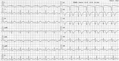
A 49-year-old man with a history of smoking was admitted to the hospital for recurrent chest pain for 1 day that had exacerbated in the previous 5 h. The patient reported having chest pain at 11 a.m. the previous day, mainly under the xiphoid process, with chest tightness. Five hours before his presentation, his chest pain recurred. The degree of the recurrence was worse than his previous chest pain and could not be relieved. It was accompanied by diaphoresis. The patient then presented to our hospital's emergency department. He was admitted to the coronary care unit (CCU) with acute coronary syndrome. After admission, his myocardial enzymes were urgently reexamined. These showed that TnI was 0.38 μg/L, myoglobin (Mb) > 900 μg/L, creatine kinase (CK) was 414 U/L, creatine kinase MB (CKMB) was 60 U/L, lactate dehydrogenase (LDH) was 274 U/L, potassium was 4.35 mmol/L, and he had mild hyperlipidaemia. The ECG showed upsloping ST-segment depression at the J-point with tall and symmetrical T waves from V2 to V6 leads, as well as ST-segment elevation in the aVR lead (Figure 1).
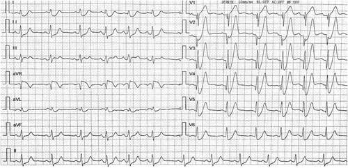
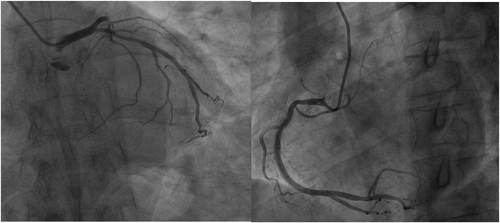
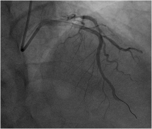
The ECG revealed a de Winter's pattern, the equivalent of an anterior STEMI. The clinicians considered a diagnosis of acute myocardial infarction, due to the combination of chest pain and ECG changes. Both aspirin and ticagrelor were given. The patient's family was informed. The patient was then sent to the catheterization laboratory. Emergency coronary artery angiography (CAG) revealed complete occlusion of the proximal left anterior descending (pLAD) artery after giving off the first diagonal branch. Moreover, diffuse lesions in the proximal left circumflex artery (LCX), 50–60% stenosis at the most severe point, and 50–60% stenosis in the proximal section of the right coronary artery (RCA) were detected (Figure 2). After consulting the patient's family, a left anterior descending percutaneous coronary intervention (PCI) was performed. Two drug-eluting stents (3.0*30 mm, 3.0*18 mm) were successfully placed in the pLAD artery. A repeat angiography showed that the thrombolysis in myocardial infarction (TIMI) blood flow in the distal anterior descending artery was grade three (Figure 3). The patient had no complications during the operation and returned to the CCU. The elevation of the post-procedural ECG showed persistent negative T waves in V2 to V6. This is commonly seen in patients that have had an acute myocardial infarction (Figure 4). The left ventricular ejection fraction (LVEF) of the patient in the emergency department prior to the intervention was 46%. The patient was discharged 6 days after the PCI with an LVEF of 58%. No complications occurred during hospitalization. The patient did not report any recurrence of chest pain or suffer from heart failure during the follow-up.
Discussion
First explained by de Winter et al. in 2008, de Winter syndrome is a rare pattern that can appear on an ECG. de Winter found this ECG pattern in 2% of 1532 patients with anterior myocardial infarctions. The diagnostic criteria for de Winter syndrome include (i) a 1 to 3 mm upsloping ST-segment depression at the J point in leads V1 to V6 that continues into tall, positive symmetrical T waves; (ii) QRS complex usually not wide or only slightly widened; (iii) in some patients, a loss of precordial R wave progression; (iv) a 1 to 2 mm ST-segment elevation in aVR.1 The underlying mechanism of this ECG pattern in acute myocardial infarctions is unknown. The loss of ST-elevation may be related to the lack of activation of the muscular ATP-sensitive potassium (KATP) channels caused by ischaemic ATP depletion.2 When the de Winter pattern is recorded, the myocardium is still in the ischaemic phase and has not progressed to complete myocardial infarction.3 In the current case, when the patient's ECG showed de Winter syndrome, myocardial enzymes such as TnI were only slightly increased, but the indicators of myocardial injury still showed an upward trend after PCI. The maximum troponin I and CKMB levels were 9.2 μg/L and 481 U/L, respectively.
Conclusions
These findings demonstrate that de Winter syndrome is likely to be an early ECG manifestation of acute myocardial infarction. Therefore, our case indicates that early identification and diagnosis of such ECGs and the timely reperfusion therapy of de Winter syndrome as a STEMI equivalent are required to improve the prognosis of such patients.
Acknowledgements
This study was supported by the National Natural Science Foundation of China (grant nos 81800324 and 81960061), Key Project of Science and Technology Research Funds of Department of Education of Jiangxi Province(grant no. GJJ170005), Youth Key Project of Department of Science and Technology of Jiangxi Province (grant no. 20192ACBL21040), and Science Project of Department of Health Commission of Jiangxi Province (grant no. 20203094).
Conflict of interest
None declared.
Consent
The author/s confirm that written consent for submission and publication of this case report, including image(s) and associated text, has been obtained from the patient in line with COPE guidance.


