Rare calcifying fibrous pseudotumor of the peritoneum in an adult woman as a random finding during laparoscopic surgery
Spyridon Topis and Menelaos Samaras contributed equally to this work.
Key Clinical Message
Calcifying fibrous pseudotumors (CFT) are rare benign lesions diagnosed by histological and immunohistochemical studies. Our case presents a rare detection of a CFT in the parietal peritoneum. These lesions can be falsely interpreted as myomas or adnexal masses and thus gynecologists should be aware of the existence of CFTs.
1 CASE IMAGE
Calcifying fibrous pseudotumor (CFT) is a rare benign lesion that was first described in children.1 Since 2016, 157 cases were identified and less than 10 cases are associated with the peritoneum.2 Most of the cases are identified in the upper gastrointestinal tract and the thorax. CFT is a dense and distinct hyalinized collagen tissue with lymphoplasmatic infiltration and psammomatous or dystrophic calcifications.3
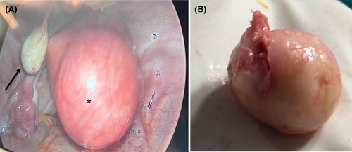
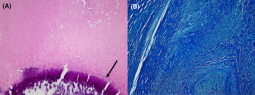
A 43-year-old nullgravida woman was presented to our laparoscopic unit complaining for intense pelvic pain the last 6 months. No other symptoms were reported. Her medical history included a diagnostic hysteroscopy and one resectoscopy for an intrauterine myoma in 2020. Clinical examination revealed an enlarged and palpable uterus. Vaginal examination and laboratory examinations were all normal. The sonographic examination revealed a bulky uterus with an intramural myoma 6 cm in diameter in the fundus. A laparoscopic myomectomy was offered to the patient. The laparoscopic myomectomy under general anesthesia was uneventful. During the surgery, a solid, pale white, lesion of 1.6 × 1.2 × 1.0 cm was identified hanging in the left posterior parietal peritoneum (Figure 1A). The aforementioned lesion was also excised at the end of the operation (Figure 1B). Histological diagnosis was suggestive of a calcifying fibrous pseudotumor with calcifications in the lower field (Figure 2A,B). The postoperative status of the patient was uneventful and she was discharged the following day.


CFTs are rare and benign tumors of the soft tissue and their treatment is mainly surgical. The World Health Organization established the calcifying tumors as an entity in 2002.2 The gold standard for diagnosis of CFTs are histological and immunohistochemical studies due to its unique findings in these examinations. These lesions can be falsely interpreted as myomas or adnexal masses and thus gynecologists should be aware of the existence of CFTs and their rare presence in the pelvis.
AUTHOR CONTRIBUTIONS
Spyridon Topis: Writing – original draft. Menelaos Samaras: Data curation; writing – original draft. Anastasios Potiris: Visualization. Nikolaos Machairiotis: Data curation. Peter Drakakis: Supervision; validation. Sofoklis Stavros: Conceptualization; project administration.
ACKNOWLEDGMENTS
The authors have nothing to report.
FUNDING INFORMATION
There is no funding for this article.
CONFLICT OF INTEREST STATEMENT
The authors declare that they have no competing interest.
ETHICS STATEMENT
This research was conducted ethically in accordance with the World Medical Association Declaration of Helsinki. The patient has given her written informed consent to publish the case (including the publication of images).
CONSENT
Written informed consent was obtained from the patient to publish this report in accordance with the journal's patient consent policy.
Open Research
DATA AVAILABILITY STATEMENT
The data that support the findings of this study are available from the corresponding author upon reasonable request.


