Spontaneous Rupture of the Rectus Femoris Masquerading as a Pseudotumor in a 60-Year-Old Male Patient: A Case Report
Funding: The authors received no specific funding for this work.
ABSTRACT
Quadriceps tendon ruptures (QTR), often involving the rectus femoris due to its superficial position, primarily affect men aged 50–60 with comorbidities weakening tendon collagen. Tears typically occur 1–2 cm above the patella or at the osteotendinous junction in older adults. Rupture of the rectus femoris muscle can sometimes mimic a pseudotumor, presenting as a soft tissue mass in the anterior thigh, with or without a clear history of trauma. In chronic cases, unrecognized or repetitive microtrauma often leads to fibrosis and incomplete healing. Isolated distal ruptures of the rectus femoris are rare, especially in young athletes. Differential diagnosis should rule out soft tissue tumors or sarcoma. Systemic diseases, obesity, or long-term steroid/quinolone use may contribute to ruptures, though in our cases, no such factors were found, suggesting old age is a potential risk. Nonsurgical healing is slow, leading to scar tissue and hindering recovery. Surgical repair is critical to prevent poor outcomes in untreated or chronic ruptures and to restore knee extensor function.
Summary
- Isolated distal rectus femoris ruptures are rare and can mimic soft tissue tumors.
- While conservative management is typically the first-line treatment, surgical intervention may be warranted in cases of complete rupture or when there is a significant reduction in knee extension power.
- Immediate surgery is not always required; however, timely evaluation and intervention—when indicated—along with a structured rehabilitation program are essential for optimal functional recovery.
1 Introduction
The rectus femoris muscle is more likely to be injured in the rare quadriceps muscle injury because of its superficial placement, muscle fiber composition, and extension across two joints [1]. Most ruptures occur in men between 50 and 60, with certain comorbidities affecting tendon collagen structure. The most common sites of tear were noted between 1 cm and 2 cm of the superior pole of the patella and, in older people, at the osteotendinous junction [2]. The rectus femoris is most commonly injured during eccentric loading [3]. Rectus femoris ruptures can be challenging to diagnose due to their atypical location and vague clinical features. MRI is essential for excluding soft tissue neoplasms, and biopsy should only be considered in exceptional cases [4].
There have been reports of quadriceps tendon tears in individuals on long-term drugs such as quinolones, statins, anabolic steroids, and intranasal steroids, as well as chronic disorders such as hyperparathyroidism, gout, chronic renal failure, and systemic lupus erythematosus. Additional contributing factors include obesity, prior trauma, and fibrotic changes resulting from arteriosclerosis [5-7].
Surgical repair can be an effective treatment for chronic rectus femoris tears unresponsive to conservative management, enabling full functional recovery [8]. To our knowledge, only a few literatures have reported postoperative outcomes following surgical intervention for complete rectus femoris rupture with reduced knee extension. This case report highlights the positive surgical outcome of rectus femoris rupture initially misinterpreted as a pseudotumor, demonstrating effective restoration of knee extension.
2 Case Report/Examination
A 60-year-old male presented to the outpatient department of Orthopedics with chief complaints of swelling on the anterior aspect of the mid-thigh for 3 weeks which arose spontaneously and is non-progressive. The patient did not experience sudden acute pain in the anterior thigh that caused him to stop his activity, nor did the pain impair his function at that time. There is no preceding history of trauma, fever, night sweats, or intake of chronic steroid use. There is no history of chronic diseases like chronic renal disease, COPD, endocrine diseases like Diabetes mellitus, hyperparathyroidism, acromegaly, etc. There is a history of Hypertension for 1 and a half years.
The patient first sought treatment at a local hospital nearby where an ultrasound examination led to a misdiagnosis of intramuscular cysticercosis. Then the patient was subsequently referred to our centre, and further evaluation, investigation, and management were done.
On inspection, a swelling measuring 8 × 4 cm was noted over the anterior aspect of the mid-thigh of the left leg, without any visible deformity or discoloration of the overlying skin. Palpation revealed a soft, regular, and non-tender lump that moved with knee movements. There was no local rise in temperature, and the range of motion of adjacent joints was full and unrestricted. The patient could ambulate without significant discomfort or functional limitations. However, a detailed functional assessment revealed a Grade III muscle strength in left knee extension, indicating a noticeable reduction in power compared to the right side, based on the manual muscle testing (MMT) scale, suggesting possible involvement of the extensor mechanism, potentially due to mechanical interference or secondary muscle weakness. Despite this functional deficit, the patient reported no significant discomfort or gait abnormality while walking, particularly on flat surfaces.
3 Differential Diagnosis, Investigations, and Treatment
The clinical features are suggestive of a benign lesion, with lipoma being the most likely diagnosis. However, differential diagnoses, including variants such as fibrolipoma or angiolipoma, and benign tumors like fibroma or neurofibroma, must be considered. Although less likely, the possibility of a malignant tumor, such as liposarcoma or other soft tissue sarcomas, cannot be entirely excluded, warranting further evaluation.
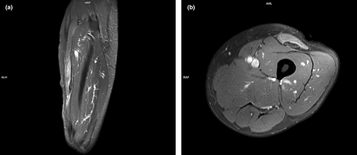
Routine blood investigation and serology were sent and were within normal limits. MRI of the left leg was done. On MRI findings, there is a complete tear of the distal musculotendinous junction of the rectus femoris muscle. The torn muscle belly is retracted proximally with a gap of approximately 3.7 cm. Minimal fluid is noted at the tear site. Edema is seen in the belly of the rectus femoris in both the direct and indirect heads. Fluid is noted in the tear site. The posterior fascia of the muscle is thickened (Figures 1a,b).

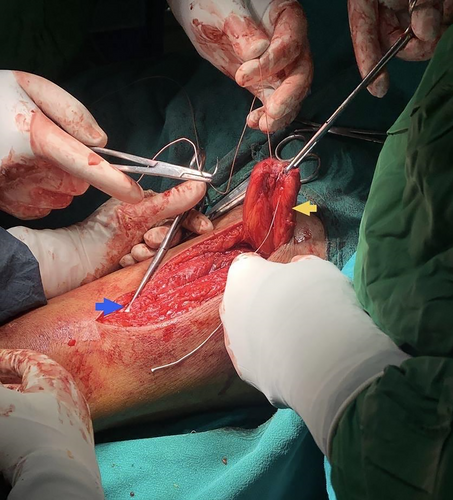
Although literature indicates that isolated distal rectus femoris ruptures typically do not require surgical intervention, in our case, surgery was necessary due to a complete rupture at the musculotendinous junction of the rectus femoris, resulting in a significant reduction in knee extension power. The patient underwent operative treatment to address the swelling and associated functional deficit in the left knee. Under strict aseptic technique, the area from the iliac crest to the mid-leg is prepared and draped. A 5 cm incision is made along the midline, just below the bulging or palpable part of the rectus femoris. Dissection is carried down to the level of the extensor mechanism, where the musculotendinous portion of the distal rectus femoris is identified. It is carefully teased out using forceps and Metzenbaum scissors. The distal tendon is then whip-stitched using a No. 2 FiberLoop suture (Figure 2). The tendon's mobility is assessed, and with the knee in full extension, a free needle is used to pass one limb of the FiberLoop suture as distally as possible, entering the common extensor tendon approximately 8 cm away. The other limb of the FiberLoop is passed in a Mason-Allen fashion. Using a tension-slide technique, knots are tied to maximize the lengthening of the rectus femoris tendon. An epitendinous stitch using small suture tape is run along both sides of the fascia for secondary reinforcement to the advanced muscle. Before closure, the knee is gently flexed to assess the achieved knee flexion, ensuring no excessive tension is placed on the repair. Post-operatively, the leg was placed in hyperextension with a hinged knee brace that restricted movement from 0° to 30° to provide support and promote proper healing while restricting movement. Pain management was achieved with analgesics, and the patient was subsequently discharged with the knee immobilizer in place for 8 weeks. A structured physiotherapy program was initiated, beginning with non-weight-bearing exercises during the immobilization phase to maintain joint mobility, enhance circulation, and prevent muscle atrophy. After 8 weeks, the patient transitioned to weight-bearing activities. Over time, consistent improvement was noted, and follow-up functional assessment using the Manual Muscle Testing (MMT) scale demonstrated Grade V strength, indicating full recovery of knee extension power and overall functional capability. The pre-operative Lysholm knee score was 68, which improved to 81 after 8 weeks, and further increased to 95 after 6 months. Regular follow-ups up to 6 months ensured close monitoring of progress, contributing to a successful recovery and return to normal activities.

4 Conclusion and Results (Outcome and Follow-Up)
Distal rectus femoris ruptures are rare but should be considered in cases of anterior thigh swelling with knee extension deficits. Accurate diagnosis through clinical evaluation and imaging, followed by timely surgical repair and structured rehabilitation, is essential for restoring function and preventing long-term disability. This case underscores the importance of prompt and multidisciplinary management for optimal recovery. In our case, patient rehabilitation is done stepwise as mentioned above, and functional recovery of the rectus femoris muscle is achieved gradually over 4–5 months. Isolated ruptures of the rectus femoris muscle often result in slow healing, fibrosis, or incomplete recovery without surgical intervention. Accurate diagnosis and surgical intervention are essential to achieve optimal function.
5 Discussion
While young athletes frequently sustain strains to their quadriceps muscles, isolated distal ruptures of the rectus femoris muscle are uncommon and have only been reported in a small number of cases [9]. It is widely acknowledged that the best way to restore knee extensor function is to repair quadriceps tendon ruptures very away [10]. A traumatic experience is frequently forgotten by patients who have a persistent injury to the rectus femoris muscle [11]. A complete rectus femoris muscle tear is a challenging injury, with natural nonsurgical healing being slow and often leading to fibrosis, incomplete recovery, or degeneration. Rehabilitation is challenging, and the healing process is often hindered by the formation of scar tissue within the muscle [12].
A soft tissue tumor or sarcoma should be ruled out in the differential diagnosis of a chronic rectus femoris muscle injury. It is possible to establish the diagnosis with meticulous history taking, physical examination, and selective radiological investigations, particularly magnetic resonance imaging, and to predict a full functional recovery. These tears may be the result of repeated microtrauma from misuse, or they may develop abruptly [11]. Chronic systemic diseases, obesity, or chronic steroid or quinolone use may be the precipitating factors for the rupture of the rectus femoris. In our cases, there are no such precipitating factors; old age may be the risk factor.
The rehabilitation process after surgical repair involves four phases. During the Post-Surgical Phase (0–6 weeks), knee immobilization stabilizes the repair, and physiotherapy focuses on preventing muscle atrophy, maintaining circulation, and avoiding stiffness. In the Gradual Passive Mobilization Phase (6–8 weeks), passive range-of-motion exercises improve joint mobility while protecting the surgical repair. The Active Movement Phase (8–10 weeks) introduces active, non-weight-bearing exercises, such as straight leg raises and quadriceps sets, to safely activate muscles. Finally, in the Weight-Bearing Phase (10–12 weeks), patients progress from partial to full weight-bearing with assistive devices, gradually building tolerance and muscle control [12, 13].
In our case, surgical intervention was chosen to restore knee extension power. While surgery is not always necessary, the patient was thoroughly counseled regarding both conservative and surgical treatment options. He opted for surgical management, considering the longer healing time, potential for fibrosis, and slower recovery associated with conservative treatment. Although surgical repair is generally advised for complete rectus femoris muscle tears, there is a lack of published data on the functional outcomes of these procedures [14]. In our case, follow-up over 6 months demonstrated significant improvement in knee extension power. The outcome suggests that surgical intervention can facilitate a faster and more complete functional recovery of knee extension compared to conservative management. These findings support considering surgical treatment, especially in cases of complete rupture with substantial loss of knee extension strength.
Author Contributions
Rishi Ram Banjade: conceptualization, data curation, formal analysis, investigation, project administration, software, writing – original draft. Sandesh Dhakal: conceptualization, investigation, project administration, writing – original draft. Dependra Bhandari: conceptualization, project administration, supervision, validation, visualization. Niraj Kumar Sharma: data curation, formal analysis, investigation, project administration, writing – original draft.
Acknowledgments
The authors have nothing to report.
Ethics Statement
The authors have nothing to report.
Consent
Written informed consent was obtained from the patient to publish this report in accordance with the journal's patient consent policy.
Conflicts of Interest
The authors declare no conflicts of interest.
Open Research
Data Availability Statement
The datasets used and/or analyzed during the current study are available from the corresponding author upon reasonable request.


