An odontoid fracture and vertebral artery injury in fast-track
Abstract
While CT scans without IV contrast are obtained commonly to evaluate vertebral injuries, CT angiography scans should be considered whenever a fracture site approaches known vasculature.
A 53-year-old man presented to the emergency department 3 days after an MVC. He complained of neck pain, and while his cervical spine was tender to palpation, he had no neurological deficits and an otherwise unremarkable exam. Through various imaging modalities an odontoid fracture and vertebral artery pseudoaneurysm were discovered.
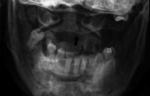
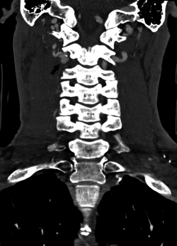
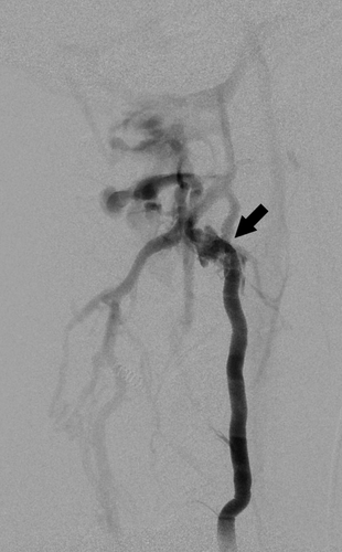
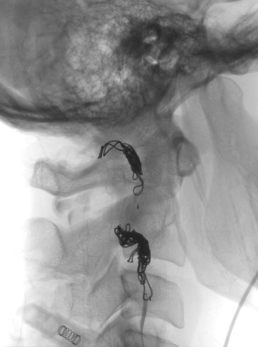
A 53-year-old man presented to the emergency department 3 days after he was the belted driver in a motor vehicle accident in which his vehicle was struck from behind. He complained primarily of posterior neck pain, and while his cervical spine was tender to palpation, he had no neurological deficits and an otherwise unremarkable physical exam. An open mouth odontoid view x-ray (Figure 1) and computed tomography (CT) scan (Figure 2) revealed a comminuted type III odontoid fracture with extension to the right pars interarticularis and C2/C3 transverse foramen. This fracture pattern prompted a CT angiogram (Figure 3) revealing a lobulated right vertebral artery pseudoaneurysm and concern for vertebral arteriovenous fistula adjacent to the fracture. A complex high flow fistula was confirmed at the level of the C2 fracture by digital subtraction angiography (Figure 4) and repaired emergently by endovascular embolization (Figure 5).

Fracture-associated vascular injuries require timely intervention. Vessel injury patterns can range from wall injury causing mild stenosis to transection. Plain radiographs or CT scans may identify bony abnormalities, but a low threshold to obtain vascular imaging should be maintained whenever a fracture site approaches known vasculature and there are several tools to help guide decision making.1, 2
CONFLICT OF INTEREST
None declared.
AUTHOR CONTRIBUTIONS
TAW: cowrote the manuscript and coconducted the literature review. SZT: coordinated the writing of the manuscript and edited the manuscript. KMB: treated the patient and edited the manuscript. ACR: treated the patient, cowrote the manuscript, and coconducted the literature review.


