Snapshot in surgery: brain abscess as a complication of a recurrent sigmoid diverticular abscess
Key Clinical Message
A 35-year-old man was found to have a cerebral abscess secondary to a recurrent sigmoid diverticular abscess. Both cultures grew Streptococcus anginosus. Brain abscess is a rare but potential complication of sigmoid diverticulitis. Streptococcus anginosus, which is found in human gut flora, is a common cause of brain abscess.
Snapshot quiz What do these images show and how should it be treated?


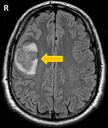
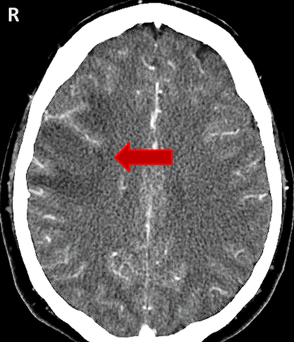
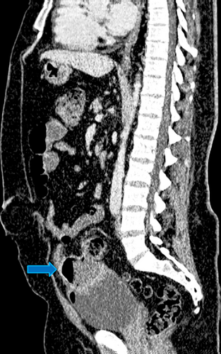
Answer: A 35-year-old man presented with one day history of dysarthria, dysphagia, and right temporal headache. He had a percutaneous drainage and antibiotic treatment of a sigmoid diverticular abscess 7 months previously. MRI brain showed a right posterior frontal motor cortex lesion (yellow arrow) which was initially diagnosed as glioma. A CT angiogram was then performed, but showed no vascular abnormality in relation to this lesion (red arrow). The images were subsequently discussed in the Neurosurgical MDT. A brain abscess with cerebritis and cerebral edema was diagnosed. A CT scan of abdomen and pelvis later showed an abscess formation (blue arrow) in the anterior pelvis which was closely related to the bladder doom and the distal sigmoid colon. The patient underwent a right frontoparietal mini-craniectomy and evacuation of cerebral abscess, followed by Hartmann's procedure. The abscess cultures both grew Streptococcus anginosus. He made a full neurological recovery and was discharged with outpatient parenteral antibiotic therapy.
Conflict of Interest
None declared.


