An unusual case of solid appendicular mass in a young female
Key Clinical Message
Endometriosis should be entertained as part of differential diagnosis in females in child-bearing age group when there is an incidental finding of solid neoplasm on imaging. It helps to guide physicians for appropriate management. It is important to emphasize that no radiological or imaging finding is pathognomonic for endometriosis.
Question
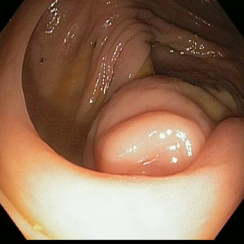
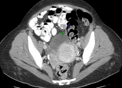
A 34-year-old female was evaluated for chronic constipation with intermittent rectal bleeding. Colonoscopy revealed a deformed appendicular orifice with a mass causing indentation of the cecum (Figure 1). Computerized tomography (CT) of abdomen showed solid mass close to 2 cm involving the base of appendix (Figure 2). How to best intervene?


Discussion
The initial diagnosis was a solid neoplasm of appendix, presumably carcinoid tumor. Considering the size and tumor bulging into cecum, patient underwent laparoscopic right hemicolectomy with tumor removal. Histopathology showed endometrioma.
Endometriosis can affect various parts of gastrointestinal system from small intestine to anus. It can present as acute appendicitis, invagination, colic, melena, or asymptomatic 1. Often Endometriotic foci were inaccessible to endoscopic biopsy due to the focality as in this case. As the surgical management varies widely from simple appendectomy to right hemicolectomy, there is a need to devise an evaluation method to better differentiate benign versus neoplastic disease preoperatively.
Conflicts of Interest
None declared.


