Indirect targeting of MYC sensitizes pancreatic cancer cells to mechanistic target of rapamycin (mTOR) inhibition
Christian Schneeweis, Zonera Hassan, and Katja Ascherl equally contributing first authors
Abbreviations
-
- 7-AAD
-
- 7-Aminoactinomycin D
-
- AKT
-
- protein kinase B
-
- ANOVA
-
- analysis of variance
-
- BET
-
- bromodomain and extra-terminal motif
-
- BETi
-
- BET inhibitor
-
- BrdU
-
- 5-bromo-2'-deoxyuridine
-
- CI
-
- Combination Index
-
- CNV
-
- Copy Number Variation
-
- E2F
-
- E2 factor (E2F) family of transcription factors
-
- EGFR
-
- epidermal growth factor receptor
-
- ERBB
-
- Erb-B receptor tyrosine kinases (EGFR family)
-
- FDR
-
- False Discovery Rate
-
- GI50
-
- Half-maximal growth inhibitory (GI50) concentration
-
- GSEA
-
- gene set enrichment analysis
-
- KEGG
-
- Kyoto Encyclopedia of Genes and Genomes
-
- MAPK
-
- Mitogen-activated protein kinase
-
- MEK
-
- MAPK/ERK kinase
-
- mRNA
-
- messenger ribonucleic acid
-
- mTOR
-
- mechanistic target of rapamycin
-
- mTORi
-
- mTOR inhibitor
-
- MYC
-
- myelocytomatosis oncogene
-
- PDAC
-
- pancreatic ductal adenocarcinoma
-
- PI3K
-
- Phosphoinositide 3-kinase
-
- PLK1
-
- Serine/threonine-protein kinase PLK1
-
- qPCR
-
- quantitative Polymerase Chain Reaction
-
- RNA
-
- ribonucleic acid
-
- RNA-seq
-
- RNA-sequencing
-
- SNP
-
- single-nucleotide polymorphism
-
- ZIP
-
- Zero interaction potency
Dear Editor,
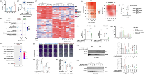
Pancreatic ductal adenocarcinoma (PDAC) remains a significant health problem with an increase in the incidence and a five-year survival rate of only 10% [1]. The Phosphoinositide 3-kinase-protein kinase-B-mechanistic target of rapamycin (PI3K-AKT-mTOR) pathway is a driver pathway in PDAC and an important therapeutic target [2]. We [3] and others [4-6] have demonstrated that the mTOR kinase is a therapeutic target in PDAC, and rationally designed mTOR inhibitor (mTORi)-based combination therapies are emerging [2]. However, clinical success has not been satisfactory so far [2] due to tumor adaption, resistance, and lack of predictive biomarkers. This implicates the need to decipher resistance mechanisms, develop rationally defined combination therapies, and find reliable biomarkers. Therefore, we aimed to understand the molecular underpinnings of mTORi resistance and treated 20 well-characterized (transcriptomics, single nucleotide polymorphisms [SNPs], copy number variations [CNVs]) murine PDAC cell lines [7] with the potent mTORi INK128 (Sapanisertib) to determine the half-maximal growth inhibitory concentration (GI50) (Figure 1A). The detailed Methods of this study can be found in the Supplementary Methods.

Resistant cell lines showed enrichment of myelocytomatosis oncogene (MYC) signatures (Figure 1B). Similar signatures were enriched in human INK128-resistant PDAC cell lines (Supplementary Figure S1A and B). Data of a CRISPR/Cas-drop out screening of PDAC lines showed a significant correlation between mTOR and MYC gene effect scores (Supplementary Figure S1C), demonstrating that some PDACs were co-addicted to MYC and mTOR. In PPT-9091MYCER cells, a conditional MYC gain-of-function model [8], activation of MYC led to a doubling of the INK128 GI50 value (Figure 1C), corroborating that MYC confers mTORi resistance, which is consistent with a recent report [9]. However, considering the heterogeneity of human cancers and complexity of the MYC network, additional pathways might also contribute to mTORi resistance.
Next, we generated a murine PDAC cell line that allows for the Cre-mediated deletion of exon 3 of the Mtor gene, called PPT4-ZH363-Mtor∆E3/lox [3]. We used Mtor-proficient (n = 4) and -deficient (n = 3) single-cell clones to find vulnerabilities associated with genetic mTOR inhibition (Supplementary Figure S2A, B). Downstream signaling, as measured by investigating the phosphorylation of the mTOR target Eukaryotic translation initiation factor 4E-binding protein 1 (4E-BP1), was distinctly reduced in the deficient clones (Supplementary Figure S2B). Stable Mtor knockouts showed increased phosphorylation of AKT (Supplementary Figure S2B and C). Functionally, inactivation of Mtor was associated with reduced proliferation (Supplementary Figure S2D). Analysis of transcriptomes demonstrated depletion of mTOR signatures in deficient clones (Supplementary Figure S2E). Further proteomics analyses revealed that proteins corresponding to MYC target genes (Supplementary Figure S2F) and metabolic pathways were the main downregulated signatures (Supplementary Figure S2G). As measured by phospho-proteomics, signaling by Erb-B receptor tyrosine kinases/epidermal growth factor receptors (ERBB/EGFR), Insulin, Mitogen-activated protein kinase (MAPK), and, as expected, mTOR were inhibited in deficient clones and therefore, vice versa enriched in proficient clones. (Figure 1D and E, Supplementary Table S1). Phosphorylation of proteins corresponding to MYC target genes were also downregulated upon Mtor deletion with borderline significance (FDR = 0.06) (Figure 1D and E).
Since we and others have demonstrated synergism of mTORi with MAPK/ERK kinase (MEK) and AKT inhibitors [3], we tested such inhibitors. The efficacy of MEKi at a dose of 5.5 nmol/L (Supplementary Figure S2H) and AKTi (Supplementary Figure S2I) was higher in Mtor-knockout clones, underscoring the value of the model to define vulnerabilities associated with genetic inhibition of mTOR.
Direct and indirect modes to inhibit MYC have been described [10]. MYC was efficiently blocked by inhibitors of bromodomain and extra-terminal motif (BET) proteins (Supplementary Figure S3A). We used the BET inhibitors OTX015 and JQ1, and the dual Serine/threonine-protein kinase PLK1/BET inhibitor BI2536 to evaluate the potential synergism between BETi and mTORi. We observed a strong significant reduction of clonogenic growth induced by all BETi in Mtor-deficient clones (Figure 1F-H, Supplementary Figure S3B). Note that the parental cell line, marked as blue dots (Figure 1G and H), responded like the proficient clones. We interpreted from the data of the genetic model that blocking mTOR signaling and MYC might be synergistic in PDAC cells. To directly test the synergism, we used parental PPT4-ZH363-Mtor∆E3/lox cells and observed, indeed, a synergistic reduction of clonogenic growth upon combined INK128 and JQ1 treatment (Supplementary Figure S3C). We extended this finding to a larger panel of murine and human PDAC models using the BET degrader ARV771 and the BET inhibitors OTX015 or JQ1. All inhibitors increased the INK128 sensitivity (Supplementary Figure S3D and E). We calculated the combination index (CI) in 23 human and murine PDAC cell lines using different dose combinations of INK128 and OTX015, and observed values below 1 for most combinations, demonstrating a synergistic effect (Supplementary Figure S3F). Cell lines with the lowest CI values displayed enriched MYC signatures (Supplementary Figure S3G), pointing to the possibility to stratify for combination therapy responders. In addition, we used different assays, treatment periods, and dose matrices via the Synergy Finder platform (https://synergyfinder.fimm.fi/) to calculate a zero-interaction potency (ZIP) score. High ZIP scores, which indicate synergism of both inhibitors, were found in a proportion of established, two-dimensional human PDAC cell lines (Supplementary Figure S3H), primary murine PDAC cells (Supplementary Figure S3I), and primary human, three-dimensional organoid PDAC models (Figure 1I and J). These data demonstrate the existence of a PDAC subtype sensitive for the combination of an mTOR and BET inhibitor across models and species (Figure 1J).
Using live-cell imaging over a period of 114 hours, we observed a profound growth arrest in the INK128 and OTX015 combination (Supplementary Figure S4A). Consistently, BrdU incorporation was synergistically reduced by the combination therapy in PPT-53631 (Figure 1K) and PaTu8988T cells (Figure 1L). 7AAD-BrdU flow cytometric analysis demonstrated that cells treated with the combination therapy were arrested in the G1-phase of the cell cycle (Figure 1M), a finding corroborated by flow cytometric analysis of propidium iodide-stained cells (Supplementary Figure S4B and C).
To define the contribution of MYC to growth inhibition, we investigated MYC expression over time. In INK128 treated cells, MYC was maintained with even a trend of increased expression (Figure 1N and O). In contrast, the INK128 and OTX015 combination reduced MYC expression in cells compared to the INK128 monotherapy (Figure 1N and O), pointing to an explanation of the synergistic growth defect. We hypothesized that a more profound inhibition of the mTOR kinase would increase the need of the cells to restore MYC expression. Therefore, we repeated the kinetic analysis using an increased dose of INK128, which was approximately 6–7 fold over the GI50 values. Indeed, at this dose, INK128 increased MYC expression, especially after 72 hours of treatment (Supplementary Figure S4D and E). To different extents, compared to the OTX015 monotherapy, MYC expression was less downregulated by the INK128 and OTX015 combination treatment (Supplementary Figure S4D and E). We analyzed RNA-seq data in the high-dose setting. Even if the results were not identical in murine PPT-53631 and human PaTu-8988T cells, the combination of INK128 and OTX015 had a profound impact on pro-proliferative transcriptional networks (Supplementary Figure S4F). This was also evident in mRNA expression profiles of INK128-treated cells compared to those treated with the combination of OTX015 and INK128. Pro-proliferative networks, including E2F and MYC, were distinctly inhibited by the combination treatment (Supplementary Figure S4G and H). These results are thus in agreement with the arrest in the G1 phase of the cell cycle and the reduced BrdU incorporation. Furthermore, the partial block in the transcriptional output of E2F and MYC might explain the growth defect also in the INK128 high dose setting, even though MYC protein expression remains. Molecular details of the mechanism that are responsible for the described regulatory circuits remain to be deciphered. Furthermore, heterogeneity of PDACs might impact the circuits activated by the BET and mTOR inhibitor combination therapy.
In summary, we developed a concept to enhance the anti-tumor activity of mTORi in a subtype of PDACs. Biomarker-stratified mTORi-based combination therapies seem promising for further pre-clinical and clinical development.
ACKNOWLEDGMENTS
We thank all the patients providing tumor tissue and the Hubrecht Institute for providing engineered cell lines. We thank all colleagues for providing vectors via the Addgene platform.
FUNDING STATEMENT
This work was supported by the Deutsche Forschungsgemeinschaft (DFG): SFB824 C9 to D.S. and G.S.; SCHN 959/3-2 to G.S.; SFB1321 (Project-ID 329628492) P13 to G.S.; SFB1321 (Project-ID 329628492) P11 to D.S and M.S.R.; SFB1321 S01 and S02 to G.S., M.R., D.S., and R.R; SCHN 959/6-1 to G.S.; RE 3723/4-1 to M.R., SFB1361 (Project-ID ID 393547839) to O.H.K; DFG KR2291-9-1/12-1/14-1 to O.H.K., Wilhelm-Sander-Stiftung (2017.048.2 to G.S. and 2019.086.1 to G.S. and O.H.K.); Deutsche Krebshilfe (70113760 to G.S.; Max Eder Program 111273 to M.R.) and Brigitte und Dr. Konstanze Wegener-Stiftung (Projekt 65) to O.H.K. This research project/publication was funded by LMU Munich‘s Institutional Strategy LMU excellent within the framework of the German Excellence Initiative to M.S.R.
CONFLICT OF INTEREST
The authors declare no competing interests.
ETHICS APPROVAL AND PATIENT CONSENT STATEMENT
The primary human PDAC cellular organoid models were established and analyzed in accordance with the declaration of Helsinki. The study was approved by the local ethical committee TUM, Klinikum rechts der Isar (Project 207/15, 1946/07, 330/19S), and written informed consent from the patients for research use was obtained prior to the investigation.
PERMISSION TO REPRODUCE MATERIAL FROM OTHER SOURCES
Not applicable.
CLINICAL TRIAL REGISTRATION
Not applicable.
AUTHORS' CONTRIBUTION
Conceptualization and design of the study: C.S., Z.H., K.A. and G.S. Data collection and/or analysis and interpretation were performed by: C.S., Z.H., K.A., M.W., S.K., F.O., L.K., C.S., R.Ö., O.H.K., R.R., M.R., M.S.R., D.S. and G.S. The manuscript was drafted by: C.S., Z.H., K.A. and G.S. All authors revised the manuscript for important intellectual content and approved the final version submitted for publication.
Open Research
DATA AVAILABILITY STATEMENT
Proteome and phosphoproteome data: ProteomeXchange Consortium via the PRIDE partner repository: PXD027779; RNA-seq via ENA: PRJEB43040 and PRJEB47050.


