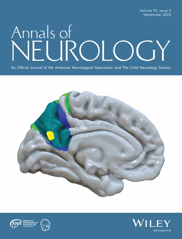
Network Localization of Awareness in Visual and Motor Anosognosia
Abstract
Objective
Unawareness of a deficit, anosognosia, can occur for visual or motor deficits and lends insight into awareness itself; however, lesions associated with anosognosia occur in many different brain locations.
Methods
We analyzed 267 lesion locations associated with either vision loss (with and without awareness) or weakness (with and without awareness). The network of brain regions connected to each lesion location was computed using resting-state functional connectivity from 1,000 healthy subjects. Both domain specific and cross-modal associations with awareness were identified.
Results
The domain-specific network for visual anosognosia demonstrated connectivity to visual association cortex and posterior cingulate while motor anosognosia was defined by insula, supplementary motor area, and anterior cingulate connectivity. A cross-modal anosognosia network was defined by connectivity to the hippocampus and precuneus (false discovery rate p < 0.05).
Interpretation
Our results identify distinct network connections associated with visual and motor anosognosia and a shared, cross-modal network for awareness of deficits centered on memory-related brain structures. ANN NEUROL 2023;94:434–441
Potential Conflicts of Interest
M.D.F. is a consultant for Magnus Medical and Soterix and holds intellectual property on using connectivity imaging to guide brain stimulation. The other authors report no competing interests.
Open Research
Data Availability Statement
The functional connectivity data equivalent to that employed in this study is available online through the Harvard Dataverse at: https://doi.org/10.7910/DVN/ILXIKS and the pipeline used to prepare the functional connectivity data is available at: https://github.com/bchcohenlab/BIDS_to_CBIG_fMRI_Preproc2016. Lesion data used in this study is publicly available and obtained from published medical literature (see Supplementary Table S2, Kletenik et al 202211 and Pacella et al eLife 20191). Statistical neuroimaging analyses, specifically the voxelwise ANOVA was performed in MatLab (version 2019b) and FSL (version 5.0.10). The maps for our primary analyses can be accessed on NeuroVault at the following link: https://identifiers.org/neurovault.collection:13792.


