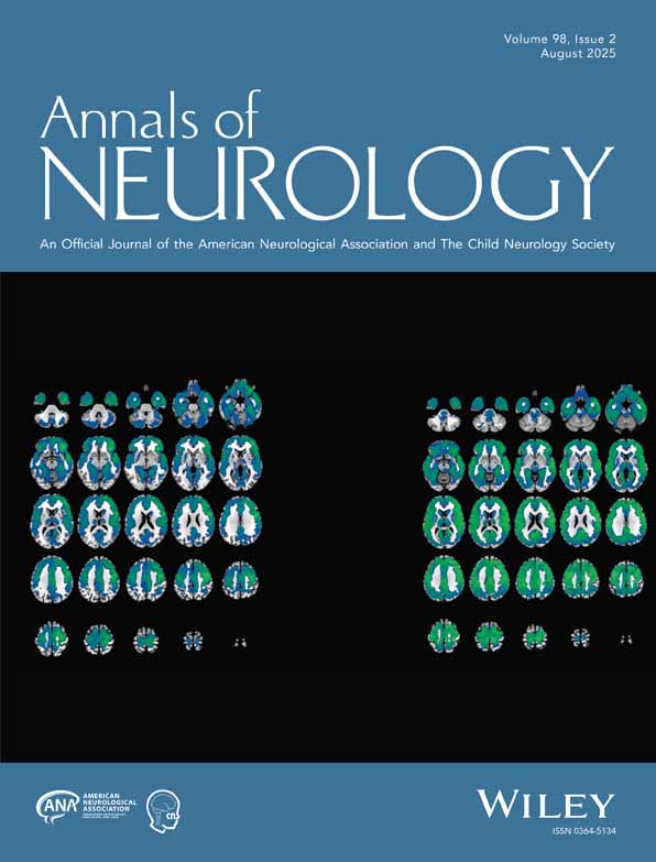
Glutathione peroxidase activity modulates recovery in the injured immature brain†
Kyoko Tsuru-Aoyagi MD
Department of Neurological Surgery, University of California, San Francisco, San Francisco, CA
Search for more papers by this authorMatthew B. Potts MD
Department of Neurological Surgery, University of California, San Francisco, San Francisco, CA
Search for more papers by this authorAlpa Trivedi PhD
Department of Neurological Surgery, University of California, San Francisco, San Francisco, CA
Search for more papers by this authorTimothy Pfankuch BS
Department of Behavioral Neuroscience, Oregon National Primate Research Center, Oregon Health and Science University, Portland, OR
Search for more papers by this authorJacob Raber PhD
Department of Behavioral Neuroscience, Oregon National Primate Research Center, Oregon Health and Science University, Portland, OR
Department of Neurology, Oregon National Primate Research Center, Oregon Health and Science University, Portland, OR
Division of Neuroscience, Oregon National Primate Research Center, Oregon Health and Science University, Portland, OR
Search for more papers by this authorMichael Wendland PhD
Department of Radiology, University of California, San Francisco, San Francisco, CA
Search for more papers by this authorCatherine P. Claus BS
Department of Neurological Surgery, University of California, San Francisco, San Francisco, CA
Search for more papers by this authorSeong-Eun Koh MD
Department of Neurological Surgery, University of California, San Francisco, San Francisco, CA
Search for more papers by this authorDonna Ferriero MD
Department of Neurology, University of California, San Francisco, San Francisco, CA
Department of Pediatrics, University of California, San Francisco, San Francisco, CA
Search for more papers by this authorCorresponding Author
Linda J. Noble-Haeusslein PhD
Department of Neurological Surgery, University of California, San Francisco, San Francisco, CA
Department of Physical Therapy and Rehabilitation Science, University of California, San Francisco, San Francisco, CA
Departments of Neurological Surgery and Physical Therapy and Rehabilitation Science, University of California, San Francisco, 521 Parnassus Avenue, Room C-224, San Francisco, CA 94143-0520Search for more papers by this authorKyoko Tsuru-Aoyagi MD
Department of Neurological Surgery, University of California, San Francisco, San Francisco, CA
Search for more papers by this authorMatthew B. Potts MD
Department of Neurological Surgery, University of California, San Francisco, San Francisco, CA
Search for more papers by this authorAlpa Trivedi PhD
Department of Neurological Surgery, University of California, San Francisco, San Francisco, CA
Search for more papers by this authorTimothy Pfankuch BS
Department of Behavioral Neuroscience, Oregon National Primate Research Center, Oregon Health and Science University, Portland, OR
Search for more papers by this authorJacob Raber PhD
Department of Behavioral Neuroscience, Oregon National Primate Research Center, Oregon Health and Science University, Portland, OR
Department of Neurology, Oregon National Primate Research Center, Oregon Health and Science University, Portland, OR
Division of Neuroscience, Oregon National Primate Research Center, Oregon Health and Science University, Portland, OR
Search for more papers by this authorMichael Wendland PhD
Department of Radiology, University of California, San Francisco, San Francisco, CA
Search for more papers by this authorCatherine P. Claus BS
Department of Neurological Surgery, University of California, San Francisco, San Francisco, CA
Search for more papers by this authorSeong-Eun Koh MD
Department of Neurological Surgery, University of California, San Francisco, San Francisco, CA
Search for more papers by this authorDonna Ferriero MD
Department of Neurology, University of California, San Francisco, San Francisco, CA
Department of Pediatrics, University of California, San Francisco, San Francisco, CA
Search for more papers by this authorCorresponding Author
Linda J. Noble-Haeusslein PhD
Department of Neurological Surgery, University of California, San Francisco, San Francisco, CA
Department of Physical Therapy and Rehabilitation Science, University of California, San Francisco, San Francisco, CA
Departments of Neurological Surgery and Physical Therapy and Rehabilitation Science, University of California, San Francisco, 521 Parnassus Avenue, Room C-224, San Francisco, CA 94143-0520Search for more papers by this authorPotential conflict of interest: Nothing to report.
Abstract
Objective
Mice subjected to traumatic brain injury at postnatal day 21 show emerging cognitive deficits that coincide with hippocampal neuronal loss. Here we consider glutathione peroxidase (GPx) activity as a determinant of recovery in the injured immature brain.
Methods
Wild-type and transgenic (GPxTg) mice overexpressing GPx were subjected to traumatic brain injury or sham surgery at postnatal day 21. Animals were killed acutely (3 or 24 hours after injury) to assess oxidative stress and cell injury in the hippocampus or 4 months after injury after behavioral assessments.
Results
In the acutely injured brains, a reduction in oxidative stress markers including nitrotyrosine was seen in the injured GPxTg group relative to wild-type control mice. In contrast, cell injury, with marked vulnerability in the dentate gyrus, was apparent despite no differences between genotypes. Magnetic resonance imaging demonstrated an emerging cortical lesion during brain maturation that was also indistinguishable between injured genotypes. Stereological analyses of cortical volumes likewise confirmed no genotypic differences between injured groups. However, behavioral tests beginning 3 months after injury demonstrated improved spatial memory learning in the GPxTg group. Moreover, stereological analysis within hippocampal subregions demonstrated a significantly greater number of neurons within the dentate of the GPx group.
Interpretation
Our results implicate GPx in recovery of spatial memory after traumatic brain injury. This recovery may be attributed, in part, to a reduction in early oxidative stress and selective, long-term sparing of neurons in the dentate. Ann Neurol 2009;65:540–549
Supporting Information
Additional Supporting Information may be found in the online version of this article.
| Filename | Description |
|---|---|
| ANA_21600_sm_SupFig1.tif35.2 MB | Supplemental Figure 1. TUNEL positive nuclei in the hippocampus at 24 hours postinjury. A–D) Representative photomicrographs of TUNEL staining, an indicator of irreversible cell injury. TUNEL positive cells are prominent in the granule cell layer of the dentate gyrus (arrows) in both genotypes. Scale bar, 100μm. E) The percentage of TUNEL-positive cells [(number of TUNEL-positive nuclei/number of Hoechst-positive nuclei) × 100%] was determined within 5 regions of the hippocampus for each genotype. The numbers of labeled nuclei are more prominent in caudal hippocampus relative to rostral. However, no differences in TUNEL labeling are noted between genotypes. |
| ANA_21600_sm_SupFig2.tif19 MB | Supplemental Figure 2. Temporal changes in the cortical lesion as assessed by magnetic resonance imaging. A) Representative T2WI and DWI from a WT injured animal. At 1 day postinjury, the site of injury exhibits relative high signal intensity on T2 images, suggesting vasogenic edema, and high signal intensity on DWI. Calculated ADCs are abnormally low for the injured region, suggesting cytotoxic edema was also present. At 7 days postinjury the lesion consists of both a hypointense region on T2 (arrows), consistent with aged clot, and hyperintense regions on T2 whose signal are almost nullified on DWI (not present in selected case) consistent with liquified tissue. At 14 and 24 days postinjury, the lesion consists of liquified tissue. B) Time course of signal increase in injured brain over 30 min after contrast administration at 1 day postinjury. This graph shows a relatively steady signal increase over time after contrast administration consistent with leakage of contrast material into the injured tissue. Minor but non-significant differences between WT and GPxTg groups can be ascribed to somewhat smaller injury among the GPxTg animals examined. |
| ANA_21600_sm_SupFig3.tif34.5 MB | Supplemental Figure 3. Exploratory activity and anxiety levels after TBI or sham surgery. Exploratory activity and anxiety levels were assessed in the open field. A) GPxTg animals spend more time in the center of the open field (# p<0.05), indicating lower levels of anxiety than WT mice. B) There is no difference in total distance moved, an indication of total activity levels. C, D) Within the GPxTg group, injured mice enter the center less (C, * p<0.05) and move less in the center (D, * p<0.05). |
| ANA_21600_sm_SupFig4.tif27.5 MB | Supplemental Figure 4. Sensorimotor learning after TBI or sham surgery. Sensorimotor function was assessed using the rotorod. A) All groups exhibit improvement with training. B) The GPxTg animals that received TBI show significantly less improvement in rotorod performance with training than GPxTg sham mice (difference in time between trial 9 and trial 1). * p<0.05 |
| ANA_21600_sm_SupFig5.tif34.4 MB | Supplemental Figure 5. Cortical and hippocampal lesion volumes at 4 months postinjury. A, B) Cresyl violet staining demonstrates how a cortical cavitation replaces the frontal and parietal gray matter and subcortical white matter. Scale bars, 10μm. C, D) Regional volumes of ipsilateral cortex and hippocampus, estimated by the Cavalieri method, are reduced after TBI. However, this reduction is similar between genotypes (unpaired T-tests; p=0.622 and P=0.512 for the cortex and hippocampus, respectively). |
| ANA_21600_sm_SupMethods.doc44.5 KB | Supplemental methods |
Please note: The publisher is not responsible for the content or functionality of any supporting information supplied by the authors. Any queries (other than missing content) should be directed to the corresponding author for the article.
References
- 1 Kraus JF, Rock A, Hemyari P. Brain injuries among infants, children, adolescents, and young adults. Am J Dis Child 1990; 144: 684–691.
- 2 Langlois JA, Rutland-Brown W, Thomas KE. The incidence of traumatic brain injury among children in the United States: differences by race. J Head Trauma Rehabil 2005; 20: 229–238.
- 3 Levin HS, Eisenberg HM, Wigg NR, Kobayashi K. Memory and intellectual ability after head injury in children and adolescents. Neurosurgery 1982; 11: 668–673.
- 4 Ewing-Cobbs L, Miner ME, Fletcher JM, Levin HS. Intellectual, motor, and language sequelae following closed head injury in infants and preschoolers. J Pediatr Psychol 1989; 14: 531–547.
- 5 Koskiniemi M, Kyykka T, Nybo T, Jarho L. Long-term outcome after severe brain injury in preschoolers is worse than expected. Arch Pediatr Adolesc Med 1995; 149: 249–254.
- 6 Luerssen TG, Klauber MR, Marshall LF. Outcome from head injury related to patient's age. A longitudinal prospective study of adult and pediatric head injury. J Neurosurg 1988; 68: 409–416.
- 7 Pullela R, Raber J, Pfankuch T, et al. Traumatic injury to the immature brain results in progressive neuronal loss, hyperactivity and delayed cognitive impairments. Dev Neurosci 2006; 28: 396–409.
- 8 Tong W, Igarashi T, Ferriero DM, Noble LJ. Traumatic brain injury in the immature mouse brain: characterization of regional vulnerability. Exp Neurol 2002; 176: 105–116.
- 9 Yager JY, Thornhill JA. The effect of age on susceptibility to hypoxic-ischemic brain damage. Neurosci Biobehav Rev 1997; 21: 167–174.
- 10 Lewen A, Matz P, Chan PH. Free radical pathways in CNS injury. J Neurotrauma 2000; 17: 871–890.
- 11 Dringen R. Metabolism and functions of glutathione in brain. Prog Neurobiol 2000; 62: 649–671.
- 12 Khan JY, Black SM. Developmental changes in murine brain antioxidant enzymes. Pediatr Res 2003; 54: 77–82.
- 13 Fan P, Yamauchi T, Noble LJ, Ferriero DM. Age-dependent differences in glutathione peroxidase activity after traumatic brain injury. J Neurotrauma 2003; 20: 437–445.
- 14 Chan PH, Kawase M, Murakami K, et al. Overexpression of SOD1 in transgenic rats protects vulnerable neurons against ischemic damage after global cerebral ischemia and reperfusion. J Neurosci 1998; 18: 8292–8299.
- 15 Mikawa S, Kinouchi H, Kamii H, et al. Attenuation of acute and chronic damage following traumatic brain injury in copper, zinc-superoxide dismutase transgenic mice. J Neurosurg 1996; 85: 885–891.
- 16 Ditelberg JS, Sheldon RA, Epstein CJ, Ferriero DM. Brain injury after perinatal hypoxia-ischemia is exacerbated in copper/zinc superoxide dismutase transgenic mice. Pediatr Res 1996; 39: 204–208.
- 17 Fullerton HJ, Ditelberg JS, Chen SF, et al. Copper/zinc superoxide dismutase transgenic brain accumulates hydrogen peroxide after perinatal hypoxia ischemia. Ann Neurol 1998; 44: 357–364.
- 18 McLean CW, Mirochnitchenko O, Claus CP, et al. Overexpression of glutathione peroxidase protects immature murine neurons from oxidative stress. Dev Neurosci 2005; 27: 169–175.
- 19 Sheldon RA, Jiang X, Francisco C, et al. Manipulation of antioxidant pathways in neonatal murine brain. Pediatr Res 2004; 56: 656–662.
- 20 Schmued LC, Stowers CC, Scallet AC, Xu L. Fluoro-Jade C results in ultra high resolution and contrast labeling of degenerating neurons. Brain Res 2005; 1035: 24–31.
- 21 Bonthius DJ, McKim R, Koele L, et al. Use of frozen sections to determine neuronal number in the murine hippocampus and neocortex using the optical disector and optical fractionator. Brain Res 2004; 14: 45–57.
- 22 Gavrieli Y, Sherman Y, Ben-Sasson SA. Identification of programmed cell death in situ via specific labeling of nuclear DNA fragmentation. J Cell Biol 1992; 119: 493–501.
- 23
Lebesgue D,
LeBold DG,
Surles NO, et al.
Effects of estradiol on cognition and hippocampal pathology after lateral fluid percussion brain injury in female rats.
J Neurotrauma
2006;
23:
1814–1827.
10.1089/neu.2006.23.1814 Google Scholar
- 24 West MJ, Slomianka L, Gundersen HJ. Unbiased stereological estimation of the total number of neurons in the subdivisions of the rat hippocampus using the optical fractionator. Anat Rec 1991; 231: 482–497.
- 25 Witgen BM, Lifshitz J, Grady MS. Inbred mouse strains as a tool to analyze hippocampal neuronal loss after brain injury: a stereological study. J Neurotrauma 2006; 23: 1320–1329.
- 26 Benice TS, Rizk A, Kohama S, et al. Sex-differences in age-related cognitive decline in C57BL/6J mice associated with increased brain microtubule-associated protein 2 and synaptophysin immunoreactivity. Neuroscience 2006; 137: 413–423.
- 27 Lister RG. Ethologically-based animal models of anxiety disorders. Pharmacol Ther 1990; 46: 321–340.
- 28 Igarashi T, Potts MB, Noble-Haeusslein LJ. Injury severity determines Purkinje cell loss and microglial activation in the cerebellum after cortical contusion injury. Exp Neurol 2007; 203: 258–268.
- 29 Ferriero DM, Holtzman DM, Black SM, Sheldon RA. Neonatal mice lacking neuronal nitric oxide synthase are less vulnerable to hypoxic-ischemic injury. Neurobiol Dis 1996; 3: 64–71.
- 30 Graham EM, Sheldon RA, Flock DL, et al. Neonatal mice lacking functional Fas death receptors are resistant to hypoxic-ischemic brain injury. Neurobiol Dis 2004; 17: 89–98.
- 31 Sheldon RA, Hall JJ, Noble LJ, Ferriero DM. Delayed cell death in neonatal mouse hippocampus from hypoxia-ischemia is neither apoptotic nor necrotic. Neurosci Lett 2001; 304: 165–168.
- 32
Payton KS,
Sheldon RA,
Mack DW, et al.
Antioxidant status alters levels of Fas-associated death domain-like IL-1B-converting enzyme inhibitory protein following neonatal hypoxia-ischemia.
Dev Neurosci
2007;
29:
403–411.
10.1159/000105481 Google Scholar
- 33 Deng Y, Thompson BM, Gao X, Hall ED. Temporal relationship of peroxynitrite-induced oxidative damage, calpain-mediated cytoskeletal degradation and neurodegeneration after traumatic brain injury. Exp Neurol 2007; 205: 154–165.
- 34 Souza JM, Choi I, Chen Q, et al. Proteolytic degradation of tyrosine nitrated proteins. Arch Biochem Biophys 2000; 380: 360–366.
- 35 Tweedie D, Milman A, Holloway HW, et al. Apoptotic and behavioral sequelae of mild brain trauma in mice. J Neurosci Res 2007; 85: 805–815.
- 36 Hall ED, Detloff MR, Johnson K, Kupina NC. Peroxynitrite-mediated protein nitration and lipid peroxidation in a mouse model of traumatic brain injury. J Neurotrauma 2004; 21: 9–20.
- 37 Assaf Y, Holokovsky A, Berman E, et al. Diffusion and perfusion magnetic resonance imaging following closed head injury in rats. J Neurotrauma 1999; 16: 1165–1176.
- 38 Bao F, Liu D. Peroxynitrite generated in the rat spinal cord induces apoptotic cell death and activates caspase-3. Neuroscience 2003; 116: 59–70.
- 39 Onyszchuk G, Al-Hafez B, He YY, et al. A mouse model of sensorimotor controlled cortical impact: characterization using longitudinal magnetic resonance imaging, behavioral assessments and histology. J Neurosci Methods 2007; 160: 187–196.
- 40 Vasa RA, Grados M, Slomine B, et al. Neuroimaging correlates of anxiety after pediatric traumatic brain injury. Biol Psychiatry 2004; 55: 208–216.
- 41 Camargo EE. Brain SPECT in neurology and psychiatry. J Nucl Med 2001; 42: 611–623.
- 42 Panksepp J, Siviy S, Normansell L. The psychobiology of play: theoretical and methodological perspectives. Neurosci Biobehav Rev 1984; 8: 465–492.
- 43 Hovatta I, Tennant RS, Helton R, et al. Glyoxalase 1 and glutathione reductase 1 regulate anxiety in mice. Nature 2005; 438: 662–666.
- 44 Kesner R. Behavioral functions of the CA3 subregion of the hippocampus. Learn Mem 2007; 14: 127–136.


