Long-Term Humoral Immune Response After West Nile Virus Convalescence in Horses in a Geographic Area of Multiple Orthoflavivirus Co-Circulation
Funding: This work was supported by University of Veterinary Medicine Budapest, Grant No. SRF-001. National Research, Development and Innovation Office, RRF 2.3.1-21-2021-00006.
ABSTRACT
Background
In the last three decades, West Nile virus (WNV, Flaviviridae, Orthoflavivirus genus) has become one of the most important encephalitic agents worldwide, causing substantial numbers of cases in humans and horses every year by re-emerging in endemic areas and emerging in new territories. It is considered that after natural WNV infection, humans and birds develop long-term immunoprotection, but data on immunoprotection in horses is scarce.
Hypothesis
West Nile virus infection provides long-term humoral immunity in subclinically infected horses.
Animals
Client-owned, naturally WNV subclinically infected non-WNV-vaccinated, healthy horses.
Methods
In this prospective cohort study, anti-WNV neutralizing antibody (nAb) titers of 25 horses were monitored for 5 consecutive years in Hungary. Serum samples were collected annually. First, a WNV immunoglobulin G (IgG) ELISA was performed, followed by virus neutralization tests (VNT) for endemic orthoflaviviruses. A VNT titer > 8 was considered positive.
Results
The mean WNV titer of horses was 260.64 ± 336.74 in 2019, 114.32 ± 107.36 in 2020, 95.38 ± 115.56 in 2021, 22.53 ± 25.71 in 2022 and 6.31 ± 5.15 in 2023. A significant decrease (p < 0.001) in the nAb titers occurred over time. In 2023, 88% of the horses had WNV VNT titers below the cut-off value.
Conclusion and Clinical Relevance
Our results showed a significant decrease in WNV titers over time. Because nAbs correlate best with orthoflavivirus protection, our findings suggest that horses might not be protected against re-infection. We recommend regular nAb titer testing or vaccination in endemic areas.
Abbreviations
-
- ADE
-
- antibody-dependent enhancement
-
- CPE
-
- cytopathic effect
-
- DENV
-
- dengue virus
-
- DMEM
-
- Dulbecco's modified eagle medium
-
- E
-
- envelope protein
-
- ECDC
-
- European Centre for Disease Control
-
- EIA
-
- equine infectious anemia
-
- IP
-
- inhibitory percentage
-
- JEV
-
- Japanese encephalitis virus
-
- mAb
-
- monoclonal antibody
-
- MBC
-
- memory B-cell
-
- MW-NT
-
- micro-well neutralization test
-
- OD
-
- optical density
-
- PFU
-
- plaque forming unit
-
- PI
-
- post-infection
-
- PRNT
-
- plaque reduction neutralization test
-
- TBE
-
- tick-borne encephalitis
-
- TBEV
-
- tick-borne encephalitis virus
-
- TCID50
-
- tissue culture infective doses of 50%
-
- USUV
-
- Usutu virus
-
- VNT
-
- virus neutralization test
-
- WNND
-
- West Nile neuroinvasive disorder
-
- WNV
-
- West Nile virus
-
- WOAH
-
- World Animal Health Organization
1 Introduction
West Nile virus is a mosquito-transmitted member of the Orthoflavivirus genus [1]. It was first isolated in 1937 from a febrile woman in Uganda [2]. At the beginning of the 21st century, the virus emerged in the European and American continents and caused major outbreaks of thousands of cases [3, 4]. The Orthoflavivirus genus belongs to the Flaviviridae family and includes several other relevant pathogens of humans and horses [5]. Presently, three orthoflaviviruses are endemic in Hungary: West Nile virus (WNV), Usutu virus (USUV), and tick-borne encephalitis virus (TBEV) [6]. Because of climate change and various socioeconomic factors, the co-circulation of orthoflaviviruses has become an increasing global problem in recent years [7-9].
West Nile virus is maintained in an enzootic cycle between birds and ornithophilic mosquitoes. Horses and humans serve as incidental hosts. After inoculation, horses develop viremia 1–6 days post-infection (PI), lasting for 1–3 days with viral titers ranging between 10 and 103 plaque-forming units/mL [10]. It is estimated that 10%–20% of horses develop neurological signs, with ataxia of different grades, weakness, muscle fasciculations, tremors, and cranial nerve deficits being reported most frequently [10-14]. Because of the long incubation period (7–10 days) but short-term, low-titer viremia in horses, the diagnosis of WNV infections relies on serological methods. Virus-specific immunoglobulin M (IgM) becomes detectable 5–7 days PI and often is used to verify recent WNV infection. Virus-specific immunoglobulin G (IgG) can be detected from 10 days PI to at least 1 year PI [15]. The virus-specific neutralizing antibody response is reported to target the viral surface envelope (E) glycoprotein. However, this protein shares different levels of amino acid sequence homology among individual members of the genus, and consequently, an orthoflavivirus infection elicits not only virus type-specific but also genus cross-reactive antibodies [16, 17]. The latter can hamper the diagnosis of the viral agents and influence the clinical manifestation of secondary infections [15]. The overlapping geographic presence of orthoflaviviruses and their cross-reactive antibodies necessitate understanding of the long-term immune response and its implications in protection against homologous or heterologous members of the genus.
Currently, the concentration of neutralizing antibodies is the best indicator of protection against orthoflaviviruses [18, 19]. After orthoflaviviral infections, birds and humans develop long-term immune protection against the pathogens, even in the case of subclinical seroconversion [20, 21]. However, some conflicting results regarding long-term antibody concentrations after WNV infection in humans have been observed [22-24]. We investigated the humoral immune response of WNV in naturally subclinically infected horses over a five-year period in Hungary, where other endemic orthoflaviviruses are present. The primary objective of our prospective cohort study was to assess the long-term humoral immune response of horses naturally and subclinically infected with WNV and other endemic orthoflaviviruses.
2 Materials and Methods
2.1 Study Design
After the major WNV outbreak in 2018 in Hungary, a WNV serosurvey was performed in horses in 2019. Blood samples for the study were obtained as part of annual obligatory active equine infectious anemia (EIA) surveillance of registered horses in Hungary. First, a commercially available WNV ELISA was performed. Because only healthy animals with no prior history of neurological signs were enrolled in the study, in the case of a positive WNV IgG result, the owner and the treating veterinarian of the horse were contacted. After confirming the animal's health status, owner consent was obtained, and serum samples were further evaluated using micro-well virus neutralization tests (MW-NT) for WNV, USUV, and TBEV. A detailed description of these procedures is given below. Blood samples from the selected horses were taken each year before the mosquito season (from June to October) from 2019 to 2023. Samples of the same year were tested simultaneously for all three orthoflaviviruses each year.
2.2 Animals
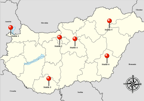
Twenty-five client-owned horses with positive initial WNV IgG ELISA results were chosen. The inclusion criteria for animals were: adult, healthy horses with no prior history of neurological signs, and non-vaccinated for WNV. The animals were kept in 6 different stables: 2 horses in Stable 1 in Győr-Moson-Sopron County, 8 horses in Stable 2 in Pest County, 5 horses in Stable 3 in Baranya County, 2 horses in Stable 4 in Pest County, 6 horses in Stable 5 in Hajdú-Bihar County, and 2 horses in Stable 6 in Békés County. The location of each stable is represented in Figure 1. All animals remained within the same facility during the study. These horses were followed for 5 consecutive years, and their health status and absence of neurological signs were monitored annually.

2.3 Sera Samples
Blood samples were taken by jugular venipuncture into tubes without anticoagulants. Samples were transported to the laboratory at 4°C, where serum was separated. Blood samples were centrifuged at 1860 rpm for 10 min, and then sera were transferred to microcentrifuge tubes. Serum samples were stored at −20°C.
2.4 Cell Lines and Viruses
African green monkey kidney Vero cells (ATCC, United States) were used for neutralization assays. Cells were grown in Dulbecco's modified eagle medium (DMEM; Capricorn Scientific GmbH, Germany) supplemented with 10% fetal bovine serum (FBS; Capricorn Scientific GmbH, Germany) and 1% penicillin/streptomycin (Sigma-Aldrich Co., United States) to make complete media. Cells were maintained in a humidified atmosphere at 37°C in 5% CO2 and were used to grow virus stocks and determine virus titrations.
West Nile virus strain 578/10 lineage II (GenBank accession number KC496015.1) was used [25]. Usutu virus Budapest-2005 strain (GenBank accession number EF206350.1) was used [26]. Tick-borne encephalitis virus KEM-1 strain (GenBank accession number MW256716.1) was used [27].
All viral suspensions were previously titrated to 100 TCID50/mL (tissue culture infective doses of 50%).
2.5 Diagnostic Assays
2.5.1 WNV IgG ELISA
Horse serum samples were screened by blocking enzyme immunoassay (INgezim West Nile Compac, Ingenaza, Spain) for IgG antibodies against WNV. This test detects antibodies produced against the E protein of WNV. The ELISA was performed following the manufacturer's protocol. Results were interpreted by calculating the inhibition percentage (IP) as described in the manufacturer's guidelines. An IP ≥ 40% was considered positive, between 30% and 40% was interpreted as inconclusive, and < 30% was considered negative.
2.5.2 Micro-Well Neutralization Assay
The micro-well virus neutralization assay was performed on all serum samples according to the World Organization for Animal Health (WOAH) Manual of Diagnostic Tests and Vaccines for Terrestrial Animals (Chapter 3.1.26.), except for the level of the serial dilution. In our study, a two-fold serial dilution was made from each serum sample, instead of the recommended dilution (1:5 to 1:640) [28]. Briefly, 96-well cell culture plates (TPP Techno Plastic Products AG, Switzerland) were used, and each well was filled with 50 μL of 0% DMEM (not supplemented with FBS). After inactivation at 56°C for 30 min, a twofold serial dilution of serum samples from 1:2 to 1:2048 was performed. Then, 50 μL of 100 tissue culture infective dose-50% (TCID50/mL) viral suspension was added to the diluted serum samples. After incubation, 104 Vero cells suspended in DMEM with 10% fetal calf serum were added to each well. Plates were incubated for 1 h at 37°C with 5% CO2. Back titration of each viral suspension was performed to confirm the viral titers, and non-infected cell culture was used as a negative control. The plates were incubated and monitored daily for 5 days at 37°C with 5% CO2. The presence and extent of cytopathic effects (CPE) were evaluated on the 5th day. The serum neutralizing titer was estimated as the highest serum dilution showing 50% of CPEs. The cut-off value of the WOAH manual (1:10) was corrected to our dilution. Therefore, sera with neutralizing titers > 1:8 were identified as positive. In case of simultaneous positive results for two or all viruses, the sample was considered positive for the agent with the highest neutralization titer.
2.6 Statistical Analysis
In our statistical modeling, titer values were log-transformed using base 2. We applied a mixed effects model with the log-transformed titer as the dependent variable. The model included the year as a continuous variable in the fixed effects component. The random effects structure consisted of both a random slope and a random intercept. The random slope allowed for variability in the pattern of antibody changes across individual horses, acknowledging that the rate of change differed. The random intercept accounted for dependency within the data, recognizing that measurements from the same horse were not independent. The statistical analysis was conducted in R (version 4.2.1, 2022-06-23 ucrt) using the lme function from the nlme package.
3 Results
3.1 Animals
Altogether, 125 samples were obtained over the 5-year study period. During the study, none of the horses were reported to have any clinical signs related to orthoflavivirus infections. The mean age of horses was 11.48 ± 3.92 years (range, 3–22 years) in 2019. The study involved 13 (52%) mares and 12 (48%) geldings. There were 8 Hungarian warmbloods, 3 Hungarian sport ponies, 3 Oldenburgers, 3 Holsteiners, 2 Welsh Ponies, 2 Gidrans, 1 KWPN, 1 Trakhener, 1 Connemara, and 1 German Sport horse included. No correlations were identified between the age or sex of horses and the VNT titers.
3.2 ELISA
All horses were WNV IgG positive in the first year (2019) of the study. In the second year, one horse became and remained WNV IgG negative for the remainder of the study period. The remaining horses were seropositive for all 5 years. The ELISA optical densities (ODs) and inhibitory percentages (IPs) of each year are presented in Table S1.
3.3 Virus Neutralization Assays
The virus-specific neutralization antibody titers are presented in Table S2 and Figure 2.

3.3.1 WNV Titers
The mean WNV titer of horses was 260.64 ± 336.739 in 2019, 114.32 ± 107.36 in 2020, 95.38 ± 115.56 in 2021, 22.53 ± 25.71 in 2022, and 6.31 ± 5.15 in 2023. A significant decrease (p < 0.001) in the serum VNTs occurred over time. In the first year (2019), all horses had positive WNV VNT titers; in the second year, 1/25 (4%); in the third year, 3/25 (12%); and in the fourth year, 17/25 horses (68%) had titers below the cut-off, which increased to 22/25 (88%) in the fifth year (2023), respectively.
3.3.2 USUV Titers
The mean USUV titer was 16.56 ± 34.04 in 2019, 4.16 ± 3.31 in 2020, 4.24 ± 4.68 in 2021, 4.61 ± 4.1 in 2022 and 8.71 ± 19.51 in 2023. In the first year 5/25 (20%), in the second year 1/25 (4%), in the third year 3/25 (12%), in the fourth year 2/25 (8%) and in the last year 4/25 (16%) horses had positive USUV VNT results. The USUV titers in the first 4 years of the study were not significantly different from the WNV titers, but in the last year, Horses No. 7 (1:45), No. 14 (1:64), and No. 21 (1:64) had increases in USUV VNT titer compared with the negative titers (≤ 1:8) of the previous year.
3.3.3 TBEV Titers
Only 1 horse (Horse No. 6) had a positive TBEV titer of 1:128 in the first year of the study. This horse did not have detectable TBEV-specific neutralizing antibodies in the subsequent years. The remaining horses were TBEV-negative during the entire study period.
3.4 Correlation of ELISA and VNT Results
No correlations were found between the ELISA OD and IP results and the WNV VNT titers.
4 Discussion
In our prospective cohort study, the orthoflavivirus-specific neutralizing antibody titers of 25 WNV naturally subclinically infected horses were followed annually from 2019 to 2023 in Hungary. Orthoflavivirus infections induce long-term immunity in humans and birds [23, 24, 29]. According to our results, WNV infection might not provide long-term humoral immune protection against re-infection in naturally subclinically infected horses. We also report the first USUV seroconversion in horses in Hungary. Although VNTs are considered to be the most specific serodiagnostic methods, cross-reactions between WNV and USUV, and in one instance, between WNV and TBEV, still were observed. Therefore, our findings emphasize the necessity of evaluating the interpretation of serological diagnostic methods in regions where multiple orthoflaviviruses are co-circulating.
Changing climatic conditions provide favorable circumstances for the vectors of orthoflaviviruses, and urbanization, global transport, and travel further facilitate transmission of these agents [8]. The co-circulation of orthoflaviviruses has become an emerging problem in recent years, and the number of affected areas is increasing [30]. All orthoflaviviruses show variable degrees of antigenic relatedness because of similarities in E protein amino acid sequences, allowing their classification into eight distinct serocomplexes [17]. Presently, there are 3 endemic orthoflaviviruses in central Europe: tick-borne encephalitis virus, Usutu virus, and WNV [6, 7]. Tick-borne encephalitis virus is a member of the mammalian tick-borne virus complex. It was first described in 1952 in Hungary, and virus-induced neurological disease is being reported annually among human patients, whereas an equine TBE case or seropositivity has never been reported [6]. Austria, Germany, and Switzerland have already registered TBEV-induced neurological disease, and other European countries reported TBEV seropositivity among horses [31-33]. The Japanese encephalitis virus complex involves USUV and WNV [17]. The mosquito-transmitted USUV was first reported in 2005 in Hungary, whereas the first USUV-induced neurologic disease in humans was diagnosed in 2018 [34]. Usutu virus infection has never been described as causing clinical signs in horses, but seropositivity has already been reported in several European countries, but not in Hungary [35-37]. West Nile virus was first isolated in 2003 in Hungary, whereas the first West Nile neuroinvasive disorder (WNND) in horses was diagnosed in 2007 [38, 39]. Afterwards, a few cases were recorded annually; in 2016, there was a mild increase in the reported equine WNND cases. In 2018, a major WNV outbreak occurred in Hungary, as well as in many other European countries. In 2023 and 2024, the incidence of registered WNND cases in both humans and horses increased significantly, compared to previous years (European Centre for Disease Control, ECDC, 2024) [40, 41].
One of the major limitations of our study is the lack of WNV IgM-positive ELISA results of the horses involved in this study, and thus a precise season of infection cannot be established. However, considering the substantial WNV outbreaks in Hungary (2008, 2016, 2018), and the mean age of our study population (11.5 years), we can assume that the animals were either infected in 2016 or 2018. Although the micro-well virus neutralization test (MW-NT) was preferred over the plaque reduction neutralization (PRNT) test because MW-NT is more rapid and less labor intensive, and it was proposed to be an effective and reliable alternative to PRNT [24], sera samples from different years were evaluated separately because of the parallel testing for all three endemic orthoflaviviruses and the relatively high number of samples (75 VNT test samples per year). We believe this method did not importantly influence the results, because the assays were performed by the same individual using the same instruments, with both virus and cell control components included in each test. Additionally, freezing and thawing may have affected our results, but previous studies indicate that these processes do not notably affect the outcomes of VNTs and ELISAs [42-44].
After the clearance of the viral agent, long-lived plasma cells secrete pathogen-specific antibodies in the absence of antigen or secondary challenge for many years, whereas virus-specific memory B-cells (MBC) circulate in the body without antibody production. In case of a subsequent viral infection, MBCs are rapidly activated to secrete pathogen-specific antibodies [9, 45, 46]. In orthoflaviviral infections, the concentration of neutralizing antibodies is the principal contributing factor of long-term antiviral protection, as direct correlations were observed between in vivo neutralizing activity and in vitro protective capacity of antibodies in mice and hamster models [18, 19, 47, 48]. The neutralizing activity of antibodies is influenced by their affinity and concentration, and accessibility of viral epitopes. Strongly neutralizing antibodies have high affinity. Therefore, they can neutralize the virus in lower quantities, whereas weakly neutralizing antibodies might be unable to neutralize the agent, even in high concentrations [49]. The WNV titer of the naturally subclinically infected horses in our study decreased significantly over time (Figure 2). The primary advantage of the two-fold serial dilution used in the VNTs of our study is that it provided more detailed monitoring of the nAb titers in comparison to the serial dilution recommended by the WOAH manual. The 1:8 titer, being closest to the WOAH positive titer of 1:10, was chosen as the cut-off value for our study. Based on this criterion, it was found that 88% of the animals had negative WNV VNT results in 2023. Our findings are in agreement with a previous study that identified a progressive decrease in WNV-specific IgG antibody concentrations 12–18 months post-infection among subclinically infected human blood donors. At the same time, some patients became seronegative in a relatively short time [22]. The age of the horses in our study population is an important factor to consider regarding the decreasing antibody titers. Immunosenescence, the age-related decline in immune function, is a well-documented phenomenon of older horses [50, 51]. Studies have shown that geriatric horses often exhibit decreased antibody production in response to antigenic stimulation [51, 52]. Although we were unable to establish any direct correlations between the age of the horses and their antibody titers, it is essential to recognize that the life expectancy of horses has gradually increased over the past few decades [53, 54]. Therefore, further investigation into the immune responses of geriatric horses is warranted, particularly concerning their natural protection against WNV. The concentration of neutralizing antibodies that provides adequate protection against WNV re-infection is presently unknown. However, we assume that the antibody concentrations in the last year of our study (2023) alone cannot provide protection against re-infection [19, 55]. Furthermore, it is worth mentioning that one of the participating horses (Horse No. 5) with a WNV VNT titer of 4 in 2023 presented with mild neurological signs during the WNV transmission season in 2024. A wide range of differential diagnoses can be listed as a potential cause of mild seasonal neurological signs in horses in Europe (including TBE, equine herpes myeloencephalitis, bacterial meningoencephalitis, anaplasmosis, and neuroborreliosis) [15, 56, 57], but because this horse was kept in the north-western part of the country (Győr-Moson-Sopron County) reporting one of the highest levels of WNV activity in 2024, a secondary WNV infection is also possible [42]. Although the horse tested negative for WNV IgM, animals that have already been exposed to WNV infection (or basic immunization without booster vaccines in the following years) might not develop sufficient WNV-specific IgM during a secondary immune response to be diagnosed. The lack of or lower magnitude of virus-specific IgM response during secondary infections is a well-known phenomenon in dengue viral infections, pathogen of the Orthoflavivirus genus, important in humans [58, 59]. Concerning the antigenic relatedness of these agents, a previous orthoflavivirus infection could have hampered the diagnosis of secondary heterologous orthoflaviviral infection [17]. This possibility is supported by a study, in which horses that had previously been vaccinated against Japanese encephalitis virus (JEV) were subsequently challenged with WNV [60]. Only one horse developed a detectable WNV-specific IgM response. Similar findings were reported in humans, where a previous WNV exposure resulted in an undetectable IgM response during secondary clinical TBEV infection [61]. Another interesting fact to consider is the lack of TBE cases in horses despite the relatively high incidence of TBE in humans in Hungary. On the other hand, seasonal neurological cases are noted among horses, as in the case of Horse No. 5, in each WNV season that test negative on WNV IgM ELISA. However, approximately half of these samples test positive on WNV IgG ELISA (Tolnai et al. unpublished data). We propose that the IgM-based diagnostic approaches of WNV infection in countries with other co-circulating orthoflaviviruses might be misleading. The VNT currently is recognized as the gold standard method for identifying the infecting orthoflaviviral agent. It also has the ability to overcome the limitations of the IgM ELISAs. This assay has the highest specificity among the available serodiagnostic tests because it detects the neutralizing capacity of antibodies that is influenced by their affinity, concentration, and the accessibility of the viral epitopes [49]. However, because the immunodominant protein of orthoflaviviruses is the E-protein that possesses different levels of amino acid sequence homologies, and because WNV infections elicit a polyclonal antibody response in horses as well, cross-reactions might occur on VNTs, especially between closely related agents [17, 62]. To minimize the risk of misleading results, it is recommended to conduct parallel VNTs for co-circulating orthoflaviviruses. If positive results are obtained for two or more agents, the end-point neutralization titer can help clarify any conflicting results [63, 64]. In our study population, five horses had positive USUV VNT results in 2019. Considering the significantly higher WNV titers in these animals and the genetic relatedness of these viruses, cross-reactivity is more plausible than USUV seropositivity. In the following years, the USUV titers were insignificant in comparison to the WNV titers. In 2023, however, three horses developed a significant USUV titer increase in comparison to the previous years, as well as the WNV titer of the same year. Usutu virus is endemic in Hungary; it is regularly isolated from birds as part of a passive surveillance program [34, 65]. Neighboring countries have already reported USUV seropositivity in horses, and here, we also present evidence of subclinical USUV seroconversion in horses in Hungary [37]. The possibility of simultaneous WNV and USUV infections must be considered when interpreting the VNT results of Horses No. 1 and No. 6 in 2019, because both viruses share the same transmission cycles and vectors. However, in the case of co-infection, we would not expect such decreases in the USUV titers in the following years. On the other hand, the persistence of USUV antibodies in horses is presently unknown. Regarding the TBEV titers, only one horse (Horse No. 6) had a positive titer in 2019. However, this horse also had positive USUV titers in 2019, and the WNV titers were significantly higher than the TBEV and USUV titers. Furthermore, the TBEV titer was negative for the remainder of the study period, whereas TBEV-specific antibodies were reported to persist for at least 12–19 months in horses [66]. Therefore, cross-reactivity seems to be a more relevant alternative than a simultaneous infection. Although the VNT is the most specific method for the diagnosis of orthoflaviviral infections, cross-reactive antibodies between closely related agents such as WNV and JEV have already been reported in horses [67]. Our results support these findings and further indicate that cross-reactivity with agents in other serocomplexes also may occur. The lack of TBEV seroconversions might be explained by the location of the stables (Figure 1) where the horses in our study were kept, because the incidence of TBE in humans is the highest in the western and southeastern parts of the country, where the humid weather conditions favor the vectors [6]. Still, the majority of the animals in our study population were kept outside of these geographic regions. Based on our results and the data in the literature [15, 31, 66], we propose that TBE in horses remains misdiagnosed in Hungary and possibly in other endemic countries because of cross-reacting antibodies from a previous orthoflaviviral infection and the lack of direct diagnostic evaluation [61]. Alternatively, TBEV and USUV-positive horses previously may have been exposed to these viruses, because it was shown in studies of humans that the MBC pool generated during the primary orthoflavivirus infection could recognize the related viral antigens. Consequently, upon secondary orthoflavivirus encounter, cross-reactive antibodies to the primary agent are rapidly elicited, while the secondary virus-specific antibody titer increases gradually. As the immune response evolves, the concentration and avidity of the antibodies against the secondary infecting agent eventually exceed that of the primary agent [9]. The data regarding the MBC response of horses in orthoflaviviral infections are scarce, but in the aforementioned study [60], the JEV-vaccinated horses also had higher JEV titers in comparison to the WNV titers in the first 3 weeks after experimental WNV infection. In our study, the horses with positive TBEV and USUV titers in 2019 had higher WNV titers in this and subsequent years. Therefore, we believe that it is a cross-reaction rather than a previous exposure.
A previous orthoflaviviral infection not only impedes the diagnosis but also may influence the clinical manifestations of the secondary infection. The antibody-dependent enhancement of infections (ADE) is a well-described phenomenon in dengue viral (DENV) infections when a person with a previous DENV infection is infected with a heterologous DENV serotype [68]. Antibody-dependent enhancement of infection occurs when antibodies attach to the virus, because these are either present in a suboptimal concentration or are non-neutralizing, the non-neutralized virus is internalized by the phagocytes recruited to clear the virus-antibody complex, which leads to enhanced viral replication within these cells [16, 69]. Although both lineage I and lineage II West Nile strains are co-circulating in Europe, ADE has never been reported in clinically infected human or equine patients [70]. On the other hand, ADE was described in one study, where TBEV antibodies from a previous infection were shown to enhance the severity of WNV infection in mice [61, 71]. In contrast, mouse models did not show ADE in WNV infections [72, 73]. Furthermore, a primary USUV infection was protective against lethal WNV infection in geese and mice [74, 75]. The influence of a previous orthoflavivirus infection on the clinical manifestation of WNV infection in horses is currently unknown. The horses in our study did not have clinical signs after the suspected USUV infections. Although USUV has never been reported to induce clinical disease in horses, the incidence of USUV-related neurological disease in human patients has significantly increased in recent years in Europe [37]. As the number of geographic regions where two or more orthoflaviviruses are co-circulating increases, the influence of previous orthoflaviviral infection on the clinical manifestation of the secondary infection should be further investigated.
No correlations were found between the VNT titers and ELISA ODs or between the VNT titers and ELISA IPs. The same results were obtained from a recent study in humans and an earlier study in horses; but different ELISA kits were used [76, 77]. In our study, the INgezim WNV IgG ELISA kit was used which targets the antibodies produced against a specific epitope of the E protein of WNV. The antibodies in the serum sample compete against the recombinant monoclonal antibody (mAb) for the epitope-binding site. As already mentioned, the horse's anti-WNV antibody response is polyclonal, and therefore antibodies are produced against many epitopes on the E protein [62]. As mentioned above, the neutralization activity depends on the antibodies' affinity and concentration. Non-neutralizing or low-affinity antibodies can be attached to the epitope, which is recognized by the recombinant mAb used in the kit [78]. Hence, considering the specificity of the E-protein IgG ELISAs and the lack of correlation between the neutralizing titers and IPs, the relevance of IgG ELISA results without VNT confirmation in the evaluation of protection is not recommended.
Inactivated and recombinant canarypox-vector vaccines are available for horses for use against WNV. To date, only one study has evaluated the antibody response of WNV seropositive horses in response to immunization with one of the inactivated vaccines [79]. Seropositive horses mounted significant antibody titer increases even after the first vaccine of the base immunization, in comparison to the seronegative animals [80]. Based on the antibody titers of the horses in the fifth year of our study, we recommend consideration of immunization of naturally WNV subclinically infected horses.
In summary, WNV seroconvalescence in horses elicits a virus-specific neutralizing antibody response, but the WNV titers decrease significantly over time. Previous exposure and co-circulation of orthoflaviviruses can substantially influence the serodiagnosis of orthoflaviviral infections. Based on our findings, we advise VNTs conducted in parallel for co-circulating agents. To achieve optimal humoral immunoprotection, we recommend regular monitoring of antibody titers of WNV seroconverted horses in endemic areas and vaccination of animals with negligible antibody titers.
Acknowledgments
This study was supported by the strategic research fund of the University of Veterinary Medicine Budapest (Grant No. SRF-001.) and the National Research, Development and Innovation Office within the framework of National Laboratory for Health Security (RRF 2.3.1-21-2021-00006).
Disclosure
Authors declare no off-label use of antimicrobials.
Ethics Statement
Authors declare no institutional animal care and use committee or other approval was needed. Authors declare human ethics approval was not needed.
Conflicts of Interest
The authors declare no conflicts of interest.


