Intraosseous hibernoma: clinicopathologic and imaging analysis of 18 cases
Chiraag N. Gangahar and Carina A. Dehner contributed equally to this study.
A portion of this work was presented as a poster at the 107th USCAP Annual Meeting, Vancouver, British Columbia, Canada, 17–23 March 2018.
Abstract
Aims
Intraosseous hibernomas are rarely reported tumours with brown adipocytic differentiation of unknown aetiology, with only 38 cases documented in the literature. We sought to further characterise the clinicopathologic, imaging and molecular features of these tumours.
Methods and result
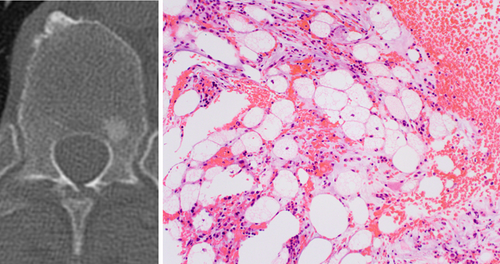
Eighteen cases were identified occurring in eight females and 10 males (median age = 65 years, range = 7–75). Imaging indication was cancer surveillance/staging in 11 patients and clinical concern for a metastasis was raised in 13 patients. The innominate bone (7), sacrum (5), mobile spine (4), humerus (1) and femur (1) were involved. Median tumour size was 1.5 cm (range = 0.8–3.8). Tumours were sclerotic (11), mixed sclerotic and lytic (4) or occult (1). Microscopically, tumours were composed of large polygonal cells with distinct cell membranes, finely vacuolated cytoplasm, central or paracentral small bland nuclei with prominent scalloping. Growth around trabecular bone was observed. Tumour cells were immunoreactive for S100 protein (15/15) and adipophilin (5/5), while negative for keratin AE1/AE3(/PCK26) (0/14) and brachyury (0/2). Chromosomal microarray analysis, performed on four cases, did not show clinically significant copy number variation across the genome or on 11q, the site of AIP and MEN1.
Conclusion
Analysis of 18 cases of intraosseous hibernoma, to our knowledge, the largest series to date, revealed that these tumours are most often detected in the spine and pelvis of older adults. Tumours were generally small, sclerotic and frequently found incidentally and can raise concern for metastasis. Whether or not these tumours are related to soft tissue hibernomas is uncertain.
Graphical Abstract
Intraosseous hibernomas are rare benign tumours most frequently detected as small sclerotic lesions in the spine and pelvis of older adults. Clinically and on imaging studies, these tumours frequently raised concern for a metastasis. Accurate classification is needed to prevent unnecessary procedures and assuage fears of an aggressive process.
Conflicts of interest
The authors have no conflicts of interest to disclose.
Open Research
Data Availability Statement
The data that support the findings of this study are available from the corresponding author upon reasonable request.


