Head and neck rhabdomyosarcoma with TFCP2 fusions and ALK overexpression: a clinicopathological and molecular analysis of 11 cases
Abstract
Aims
Primary intraosseous rhabdomyosarcoma (RMS) is a rare entity defined by EWSR1/FUS–TFCP2 or, less commonly, MEIS1–NCOA2 fusions. The lesions often show a hybrid spindle and epithelioid phenotype, frequently coexpress myogenic markers, ALK, and cytokeratin, and show a striking propensity for the pelvic and craniofacial bones. The aim of this study was to investigate the clinicopathological and molecular features of 11 head and neck RMSs (HNRMSs) characterised by the genetic alterations described in intraosseous RMS.
Methods and results
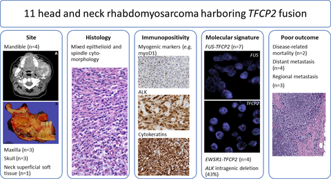
The molecular abnormalities were analysed with fluorescence in-situ hybridisation and/or targeted RNA/DNA sequencing. Seven cases had FUS–TFCP2 fusions, four had EWSR1–TFCP2 fusions, and none had MEIS1–NCOA2 fusions. All except one case were intraosseous, affecting the mandible (n = 4), maxilla (n = 3), and skull (n = 3). One case occurred in the superficial soft tissue of the neck. The median age was 29 years (range, 16–74 years), with an equal sex distribution. All tumours showed mixed epithelioid and spindle morphology. Immunohistochemical coexpression of desmin, myogenin, MyoD1, ALK, and cytokeratin was seen in most cases. An intragenic ALK deletion was seen in 43% of cases. Regional and distant spread were seen in three and four patients, respectively. Two patients died of their disease.
Conclusions
We herein present the largest series of HNRMSs with TFCP2 fusions to date. The findings show a strong predilection for the skeleton in young adults, although we also report an extraosseous case. The tumours are characterised by a distinctive spindle and epithelioid phenotype and a peculiar immunoprofile, with coexpression of myogenic markers, epithelial markers, and ALK. They are associated with a poor prognosis, including regional or distant spread and disease-related death.
Graphical Abstract
Conflict of interest
The authors state that they have no competing financial interests.


