Dynamic interplay of microRNA in diseases and therapeutic
Abstract
MicroRNAs are the major class of small non-coding RNAs, evolutionary conserved post-transcriptional regulators of gene expression. Since their discovery in 1993, they have been implicated as master regulators in numerous cellular processes. MicroRNA (miRNA)s regulate gene expression by attenuation and/or mRNA degradation and are commonly associated with cell development, differentiation, and homeostasis. Extensive research in past two decades has provided new insights into the potential implications of miRNA in the onset, progression, and therapeutic nature of miRNAs in disease manifestation. Owing to the novel discoveries, “miRNAs” would probably pave a new direction in therapeutic research. However, “micro” in length miRNAs have attracted considerable attention in numerous other fields. Understanding the functionality of miRNAs, in this review article, we discussed the mechanistic role of miRNAs in human diseases and have outlined most of the recent published work in clinical therapeutics. We have constructed different network models for miRNA and its targets which made us understand their interrelationship and association with diseases. Future research would surely overcome challenges and would introduce new strategies for the utility of miRNAs in a broader setting.
1 INTRODUCTION
Small RNAs regulate cellular processes, namely cell differentiation, cell proliferation, and transcriptional silencing. Small ncRNAs include siRNAs, snRNAs, microRNAs, and Piwi-interacting RNAs. MicroRNAs (miRNAs) are approximately 22 nucleotides long and play an important role in gene regulation by binding to the 3′untranslated regions of the target mRNA, causing translational inhibition/degradation of mRNA. The cellular functions of miRNAs vary among organisms, including pattern development in insects, leaf/flower development in plants, and hematopoietic cell differentiation in mammals. Novel microRNAs arise in several ways: random formation of hairpins in introns, inverted duplications of protein-coding sequences, and small, imperfect hairpins resulting from transcription of miniature transposable elements with inverted repeats.1 In 1993, Lee, Feinbaum, and Ambros, apparently discovered the first microRNA, “lin-4,” which represses expressin of lin-14, allowing the transition from the first to the second larval stage in Caenorhabditis elegans.2 Subsequent discoveries and research led to the establishment of the miRNA domain, which became established as a class of gene regulators. Subsequent discoveries of miRNAs led to the need for a repository tool, and thus miRBase (http://mirbase.org/), a primary online repository tool for miRNAs, was established in 2002 to provide information on published miRNAs.
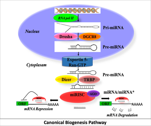
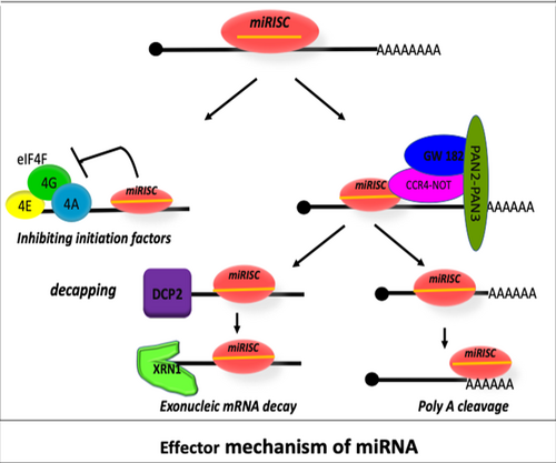
MicroRNAs originate from a dominant major pathway called the canonical biogenesis pathway, which involves cleavage of pri-miRNA by Drosha in the nucleus, followed by its export to the cytoplasm, where it is cleaved by Dicer (Figure 1). miRNAs regulate translational mechanisms by inhibiting the initiation of translation or recruiting factors for mRNA cleavage (Figure 2).
2 miRNA AND DISEASE ASSOCIATION
Studies suggest the role of miRNAs in complex biological pathways. Computational methods predict the target genes of miRNA, effectively reducing the time and cost of biological experiments by assessing the relationship between miRNA and disease. On March 27, 2019, the Human MicroRNA Disease Database HMDD v3.2 (http://cmbi.bjmu.edu.cn/hmdd) provided manually collected 35 547 miRNA-disease association records, covering 1206 miRNA genes and 893 diseases from 19 280 articles. This shows that miRNA plays an important role in clinical disease severity. In this section, we discuss the role of microRNAs in the context of haemoglobinopathies, cancer, neuronal dysfunction, autoimmune diseases, cardiomyopathies, infectious diseases, and graft-versus-host diseases.
2.1 MicroRNAs in haemoglobinopathies
Hemoglobinopathies are the most common monogenic disorders affecting the structure or production of hemoglobin. The two most common forms of disorders are sickle cell disease (SCD) and β-thalassemia. β-thalassemia is caused by the absence or decreased synthesis of the β-globin gene and has different clinical manifestations, ranging from mild anemia in the thalassemia intermedia group to severe transfusion dependence in the thalassemia major group.3 The safe and effective measure to treat hemoglobinopathies is to reactivate the γ-globin gene in patients.
Specific transcriptional regulators such as MYB, BCL11A, and KLF regulate fetal hemoglobin.4 Modulation of the expression of these regulators can effectively reduce clinical complications in patients. Gholampour et al. showed that higher miR-30a expression reduced BCL11A gene expression and thereby increased γ-globin gene expression.5 Sawant et al. showed significant association of miR-210 in inducing HBG2 expression in hydroxycarbamide-mediated SCA patients.6 Lulli et al. observed that higher expression of miR-486-3p resulted in decreased BCL11A protein level and increased γ-globin gene expression, while antagomir miR-486-3p resulted in increased BCL11A and decreased γ-globin expression.7 Srinoun et al. demonstrated that oxidative stress in SCD patients increases levels of nuclear factor erythroid 2-related factor 2 (NRF2) to control cellular ROS levels. In vitro analyzes showed that miR-144 represses the NRF2 gene and inhibits γ-globin gene transcription and HbF synthesis. Treatment with antagomiR-144 reverses this effect. In this study, the relationship between miR-144/NRF2 gene and HbF synthesis was demonstrated.8 Li et al. found that miR-326 suppresses EKLF expression in K562 cells by targeting its 3′-UTR to increase HbF levels.9 Wang et al. showed that miR-27a targets CDC25B and promotes erythroid differentiation and γ-globin gene activation in hemin-induced K562 cells.10 Ward et al. demonstrated that miR-34a regulates negative γ-globin regulators such as YY1, histone deacetylase 1, and STAT3. However, Western blot analysis of these regulators showed no change in YY1 and histone deacetylase 1 levels, but total and phosphorylated STAT3 levels were reduced in individual miR-34a-K562 cell clones. Thus, this study supports a role for miR-34a in the induction of γ-globin by STAT3 gene silencing.11
Pule et al. reported direct association of miR-26b and MYB and indirect modulation of BCL11A via activation of KLF-1, all of which together contribute to HbF induction.12 Cheng et al. showed that higher expression of miR-2355-5p suppressed the expression of KLF6 in HUDEP-2 and CD34+ cells, resulting in increased γ-globin expression. Thus, the experimental evidence demonstrates that the relationship between miR-2355-5p and KLF6 affects γ-globin expression, which may provide clinical insights for better treatment of patients with β-thalassemia or sickle cell anemia.13 Thus, overall, the studies suggest that miRNAs are used to stimulate HbF production in patients. However, further studies may be needed to achieve this goal in order for miRNAs to be more successful in treating patients with β-globin disorders.
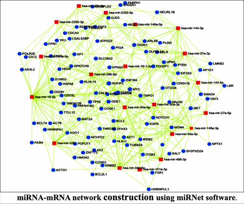
To interpret the overall biological and functional role of miRNAs in hemoglobinopathies, we generated miRNA-target network interactions using the miRNet web tool, which allowed us to identify miR target genes enriched in KEGG pathways (Figure 3). miRNet is a tool that allows identification of multiple targets of miRNAs, network construction, visualization, and functional enrichment of miRNAs. In addition, the tool enables easy customization of large networks by integrating multiple high-quality miRNA-target gene interactions. Therefore, the miRNet web tool was selected to build the miRNA-target gene network. The well-annotated databases such as miRTarBase v8.0 were used to retrieve the data of target genes searched against the input miRNAs list. The hypergeometric distribution was used to filter out less significant miRNA pairs with p > 0.05. The network analysis was based on the degree centrality and betweenness centrality of miRNAs and target genes. The network result showed the interaction of miRNA (red square node) and target genes (blue circle node). We found that miR-16 had the highest node degree and the highest centrality among all other miRNAs. The most abundant target genes of miRNAs were CCND1, MYC, MET, TAOK1, NOTCH2, HMGA2, FOXO1, TNPO1, and SLC38A2. Thus, network analysis could help researchers validate other target genes of miRNAs and investigate their role in haemoglobinopathies.
2.2 MicroRNA in cancer
Tumorigenesis is triggered by altered expression of oncogenes and tumor suppressor genes, which together drive cancer development and progression. miRNAs have been widely studied and recommended as effective biomarkers for cancer diagnosis and even as promising targets for cancer therapy. In 2008, Calin et al. discovered for the first time an association between miR-15a and miR-16-1 and chromosomal aberrations in chronic lymphocytic leukemia.14 Subsequent pioneering studies led to the discovery of several miRNAs involved in many cancer pathways. Acute myeloid leukemia is the most common type of hematologic malignancy, and although patients are treated with chemotherapy, major clinical problems still occur. Identification of novel biomarkers would help stratify early diagnosis. Li et al. showed higher expression of the chaperone protein HSPA8 in patients, which promotes cancer cell proliferation. A miRNA network study confirmed the positive correlation of hsa-mir-508-3p, hsa-mir-1269a, and hsa-mir-203a with HSPA8 expression. Thus, the study suggests a likely association between the miRNA-HSPA8 network and an effective prognostic factor.15 Breast cancer is the most common malignancy in women, which results from uncontrolled proliferation of ductal and lobular epithelial cells of the breast. Although triple test and radiological ultrasonography are routine diagnoses, there are still limitations in early detection. Diansyah et al. demonstrated higher expression of miR-21 in plasma in early stage of breast cancer compared with normal, serving as risk predictors in the early stages of breast cancer.16 Cervical cancer (CC) is caused by human papillomavirus (HPV) infection. Currently, the HPV DNA test is used to screen for CC, but it is less specific because most HPV infections do not result in cervical lesions. Therefore, biomarker identification is important for cervical lesion screening. Temimi et al. reported lower miR-432 expression in patients compared with controls, suggesting that downregulated expression of miR 432 is an indicator of CC progression.17 Hepatocellular carcinoma (HCC) is a malignant liver cancer that has an inadequate prognosis due to limited treatment options. Hu et al. demonstrated increased expression of multidrug resistance-associated protein 4 (MRP4) in liver cancer tissues. Transfection studies showed that miR-4524-5p and miR-124-3p reduced MRP4 expression at the protein level. In vivo and luciferase assays showed, that circHIPK3 binds to miR-4524-5p and miR-124-3p. Knockdown of circHIPK3 leads to downregulation of MRP4 protein, whereas its expression is restored by co-transfection of circHIPK3 siRNA miR-4524-5p and miR-124-3p or inhibitors. Thus, this study shows for the first time that miR-4524-5p downregulates MRP4 expression and circHIPK3 regulates MRP4 expression by sponging miR-124-3p and miR-4524-5p.18
Ovarian cancer has a high mortality rate in adult women. It is important to find and develop a new effective treatment. Yang et al. showed that higher expression of miR-802 suppressed the proliferation of ovarian cancer cells by silencing the expression of YWHAZ.19 Li et al. showed that overexpression of XIAP correlated with cancer cell drug resistance. Therefore, suppression of XIAP is achieved by transfection of miR-137 mimic into SKOV3 ovarian cancer cells. Disruption of miR-137 using CRISPR/Cas9 leads to an increase in XIAP protein, suggesting that miR-137 regulates XIAP in ovarian cancer.20 miRNAs appear to influence various features of cancer, so a comprehensive understanding of miRNAs in cancer biology may pave new ways for cancer therapy in the future.
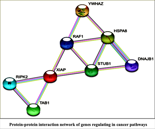
Protein structure and functional dynamics are disrupted in cancer. Therefore, the study of protein–protein interaction would help to understand the role of proteins in biological processes. The PPI network was created using 3 commonly dysfunctional cancer proteins YWHAZ, XIAP and HSPA8. YWHAZ is the nodal protein involved in tumor progression in many cancers such as HCC, colorectal cancer, gastric cancer, lung cancer, breast cancer, and prostate cancer.21 XIAP plays a central role in inhibiting apoptosis and is therefore the major target for cancer therapeutics.22 Heat shock protein chaperones (HSPAs) are known to promote cancer cell growth.23 Therefore, we constructed a protein–protein interaction network (PPI) using STRING to analyze the interaction between miRNA targets involved in cancer pathways (Figure 4). The Ensembl Ids of these targets were entered into the multi-protein identifier section of the STRING version 11.5 web tool. The result showed a PPI network with 8 prominent nodes (protein), 12 edges, average node degree (number of connections of the node in the whole network): 3, average local clustering coefficient: 0.729 and PPI enrichment p-value (p:0.0415). The additional hub proteins that interacted with our 3 entered targets were DNAJB1, STUB1, RAF1, TAB1 and RIPK2. A total of 8 targets (hubs) were enriched in biological processes with a significant false discovery rate (FDR), such as chaperone-mediated autophagy (FDR = 0.016), MAPK cascade (FDR = 0.018), GP1b- IX -V activation signaling (FDR = 0.01), NF-kappa B signaling pathway (FDR = 0.011), and so forth. Thus, the constructed PPI network of cancer hub proteins shows that most of these signaling pathways are of global importance for various cancers. The dysfunction of these additional target proteins may help researchers gain insights into the complex interphase of cancer.
2.3 MicroRNA in autoimmune disease
Autoimmune diseases are rare and complex immune disorders resulting from a loss of self-tolerance. However, the exact mechanism of autoimmune disease is not yet understood. Recently, researchers have focused on the role of miRNAs in immune regulation complexes. Systemic lupus erythematosus (SLE) is characterized by inflammation in connective tissue, cartilage, and the lining of blood vessels. Transfection studies by Shi et al. showed that inhibition of miR 29a increased IgG production by regulating CRKL protein expression in B cells.24 Rheumatoid arthritis (RA) is characterized by inflammation of the synovial membrane. Jiang et al. reported that miR-147b-3p contributes to synoviocytes by suppressing ZNF148 in experimental arthritis.25 Multiple sclerosis (MS) is a neuroinflammatory disease of the brain and spinal cord. Numerous studies have confirmed that miRNAs are valuable pathological and diagnostic prognostic markers. Mosarrezaii Aghdam et al. reported an association of miR-125a-5p and miR-218-5p with disease pathogenesis.26 Myasthenia gravis (MG) is a neuromuscular disease that results in skeletal muscle weakness. Although the determination of serum antibodies of acetylcholine receptor (AChR+) or muscle-specific tyrosine kinase (MuSK+) is often insufficient to reflect disease progression, the development of a reliable biomarker panel would help to understand disease progression. Sabre et al. analyzed the lower expression of miR-210-3p and miR-324-3p in MuSK+ MG patients compared with healthy individuals.27 Therefore, these miRNAs may serve as diagnostic markers in the future.
2.4 MicroRNA in neurodegenerative diseases
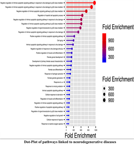
Neurodegenerative diseases (NDD) are characterized by the progressive loss of neuronal gene networks, which often results from the loss of synapses and degeneration of the motor neuron network. miRNAs have been implicated in the neurogenic process. Alzheimer's disease (AD) is a progressive NDD characterized by the deposition of amyloid-beta (Aβ) and neurofibrillary tangles. The 2012 study by Zhu et al. confirmed that miR-195 inhibits the translation of beta-site APP cleaving enzyme 1 (BACE1), resulting in reduced Aβ plaque formation.28 Zhang et al. showed that increased expression of miR-9-5p leads to decreased expression of Xenopus kinesin-like protein 2 (TPX2), while inhibition of interleukin-1β leads to suppression of Aβ-plaques in mice.29 Parkinson's disease (PD) is a common NDD characterized by selective loss of midbrain dopaminergic neurons (DA). Caggiu et al. observed upregulated miRNA-155-5p and downregulated miR-146a-5p expression in PD patients compared to controls. However, levodopa-treated patients showed downregulated miR-155-5p expression, suggesting that miRNA may be a promising candidate for disease prognosis.30 Wang et al. showed that higher expression of miR-93 inhibited STAT3, resulting in inhibition of iNOS, IL −6 and TNF, and elevation of TGF-1 and IL −10. This indicates that miR-93 may be involved in the pathogenesis of PD.31 Huntington's disease (HD) is an inherited lethal NDD caused by abnormal aggregation of mutant huntingtin protein (mHTT). A study confirms the decreased expression of miR-124, which is responsible for neurogenesis in the mouse brain HD. Thus, it plays an important role in neuronal differentiation and survival.32 Cheng et al. reported higher miR-196a expression in transgenic mice, which resulted in suppression of mutant HTT and reduction of nuclear, intranuclear and neurophilic aggregates. Thus, miR-196a suggests a potential therapeutic role in HD.33 Amyotrophic lateral sclerosis (ALS) is characterized by motor neuron degeneration. Li et al. demonstrated increased expression of miR-338-3p in mouse glycogen phosphorylase (PYGB), which is responsible for decreased glycogenolysis and resulting glycogen accumulation. Thus, the study provides a better understanding of the role of miR-338-3p in biochemical metabolic pathways in ALS.34 The overall ground breaking work allows us to understand that miRNA regulates genes involved in NDDs. To understand various pathways associated with NDD, we used Gene Ontology and the KEGG Pathway Analysis Tool. To analyze the enrichment of genes involved in NDD, we used the Shiny GO tool (Figure 5). We selected 6 target genes regulated by miRNAs for enrichment analysis: BACE1, TPX2, BCL2, SIRT1, STAT3, and iNOS. We found the top 30 GO and KEGG-enriched pathways associated with miRNAs involved in NDD. Pathway enrichment analysis is very useful for computational biologist to integrate the information about the most enriched pathways from numerous target genes. In this way, pathway enrichment imaging would improve the understanding of the complex mechanisms of NDD and help to gain in-depth knowledge of the interconnectedness of target genes in an enriched pathway.
2.5 MicroRNA in cardiomyopathies
Cardiomyopathy is a leading cause of heart failure because it hardens the heart muscle, making it difficult for the heart to collect or pump blood. Imaging techniques have been used to identify problems associated with the heart muscle by developing a genetic panel that would help understand the onset and progression of heart disease. Obradovic et al. analyzed the higher expression of miR-206 and miR-155 in plasma of patients with inflammatory cardiomyopathy compared with normal patients, so the study could provide a path to noninvasive diagnosis via advanced technology.35 Diabetic cardiomyopathy is the most common cardiac problem in patients with diabetes mellitus. Li et al. showed higher expression of miR-30d in diabetic rats, which induces pyroptosis of cardiomyocytes, while knocking down of miR-30d inhibits it.36 Myocardial hypertrophy is a major cause of heart failure. Overexpressed miRNA-20 induced cardiomyocyte hypertrophy by inhibiting mitofusin-2 (MFN2). Thus, miRNA-20 plays an important role in promoting cardiomyocyte hypertrophy.37 Park et al. demonstrated increased apoptosis in diabetic rat models when transfected with miR-34a mimic. In contrast, reduced miR-34a activates anti-apoptosis in rat models.38 Xie et al. indicated that miR-146a can regulate Toll-like receptor 4/NF κB signaling pathway, leading to improvement of inflammatory response in septic cardiomyopathy.39
2.6 MicroRNA in infection diseases
Infections are usually caused by non-human creatures and are often transmitted from an infected person to a normal person. HIV infection affects the immune system, especially the crucial T cells. Nguyen et al. propose that GAS5 regulates apoptosis of CD4+ T cells via miR-21-mediated signaling.40 Zheng et al. study shows that miR-191-5p inhibits HIV-1 replication by suppressing CCR1 and NUP50 expression.41 Hepatitis B virus (HBV) infection is one of the most common causes of liver cancer with high mortality. Li et al. demonstrated higher expression of miR-487b in serum of infected patients, thus miR-487b can be used as a diagnostic marker for HCC.42 Li et al. demonstrated that overexpression of miR-1271-5p inhibits HBV antigens and suppresses tumor growth by reducing the expression of AQP5.43 Tuberculosis (TB) is caused by Mycobacterium tuberculosis, which most commonly affects the lungs but can also affect any other part of the body. Diagnosis of TB is a major challenge that requires the development of accurate diagnostic tools. There is extensive evidence that miRNAs strongly regulate macrophage defense against mycobacterial infection. Zhang et al. demonstrated that overexpression of miR-32-5p promotes the survival of intracellular mycobacteria by negatively regulating the expression of the target protein follistatin-like protein 1 (FSTL1). While inhibition of miR-32-5p impairs mycobacterium survival.44 In late 2019, a terrible pneumonia of unknown cause occurred in Wuhan, China. Several laboratories in China identified the causative agent of this mysterious pneumonia as novel coronavirus (nCov) or severe acute respiratory syndrome coronavirus 2 (SARS-CoV2). The basic principle of coronavirus replication is largely understood. Most viruses produce their miRNAs, but the exact role of viral miRNAs in viral replication, translation, or infection is not fully understood. Ivashchenko et al. proposed the use of miRNAs regulating protein translation of coronaviruses. The miRTarget program was used to identify the binding site of miRNAs to viral mRNA. Of the total 2565 miRNAs, miR-4778-3p, miR-6864-5p, and miR-5197-3p were identified as the most suitable for binding to the genomic RNA of SARS-CoV, MERS-CoV, and COVID-19, respectively. A complete complementary miRNA-binding site (cc-miRNA) was generated based on the sequences of miR-4778-3p, miR-6864-5p, and miR-5197-3p. Interestingly, these cc-miRNAs did not bind to the human genome, indicating a lack of off-target activity of the miRNAs. The study suggests that cc-miRNAs can be introduced into exosomes or vesicles and then suppress viral replication when they enter the blood and other organs.45 An interesting study by Centa et al. identified miRNAs in postmortem lung biopsies from patients with severe respiratory and thrombotic events. Functional enrichment and miRNA mRNA network analysis revealed a close association of miR-26a-5p, miR-29b-3p, and miR-34a-5p with endothelial activity. miR-26a-5p and miR-29b-3p also correlated positively with inflammatory biomarkers such as IL-6, ICAM-1, and IL4 and IL8, respectively. These results support the importance of miRNAs in COVID-19 patients with endothelial dysfunction and inflammatory response.46 A study by Farr et al. demonstrated a host-encoded microRNA response to SARS-CoV-2 infection. Expression profiling of circulating miRNAs from 10 infected patients showed altered expression of 55 miRNAs in early disease. Machine learning analysis showed that miR-423-5p, miR-23a-3p, and miR-195-5p were detected as characteristic miRNAs for SARS-CoV-2 infection with 99.7% accuracy. Thus, the study was able to distinguish SARS-CoV-2 infection from influenza A (H1N1) infection.47
2.7 miRNA in graft versus host diseases
Graft-versus-host disease (GvHD) can occur in patients who receive a transplant from HLA-mismatched donors. Despite recent scientific advances, there is no developed biomarker panel to determine the clinical complications and progression of GvHD. miRNA have long been known to regulate T and B cell development and proliferation. Wu et al. reported that expression of miR-17-92 promotes T cells, B cells, and plasma cells. However, transfection of antimir-17 suppresses B cell activation in the cGvHD model.48 Liu et al. showed that miR-223 can inhibit the clinical syndrome of aGvHD in mice by suppressing the migration of donor T cells.49 Crossland et al. demonstrated miR-155 and miR-146a as non-invasive biomarkers for acute GvHD.50 A similar study also reported that miR-155 strongly affects the expression of alloreactive T cells in acute GvHD.51 miR-181a affects the function of T lymphocytes by downregulating the target cytokine IFN-γ, thereby attenuating the severity of acute GvHD.52 Ranganathan et al. indicated that miR-29a activates dendritic cells via TLR7 and TLR8, leading to activation of the NF-κB pathway and secretion of the proinflammatory cytokines TNF-α and IL-6. miR-29a significantly improved immune status in a mouse model of GvHD.53 Thus, overall, these studies highlight the contribution of miRNA in the clinical manifestation of GvHD.
miRNAs have gradually attracted the attention of biomedical researchers. In fact, the research would undoubtedly expand their applications and provide a new direction in clinical therapy. The main findings on miRNA in human diseases are summarized in Table SS1.
3 INSIGHTS OF MicroRNA AS CLINICAL THERAPEUTICS
MicroRNAs have an impact on gene regulation, so there is a need to expand miRNAs from laboratory research to clinical applications. Microarray profiling allows us to identify candidate miRNAs responsible for gene regulation, while their targets could be validated by in vitro studies. Therefore, miR target association study would pave a new way for the development of therapeutic strategies in the pharmaceutical industry.
3.1 miRNA-mimics and antimiRs
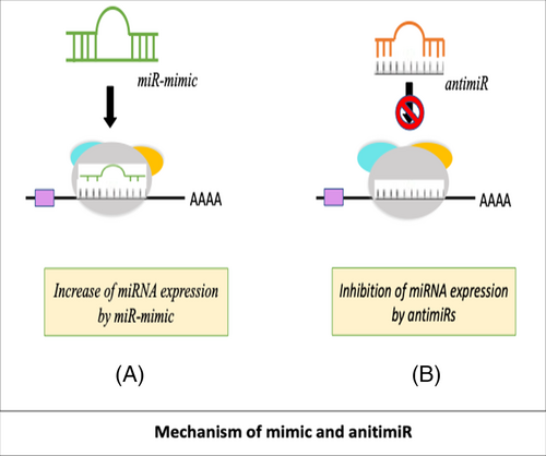
miRNA mimics and antimiRs are specialized and innovative synthetic molecules to enhance or inhibit miRNAs. miRNA mimics and antimiRs are synthetic RNA fragments that mimic or inhibit the expression of endogenous miRNA.54 The interaction of miR-mimics/antimiRs is shown in Figure 6A,B. The mimics/antimiRs are delivered into cells via a miRNA vehicle. An ideal delivery vehicle should protect the miRNA from degradation, immune testing, and off-target effects. Numerous miRNA delivery strategies have been developed that would ensure stable integrity of miRNA in cells.
3.2 miRNA vehicle or miRNA delivery system
The two most commonly used miRNA delivery systems include viral and nonviral vectors.55 A recent study validated efficient delivery of antimir-199a by adeno-associated virus (AVV) to treat cardiac hypertrophy by restoring mitochondrial function in mice.56 Suardi et al. used positively charged chitosan nanoparticles that electrostatically interacted with the poly-anionic combination of miR-155-5p and miR-324-5p. The ionic electrostatic crosslinking approach enhances the molecule delivery.57 Castaño et al. introduced nanohydroxyapatite (nHA) particles in combination with reporter miRNAs and collagen nanohydroxyapatite scaffolds to efficiently deliver miRNA to human mesenchymal stem cells.58 The liposome/microbubble formulation was used to deliver miR-133 mimics to suppress cardiac hypertrophy in vitro.59 A stearylamine-based cationic liposome (SA) was used to deliver a miR-191 inhibitor to treat breast cancer.60 A novel lipid-based nanoparticle enabled efficient delivery of anti-miR-155 to HCC using lactosylated gramicidin-containing lipid nanoparticles (Lac-GLN) in mice.61 Di Martino et al. encapsulated a miR-34a mimic in stable nucleic acid particles to treat myeloma.62 Thus, viral and non-viral miRNA delivery systems have opened a new avenue for the introduction of miRNA into clinical research.
4 miRNA AS CUSTOMIZED MEDICINE IN THE FUTURE?
The role of miRNA as a gene modulator has attracted the attention of many researchers to develop customized cell models. MRG-106′, an inhibitor of miR-155-5p, showed significant improvement in 6 mycosis fungoides patients (MF) by intratumoral injection of MRG-106, making it as the first inhuman application of miR treatment.63 Abplanalp et al. targeted endogenous miR-92a-3p with “MRG-110” for the treatment of cardiovascular disease and wound healing.64 miR-34 is known to regulate cancer-associated genes and also suppress hematologic malignancies. MRX34, a liposome-based double-stranded miR-34 mimic, was developed and was in phase I clinical trials in cancer patients.65 “Miravirsen” is a β-d-oxy-locked nucleic acid-modified phosphorothioate antisense oligonucleotide with antiviral activity in HCV infection that targets endogenous miR-122.66 miRNAs serve as therapeutic targets for the alleviation of numerous diseases. Moreover, the study of miRNA pharmacodynamics would definitely open a new avenue in therapeutic technology.
5 CONCLUSION
MicroRNA research is spreading enormously in various other fields. The studies offer a promising perspective for miRNA identification as biomarkers. MicroRNA profiling can be used for early disease diagnosis in patients, and it also improves our ability to determine their functional role in diseases and their therapeutic and clinical implications. This review article outlines the impact of miRNA in various disease groups and suggests that microRNA can be used as a novel target for improving disease severity. Therefore, in the future, it is necessary to transfer miRNAs from laboratory research to pharmaceutical industry. Future research will certainly pave the way for miRNA therapeutics.
AUTHOR CONTRIBUTIONS
Neha Kargutkar and Priya Hariharan: wrote the manuscript; Anita Nadkarni: helped in designing and writing the manuscript and data analysis.
ACKNOWLEDGMENTS
We thank ICMR-National Institute of Immunohaematology for providing a research platform. We also thank the University of Mumbai for providing an opportunity to study higher aspects of the research field.
CONFLICT OF INTEREST
The authors declare no conflicts of interest.
Open Research
DATA AVAILABILITY STATEMENT
The review article is written using the information available in the previously published papers


