Novel CDH3 variants in Brazilian families with hypotrichosis and juvenile macular dystrophy revealed by exome sequencing
Funding information: Coordenação de Aperfeiçoamento de Pessoal de Nível Superior, Grant/Award Number: Student Scholarship; Fundação da Universidade Federal do Paraná, Grant/Award Number: FUNPAR-LIGH Alliance 3301
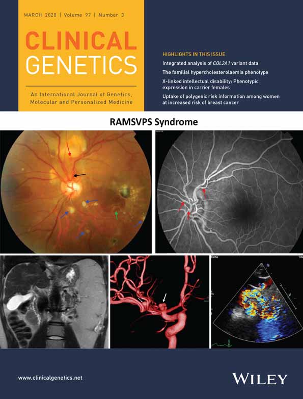
Graphical Abstract

Hypotrichosis and Juvenile Macular Dystrophy (HJMD, MIM:601553, autosomal recessive) is a rare disease, caused by mutations in the CDH3 gene (NM_001793.6, MIM:114021), with onset early in life, characterized by thin and sparse scalp hair and progressive retinal degeneration. In 2002, our group described one Brazilian family (Family #1) with macular dystrophy associated with Loose Anagen Hair Syndrome (LAHS [MIM: 600628]).1 As this association was not previously described, a new syndrome was proposed.1-4 Subsequently, two further families with macular dystrophy and alopecia were referred to us for evaluation (Families #2 and #3). Despite phenotypic variability, we hypothesized that these patients had HJMD. The Ethics Committee of Hospital de Clínicas-UFPR approved this study and informed consent was obtained from all participants.
To identify genetic variants associated with patients' phenotypes, we performed whole exome sequencing (WES) in seven individuals (6 patients, 1 control). We used SIFT, PolyPhen, MutationTaster, PhyloP and SiPhy tools for variant effect prediction and sequence-based genotyping for validation. Medical history was obtained from all families. All subjects underwent ophthalmic and dermatological evaluation. Spontaneously shed hairs assessment was performed by light and scanning electron microscopy (SEM).
Family #1 (Figure 1A-G) comprised four patients born with LAHS and progressive visual impairment.1-4 Clinical follow up showed the progressive course of macular degeneration. Genetic analysis revealed all patients as homozygotes for the variant c.160+1G>A (exon 2), which disrupts a highly conserved consensus donor splice site in CDH3 gene.5

Family #2 (Figure 1H-K) comprised two patients, both born with thin sparse scalp hair with no growth and progressive visual impairment. One patient had nystagmus and the other had inborn cleft lip and palate. Dermatological investigation did not associate clinical findings with any specific hypotrichosis. Molecular analysis in both patients identified compound heterozygosity for missense variants c.160+1G>A (exon 2) and c.1063G>T:p.Asp355Tyr (novel variant in exon 9) in CDH3 gene. The latter has been predicted to be deleterious (ClinVar SCV000493832.1).
Family #3 (Figure 1L-O) comprised two patients born with thin sparse scalp hair and progressive visual impairment. Patient III-10 is more severely affected. Hair shaft microscopy revealed abnormalities as longitudinal groove, bulb dystrophy, flattening and dystrophic anagen bulb. Both patients presented a new deleterious splicing site variation c.1795+1G>C (exon12) in the CDH3 gene (ClinVar SCV000493833.1). Patient III-10 had an additional heterozygous missense variation in the KCNV2 gene (c.1262G>A, rs563513486), likely pathogenic and probably responsible for worse visual acuity in this patient.
Electroretinography of patients showed decreased amplitude and increased implicit time in both scotopic and photopic adaptations. Patients' electrooculogram was subnormal in families #1 and #2, and normal in patient III-10 from family #3 (electroretinography and electrooculogram data from patient III-11 were not available). Ophthalmoscopy showed RPE atrophy and pigment dispersion in posterior pole.
No gene variant other than CDH3 and KCNV2 was found explaining the observed phenotype variations.
Our investigation lead to the first genetic diagnosis of HJMD in Latin America, with the description of two novel CDH3 variants. Additionally, we reported a CDH3 variant segregating with LAHS in one family.
ACKNOWLEDGEMENTS
We thank CAPES, FUNPAR, Retina e Vitreo Consultoria, CME/UFPR and LIGH/UFPR for financial and/or technical support and patients for participation.
Open Research
DATA AVAILABILITY STATEMENT
Data supporting these findings is available upon reasonable request.