Molecular genomic landscapes of hepatobiliary cancer
Abstract
Hepatocellular carcinoma (HCC) and biliary tract cancer are more frequent in East Asia including Japan than in Europe or North America. A compilation of 1340 multi-ethnic HCC genomes, the largest cohort ever reported, identified a comprehensive landscape of HCC driver genes, comprised of three core drivers (TP53, TERT, and WNT signaling) and combinations of infrequent alterations in various cancer pathways. In contrast, five core driver genes (TP53, ARID1A, KRAS, SMAD4, and BAP1) with characteristic molecular alterations including fusion transcripts involving fibroblast growth factor receptor 2 and the protein kinase A pathway, and IDH1/2 mutation constituted the biliary tract cancer genomes. Consistent with their heterogeneous epidemiological backgrounds, mutational signatures and combinations of non-core driver genes within these cancer genomes were found to be complex. Integrative analyses of multi-omics data identified molecular classifications of these tumors that are associated with clinical outcome and enrichments of potential therapeutic targets, including immune checkpoint molecules. Translating comprehensive molecular-genomic analysis together with further basic research and international collaborations are highly anticipated for developing precise and better treatments, diagnosis, and prevention of these tumor types.
Abbreviations
-
- 1,2-DCP
-
- 1,2-dichloropropane
-
- AA
-
- aristolochic acid
-
- AAV2
-
- adeno-associated virus type 2
-
- ARID
-
- AT-rich interaction domain
-
- BTC
-
- biliary tract cancer
-
- CCA
-
- cholangiocarcinoma
-
- CDKN2A
-
- cyclin-dependent kinase inhibitor 2A
-
- COSMIC
-
- Catalogue of Somatic Mutations in Cancer
-
- CTNNB1
-
- catenin β1
-
- ECC
-
- extrahepatic cholangiocarcinoma
-
- GB
-
- gallbladder cancer
-
- HBV
-
- hepatitis B virus
-
- HCC
-
- hepatocellular carcinoma
-
- HCV
-
- hepatitis C virus
-
- ICC
-
- intrahepatic cholangiocarcinoma
-
- TCGA
-
- The Cancer Genome Atlas
-
- TERT
-
- telomerase reverse transcriptase
-
- TPM
-
- TERT promoter mutation
-
- WES
-
- whole exome sequencing
-
- WGS
-
- whole genome sequencing
1 GEOGRAPHICAL AND EPIDEMIOLOGICAL DIVERSITIES OF HEPATOBILIARY CANCERS
Hepatocellular and biliary tract cancers are both dismal tumors. The frequencies of these tumors are geographically more common in East Asia including Japan and BTC is especially more frequent in Southeast Asian countries such as Thailand.
The epidemiological backgrounds of these tumor types are also unique. Hepatitis virus infection, aflatoxin B exposure, alcohol intake, and other metabolic diseases (including obesity, diabetes mellitus, and hemochromatosis) are well-known risk factors for HCC.1, 2 Hepatitis B virus infection is a worldwide health problem, whereas HCV is more prevalent in Japan. However, the number of patients infected with HCV has been rapidly increasing in developed countries, especially the USA. Biliary tract cancer encompasses three subtypes based on the location: ICC, ECC, and GB. The former two are collectively called CCA. Primary sclerosing cholangitis and bile duct anomaly are the well-known predisposing factor for ICC.3 In South-East Asia, especially in Northeast Thailand, parasite infection, particularly the endemic liver flukes such as Opisthorchis viverrini, is a major epidemiological factor for CCA.4
2 DRIVER GENE LANDSCAPE IN THE LARGEST HCC COHORT
Several landmark studies of WES and WGS, including those from the International Cancer Genome Consortium and TCGA, have already analyzed large-scale cohorts of HCC in different epidemiological and ethnic backgrounds.5-8
At present, genomic data from more than 1000 HCC cases are publicly available. We compiled these data from four previous studies that included 1340 cases from different ethnic groups, including 642 cases from a Japanese cohort, 451 cases from TCGA, and 247 cases from a French cohort (Table S1). Using the largest HCC cohort ever reported, we attempted to identify driver genes that could evaluate the most precise frequencies of mutations in HCC. We defined a driver gene as a statistically significantly mutated gene after adjusting the background mutation rate (Appendix S1). Thirty-three significantly mutated driver genes (q-value < 0.1; Table S2) are listed in Figure 1. The two most frequently mutated genes were TP53 (29.1% of total cases) and CTNNB1 (28.6%). WNT signaling regulates cell proliferation, stem cell maintenance, and other oncogenic signals, and CTNNB1 plays a central role in activating WNT signaling. Previous sequencing analyses of different ethnic groups reported that these two genes were the most frequent, confirming that they are master driver genes of HCC beyond the diversities of ethnic/epidemiological backgrounds.
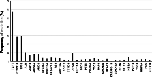
Telomerase reverse transcriptase restores telomere length during replication, and is required for cell transformation or escaping from senescence. Recently, TPMs were reported in a broad range of human cancers including HCC.9 Because TPMs were analyzed in part by studies we compiled (TPMs were analyzed in 1093 cases) and are difficult to be precisely identified by Illumina sequencing due to their presence in the highly GC rich genome area, the true frequencies of TPMs could not be established in the current study. However, four large-scale studies reported highly frequent TPMs (>50%) in Japanese, French, and US (TCGA) cohorts,5, 7-10 suggesting that TERT also belongs to the master HCC driver genes. TERT promoter mutations are reported to occur in dysplastic nodules or early HCC, suggesting that TPMs could be founding driver genes in HCC.11 Chiba et al12 reported that TPMs have a two-step mechanism in cell transformation: promoting immortalization in the initial phase, and induction of genomic instability in the later stage. Collectively TP53, CTNNB1 (WNT signal), and TERT (immortalization) are the core driver genes in HCC of all ethnic groups (Figure 2A).
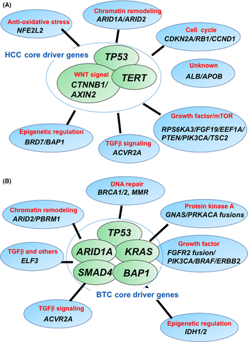
The second most frequently mutated group (>7%) of HCC driver genes included ALB (10.2%), APOB (9.8%), ARID1A (8.8%), ARID2 (8.2%), and AXIN1 (7.5%). Both ARID1A and ARID2 encode components of SWI/SNF chromatin remodeling machinery, and their genetic alterations are also reported in other tumor types.13 Because SWI/SNF machinery regulates chromatin conformation in an ATP-dependent manner, their alterations are supposed to affect epigenetic changes in cancer cells; however, the biological roles of ARID1A/ARID2 mutations in HCC remain to be clarified. Recently, Sun et al14 reported context-dependent roles of ARID1A in hepatocarcinogenesis by analyzing animal models. Axin 1 promotes degradation of CTNNB1 protein and negatively regulates WNT signaling. Therefore, its inactivation is largely complimentary to CTNNB1 activating mutations for WNT signal activation in HCC.15 In contract to these well-characterized tumor suppressor genes, the biological roles of highly frequent mutations of ALB and APOB in HCC are uncertain. Because both ALB and APOB mutations were strongly enriched in indels, these indels could be caused by replication slippage errors and may result from conflicts between the replication and transcription machineries.16
The remaining top driver genes were mutated in a fraction of cases (<5%) with various biological functions (Figure 2A). CDKN2A, RB1, and CCND1 regulated cell cycle checkpoints, whereas RPS6KA3, PTEN, PIK3CA, and TSC2 are implicated in growth factor signaling and the mTOR pathway. EEF1A1 encodes the alpha subunit of the elongation factor-1 complex, which is responsible for the enzymatic delivery of aminoacyl tRNAs to the ribosome, which has oncogenic and invasive activities.17 They also include therapeutic targets such as PIK3CA, PTEN, and CCND1. These infrequently mutated driver genes might contribute to specific cellular phenotypes such as aggressive growth, invasiveness, or metastasis. Alternatively, combined with other types of alterations or alterations of other genes involved in the same functional pathways, they could play more major roles in HCC.
There are little data regarding non-coding driver genes in HCC. Fujimoto et al7 reported the largest WGS of 300 Japanese cases of HCC and identified recurrent non-coding mutations. They found that TPM was the most frequent and significant mutation, followed by NEAT1 and MALAT1, both of which are non-coding RNAs.
3 STRUCTURAL REARRANGEMENTS AND VIRUS INTEGRATION IN HCC
Copy number alterations and structural rearrangements occur frequently in HCC.5-8 Although rare, chromothripsis was also reported in HCC.7 A broad range of cancer-related genes including oncogenes (including MYC, TERT, CCND1, and MET) and tumor suppressor genes (including TP53, RB1, ARID1A, CDKN2A, and APC) were affected by these molecular events.5, 7, 8 In addition to somatic rearrangements, HBV genome integration also affects gene expression in HCC. Recurrent targets of HBV integration include TERT, MLL4, and CCNE1, and their expressions were elevated by virus genome integrations.18, 19 Copy number alterations were also significantly increased at HBV breakpoint locations, implying that HBV genome integration was associated with chromosomal instability.18, 19
Three groups reported integration of the AAV2 genome in HCC.7, 20, 21 Integrations of the AAV2 genome were detected 5.7% of French HCC cases.20 It was reported that AAV2-positive tumors tended to occur in non-cirrhotic liver without known risk factors, suggesting that AAV2 genome integration might play an oncogenic role in some of the cases currently classified as risk-free. Integration of AAV2 was reported to be rarer (~1%) in Japanese (3/300) and Korean (2/289) cohorts.7, 21
4 MUTATIONAL SIGNATURES IN THE HCC GENOME
Endogenous and exogenous mutational processes induced by various factors, such as defects in DNA repair or exposure to smoking-associated carcinogens, leave unique nucleotide changes (substitutions, insertion, or deletions) within characteristic sequence contexts (Figure 3). Examining such patterns of somatic mutations, called “mutational signatures”, can provide an indication of the etiology of the operative mutational processes in each cancer case. Analyzing more than 7000 cancer genome sequences uncovered more than 30 distinctive mutational signatures operating in human cancers.22
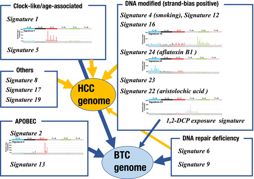
Totoki et al5 extracted mutational signatures in WES data of 503 HCC cases from both Japanese and TCGA cohorts. They isolated three mutational signatures (COSMIC signatures 1, 16, and 22) in that dataset, and reported unique associations between mutational signature and ethnic backgrounds. Although COSMIC signature 1 (NpCpG>NpTpG) was common in all ethnic groups, COSMIC signature 16 (ApTpN>ApCpN) was more frequent in Japanese male patients, and COSMIC signature 22 (AA exposure signature) (T>A substitution) was preferentially detected in Asian patients living in the USA. Surprisingly, virus status (HBV, HCV, and non-viral) did not show significant associations with any mutational signature. Fujimoto et al7 extracted six COSMIC mutational signatures (COSMIC signatures 1, 4, 9, 12, 16, and 19) and one new signature (signature W6) WGS data from 300 cases. Each mutational signature was associated with epidemiological, clinical, and driver gene status, which was revealed by multiple linear regression analysis. COSMIC signature 4 (C>A substitution) was associated with smoking status, patient age, clinical history of double cancer, and the presence of TP53 mutation. COSMIC signature 16 was associated with alcohol consumption and the presence of TPM and CTNNB1 mutation, and also male gender and older age. No association between virus status and each mutation signature was also confirmed in this study.
Schulze et al6 analyzed mutational signatures in 233 cases of HCC from Europe and Africa. They identified eight mutational signatures, including two new ones that were later classified as COSMIC signatures 23 and 24. COSMIC signature 24 was found to be an aflatoxin B1-associated signature, and was more frequent in African cases with significant enrichment of a somatic TP53 p. Arg249Ser mutation, which is a typical mutation of aflatoxin B1-exposed HCC. Letouzé et al23 further analyzed WGS of European HCC cases and identified 10 mutational signatures (COSMIC signatures 1, 4, 5, 6, 12, 17, 16, 22, 23, and 24). Of note, they affirmed a significant association between COSMIC signature 16 and epidemiological backgrounds such as male gender, alcohol intake, and tobacco consumption. They also reproduced the significant association between the presence of CTNNB1 mutation and the frequency of COSMIC signature 16.
Recently, Ng et al24 reported broad prevalence of the AA signature (COSMIC signature 22) in HCC cases in Taiwan. They undertook WES of 98 HCC cases from two hospitals in Taiwan, and found that 78% of cases harbored the AA signature. Further searching in 1400 HCCs from diverse geographic regions found that 47% of Chinese HCCs and 29% of HCCs from Southeast Asia showed the AA signature, which is somewhat consistent with exposure through known herbal medicines. The AA signature was also but infrequently detected in 13% and 2.7% of HCCs from Korea and Japan, respectively, as well as in 4.8% and 1.7% of HCCs from North America and Europe, respectively. The TCGA HCC group reported 4.6% of cases with significant contribution of the AA signature.8 Although the frequency of AA-associated HCC is low in Japan and Western countries, exposure to AA or their derivatives would play an important role in HCC globally, especially in East Asia, implying substantial opportunities for prevention and intensive screening of HCC in these endemic areas.
5 DRIVER GENE LANDSCAPE OF BTC
Nakamura et al25 reported the first large-scale WES and transcriptome sequencing of 260 BTC cases that contained three anatomical subtypes (145 ICC cases, 86 ECC cases, and 29 GB cases). Three top diver genes were TP53 (26% of all cases), KRAS (18%), and ARID1A (11%), followed by SMAD4, BAP1, PIK3CA, ARID2, and GNAS. Similar to HCC, most driver genes were altered in a fraction (<5%) of cases, suggesting of genetic heterogeneity in BTC. TERT promoter mutations were detected in 21% of GB cases, whereas only one ICC case had these mutations. This is quite a contrast with the high frequency of TPMs in HCC as described above.
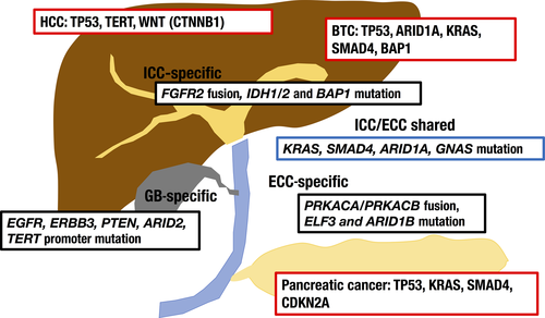
The list of frequently mutated driver genes in BTC cases looks partly similar to that of HCC (TP53, ARID1A, and ARID2) and pancreatic cancer (TP53, KRAS, and SMAD4) (Figure 4). By contrast, characteristically, alterations in WNT signaling and TERT were infrequent and IDH1 mutations (3%) were unique in BTC. Nakamura et al further classified driver genes among anatomical subtypes, and reported subtype-specific and shared driver genes (Figure 4). For example, IDH1 and BAP1 mutations were ICC-specific, whereas ELF3 and ARID1B mutations were more common in ECC. EGFR, ERBB2, PTEN, and ARID2 mutations with TPMs occurred more frequently in GB cases. The molecular mechanisms underlying the selection of specific driver genes along anatomical locations and characteristic differences between HCC and ICC remain completely unknown. It could be possible that cell type-specific expression levels or signaling cascades, or carcinogenic causes, might affect the selection of driver genes. Whole genome sequencing of liver cancer displaying biliary phenotypes revealed a mixture genetic profiles of HCC and ICC, suggesting that this category could be a marginal mixture of two tumor types.26 Activation or inactivation of some driver genes might exert cytotoxicity or a differentiation switch in specific lineages of liver and bile duct cancer, as reported in the case of IDH1 mutations.27
The Cancer Genome Atlas CCA project analyzed 34 CCA samples that were fluke-negative and HBV/HCV-negative.28 Due to the limited size of the cohort, frequencies of driver genes were variable compared to other large-scale studies; however, notably, this cohort represented a higher frequency of IDH1 (13%) and IDH2 (5%) mutations compared to Asian cohorts. Other studies also reported high frequencies of IDH1/2 mutations in Caucasian CCA cases,29 suggesting that IDH1/2 could be ethnic-preferential driver genes in BTC. The CCA cells with IDH1 mutations were reported to be more sensitive to a multikinase inhibitor dasatinib.30
Jusakul et al31 compiled molecular data of 489 CCA cases (310 ICCs and 173 ECCs) from 10 countries including Japan, Thailand, China, Korea, and Italy. This is the largest CCA cohort to date and covers liver fluke-positive and -negative cases across various regions including both Asian and Caucasian ethnicities. The top five driver genes were TP53 (32%), ARID1A (17.4%), KRAS (16.5%), SMAD4 (13%), and BAP1 (8.5%), which are the same as those identified in the Japanese cohort, followed by APC, PBRM1, and ELF3. Infrequent (<5%) but actionable mutations include those of BRAF, IDH1, and BRCA2. Because this cohort contains the largest number of fluke-positive (133 cases) and fluke-negative (356 cases) samples, more accurate fluke-associated driver genes were identified. They found that KRAS, FBXW7, and PTEN mutations and ERBB2 amplification were significantly more frequent in fluke-positive cholangiocarcinoma, whereas IDH1, PBRM1, and ACVR2A mutations occurred more frequently in fluke-negative cases. Mutations of TP53, ARID1A, and SMAD4, three of the top five driver genes, were common in both types.
There are limited genomic data of GB cancers. Li et al32 reported WES of 57 Chinese GB samples. TP53 (47.1%), ERBB3 (11.8%), and KRAS (7.8%) were the top three driver genes, and ErbB signaling (including EGFR, ERBB2, ERBB3, and ERBB4 and their downstream genes) was the most extensively mutated pathway. These observations were consistent with the above report of a Japanese cohort.32
Whole transcriptome sequencing revealed fusion genes in CCA. Among them, fusion genes containing FGFR2 tyrosine kinase and cyclic AMP-dependent PKA signaling components (PRKACA and PRKACB) they are recurrently reported in previous papers and they are also therapeutic targets.25, 31 The FGFR2 fusion genes were generally exclusive to genetic alterations of other receptor kinase signal components such as KRAS, NRAS, and BRAF.25 Protein kinase A component fusions (ATP1B1-PRKACA/PRKACB, and LINC00261-PRKACB) presented exclusiveness to other genetic alterations of PKA signaling, such as GNAS.25 Other kinase fusion genes reported in BTC include FIG-ROS1, FGFR3-TACC3, PTPRK-RSPO3, and EML4-ALK.25, 31, 33
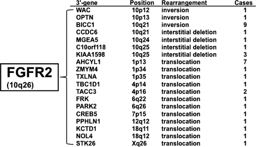
The FGFR2 fusion gene is one of characteristic molecular signatures of CCA with a frequency of approximately 10% of cases.25, 28, 34-36 Previous studies reported at least 19 different fusion partners, and various types of structural alterations, including intrachromosomal (inversion and interstitial deletion) or interchromosomal (translocation) rearrangements, are involved (Figure 5). Invariably, fusion partners replace the last exon of the FGFR2 gene, which encodes C-terminal protein and 3′-UTR. This fusion event is supposed to cause two molecular changes; one is to add a protein interaction domain for promoting ligand-independent FGFR2 dimerization, and the other is to stabilize the FGFR2 transcript, probably by deleting micro-RNA target sequences in the 3′-UTR. Interestingly, Jusakul et al31 reported interchromosomal translocations that only deleted the last exon of FGFR2 (3′-UTR loss) in two cholangiocarcinoma cases. They showed that cases with 3′-UTR-truncated FGFR2 exhibited higher expression levels compared with those of wild-type FGFR2 transcript. The FGFR2 fusion gene is also important for clinical management of CCA. In vivo and early clinical studies showed that kinase inhibitors targeting FGFR2 could be effective for these kinase fusion genes,35-37 and clinical trials of several FGFR2 inhibitors are ongoing. The response rate of a recent phase II clinical trial was 18.8% for FGFR2 fusion-positive CCA,38 and acquired resistant secondary FGFR2 mutations have been identified.39 Clinical features of FGFR2 fusion-positive CCA remain unclear, however, fluke-positive CCA was reported to be negative for FGFR2 fusion genes.31
6 MOLECULAR CARCINOGENESIS OF BTC (FIGURE 3)
Nakamura et al25 extracted two mutational signatures (COSMIC signature 1 and the APOBEC signature: COSMIC signatures 2/13) from the Japanese BTC cohort. Contributions of these signatures were significantly different among anatomical subtypes; COSMIC signature 1 was more frequent in ICC, whereas the APOBEC-associated process was more prevalent in ECC and GB. Jusakul et al31 analyzed WGS data of 71 CCA cases and extracted 10 mutational signatures comprising COSMIC signatures 1, 5, 8, 16, and 17, APOBEC signatures (C>T and C>G substitutions), mismatch-repair deficiency-associated signatures (COSMIC signatures 6 and 20), and AA-exposure signature 22. Fluke-positive cholangiocarcinoma were enriched for APOBEC signatures. In contrast, in TCGA cohort, a collection of both fluke and hepatitis-negative CCA cases showed only COSMIC signatures 6 and 1.28 Possibly chronic infection caused by fluke and other pathogen infections may be prone to induce APOBEC signatures, however, further comprehensive study will be required for determining precise associations.
Recently, an outbreak of CCA was reported among workers in an offset color proof-printing company in Japan.40 These cases had been exposed to high concentrations of chemical compounds, including 1,2-DCP and/or dichloromethane. The International Agency for Research on Cancer categorized 1,2-DCP as a Group 1 (carcinogenic to humans) carcinogen and dichloromethane as Group 2A (probably carcinogenic to humans).41 Whole exome sequencing of four CCA cases of the printer workers revealed unique and predominant mutational spectra that are distinct from known COSMIC mutational signatures and partly reproduced by in vitro 1,2-DCP exposure.42 These unique mutation spectra could be useful for understanding carcinogenesis processes by these chemicals and evaluating chemical exposure and screening.
7 INTEGRATIVE MOLECULAR CLASSIFICATION OF HEPATOBILIARY CANCER (FIGURE 6)
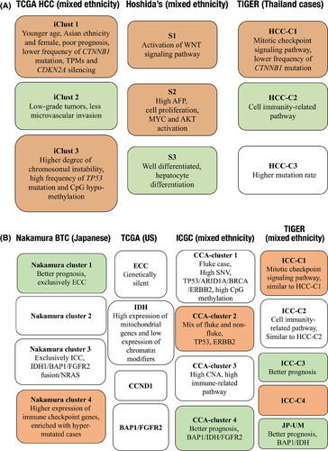
Integrating genome data (mutation, copy number, fusion gene, and mutational signature) with transcriptome, epigenome, proteome, and metabolome data will contribute to identifying unique molecular subtypes in cancer.
The Cancer Genome Atlas HCC group assembled the largest collection of genome, transcriptome, epigenome, and proteome data in 196 HCC cases.8 By integrating these omics data, they identified three subclasses (iClust 1-3). These clusters partly overlapped with the gene expression-based subtypes (S1-3) reported by Hoshida et al.43 Tumors in iClust 1 tended to have lower frequency of CTNNB1 mutation, TPMs, and CDKN2A silencing, whereas iClust3 was characterized by a higher degree of chromosomal instability with high frequency of TP53 mutation and CpG hypomethylation.
Chaisaingmongkol et al44 reported genome, transcriptome, and metabolome analyses of 199 liver cancers (130 ICC and 69 HCC) from Thailand as the TIGER consortium. Based on gene expression data, they identified three HCC subtypes (HCC C1-3), four ICC subtypes (ICC C1-4), and a Japanese-specific ICC subtype (UM subtype). By comparing expression signatures in these subtypes, they found significant similarity between HCC-C1 and ICC-C1 (common C1), and HCC-C2 and ICC-C2 (common C2). Genes highly expressed in common C1 tumors were enriched with mitotic checkpoint signaling pathway and Hoshida's S1-2-related genes, whereas common C2 tumors tended to express more cell immunity-related pathway. They further collected molecular data of 847 liver cancers (561 HCC and 286 ICC, 582 Asian and 265 Caucasian), and found that C1/C2 clusters were reproducible in Asian samples but were less stable in Caucasian cases.
Transcriptome sequencing and WES of 160 Japanese BTC identified four BTC subtypes (Nakamura clusters 1-4), that were associated with prognosis and specific driver gene profiles.25 Nakamura cluster 3 was enriched with IDH1, BAP1, and NRAS mutations and FGFR2 fusion genes, but rarely harbored TP53, KRAS, and SMAD4 mutations. This cluster partly overlaps to the UM subtype identified in the above study,44 which was enriched in IDH1 and BAP1 mutations. Nakamura cluster 4 showed higher immune checkpoint activity and was enriched with hypermutated and more neo-antigen-presenting cases, suggesting immune evasion processes were more active in this subtype.
Integration of genome, transcriptome, and epigenome data from 95 CCA cases revealed four subtypes (CCA clusters 1-4) that were associated with the presence of liver fluke infection and prognosis.31 Tumors in CCA cluster 1 were predominantly liver fluke-positive and harbored ERBB2 amplification and CpG island hypermethylation. Cholangiocarcinoma cluster 4 was enriched with liver fluke-negative cases and BAP1 and IDH1/2 mutations and FGFR alterations, which are similar to Nakamura cluster 3. Cases in CCA cluster 4 also showed CpG shore hypermethylation, partly due to IDH1 mutations, and better prognosis. Tumors in CCA cluster 1 showed a significantly higher contribution of COSMIC signature 1 and intratumoral heterogeneity. They proposed that chronic inflammation by liver fluke infection might continuously induce CpG island methylation, which resulted in accumulation of COSMIC signature 1-associated mutations and tumor heterogeneity in CCA cluster 1 cases, whereas tumors in CCA cluster 4 acquired strong driver mutations with rapid growth. The Cancer Genome Atlas CCA group more clearly isolated a subtype enriched with IDH1/2-mutated tumors, which showed a unique methylation pattern and upregulation of mitochondrial gene expression signatures.28 Their data could separate IDH1/2 mutated cases from BAP1/FGFR2 altered cases based on methylation signature.
Current integrative studies have already identified stable molecular subtypes (possibly three) in HCC, and four or more subtypes including FGFR2 fusions, IDH1/2 and BAP1-mutated group in CCA, however, further collection of trans-omics data of clinical samples with different ethnic and epidemiological backgrounds should be required. There would exist some overlap in molecular signatures of subtypes between HCC and ICC despite distinctive core driver gene sets between the two tumor types.
8 FUTURE PERSPECTIVES
Comprehensive molecular and genomic analyses of large cohorts of hepatobiliary cancers have now uncovered landscapes of driver genes, characteristic mutational signatures, and molecular classifications of these tumor types. To apply this knowledge for better treatment, diagnosis, and prevention of these intractable cancers, genetic screening of each tumor sample, so-called clinical sequencing, for optimized and individualized choice of treatment, clinical trials, and translational research of new drugs, and novel approaches for early diagnosis, such as liquid biopsy, are expected. Furthermore, deeper and broader genomic and basic research, together with international collaborations, are required to understand the biological roles of each or combinations of identified driver genes in the carcinogenesis processes of these tumor types, and to uncover intratumoral, interpatient, and inter-ethnic diversities of genomic profiles in more detail.
ACKNOWLEDGMENTS
This work was supported in part by the Practical Research for Innovative Cancer Control from the Japan Agency for Medical Research and Development (AMED), National Cancer Center Research and Development Funds (29-A-6).
CONFLICT OF INTEREST
The authors have no conflict of interest.


