Volume 50, Issue 7 pp. 903-904
CASE IMAGE

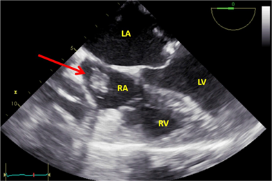
Mal-positioned coronary sinus catheter masquerading as right atrium mass – Thanks to transesophageal echocardiography!

Alisha Goel DM

Alisha Goel DM

Department of Anaesthesia & Intensive Care, Postgraduate Institute of Medical Education and Research, Chandigarh, India

Search for more papers by this author
Krishna Prasad Gourav MD, DM

Corresponding Author

Krishna Prasad Gourav MD, DM

Department of Anaesthesia & Intensive Care, Postgraduate Institute of Medical Education and Research, Chandigarh, India

Correspondence

Krishna Prasad Gourav, MD, DM, Department of Anaesthesia & Intensive Care, Room No. 4014, Level 4, Advanced Cardiac Centre, Postgraduate Institute of Medical Education and Research, Sector 12, Chandigarh 160012, India.

Email: [email protected]

Search for more papers by this author
Sujitha Reddy MD, DM

Sujitha Reddy MD, DM

Department of Anaesthesia & Intensive Care, Postgraduate Institute of Medical Education and Research, Chandigarh, India

Search for more papers by this author
Gurucharan Dasari MD

Gurucharan Dasari MD

Department of Anaesthesia & Intensive Care, Postgraduate Institute of Medical Education and Research, Chandigarh, India

Search for more papers by this author
First published: 11 May 2022

Graphical Abstract

Mid esophageal 4 chamber view showing the mass in RA (red arrow) immediately after insertion of coronary sinus catheter. LA, Left atrium; LV, Left Ventricle; RA, Right atrium; RV, Right ventricle.

CONFLICTS OF INTEREST

The authors declare no potential conflict of interest.

DATA AVAILABILITY STATEMENT

The data that support the findings of this study are available from the corresponding author upon reasonable request.

The full text of this article hosted at iucr.org is unavailable due to technical difficulties.