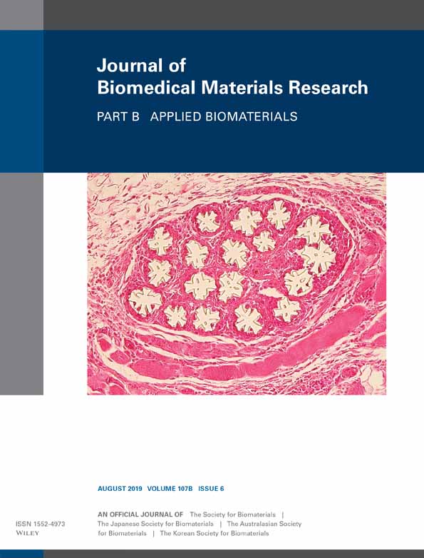
Influence of negative pressure wound therapy on peri-prosthetic tissue vascularization and inflammation around porous titanium percutaneous devices
Divya R. L. Pawar
Orthopaedic Research Laboratories, George E. Wahlen Department of Veterans Affairs Medical Center, and University of Utah Orthopaedic Center, Salt Lake City, Utah, 84148
Department of Bioengineering, University of Utah, Salt Lake City, Utah, 84112
Search for more papers by this authorSujee Jeyapalina
Orthopaedic Research Laboratories, George E. Wahlen Department of Veterans Affairs Medical Center, and University of Utah Orthopaedic Center, Salt Lake City, Utah, 84148
Department of Surgery, University of Utah School of Medicine, Salt Lake City, Utah, 84132
Search for more papers by this authorKelli Hafer
Department of Bioengineering, University of Utah, Salt Lake City, Utah, 84112
Search for more papers by this authorCorresponding Author
Kent N. Bachus
Orthopaedic Research Laboratories, George E. Wahlen Department of Veterans Affairs Medical Center, and University of Utah Orthopaedic Center, Salt Lake City, Utah, 84148
Department of Bioengineering, University of Utah, Salt Lake City, Utah, 84112
Correspondence to: K. N. Bachus; e-mail: [email protected]Search for more papers by this authorDivya R. L. Pawar
Orthopaedic Research Laboratories, George E. Wahlen Department of Veterans Affairs Medical Center, and University of Utah Orthopaedic Center, Salt Lake City, Utah, 84148
Department of Bioengineering, University of Utah, Salt Lake City, Utah, 84112
Search for more papers by this authorSujee Jeyapalina
Orthopaedic Research Laboratories, George E. Wahlen Department of Veterans Affairs Medical Center, and University of Utah Orthopaedic Center, Salt Lake City, Utah, 84148
Department of Surgery, University of Utah School of Medicine, Salt Lake City, Utah, 84132
Search for more papers by this authorKelli Hafer
Department of Bioengineering, University of Utah, Salt Lake City, Utah, 84112
Search for more papers by this authorCorresponding Author
Kent N. Bachus
Orthopaedic Research Laboratories, George E. Wahlen Department of Veterans Affairs Medical Center, and University of Utah Orthopaedic Center, Salt Lake City, Utah, 84148
Department of Bioengineering, University of Utah, Salt Lake City, Utah, 84112
Correspondence to: K. N. Bachus; e-mail: [email protected]Search for more papers by this authorAbstract
Negative Pressure Wound Therapy (NPWT) has been shown to limit downgrowth around percutaneous devices in a guinea pig model. However, the influence of NPWT on peri-prosthetic tissue characteristics leading to limited downgrowth is still unclear. In order to investigate this, 12 CD hairless rats were assigned into two groups, NPWT and Untreated (n = 6/group). Each animal was implanted with a porous coated titanium percutaneous device and was dressed with a gauze and semi-occlusive base dressing. Post-surgery, animals in the NPWT Group received a regimen of NPWT treatment (−70 to −90 mmHg). After 4 weeks, tissue was collected over the device and stained with CD31 and CD68 to quantify blood vessel density and inflammation, respectively. The device with the surrounding tissue was also collected to quantify downgrowth. NPWT treatment led to a 1.6-fold increase in blood vessel densities compared to untreated tissues (p < 0.05). NPWT treatment also resulted in half the downgrowth as the Untreated Group, although not statistically significant (p = 0.19). Additionally, the results showed a trend toward increased CD68 cell densities in the NPWT Group compared to the Untreated Group (p = 0.09). These findings suggest that NPWT may influence wound healing responses in percutaneous devices by increasing blood vessel densities, limiting downgrowth and potentially increasing inflammation. Overall, NPWT may enhance tissue vascularity around percutaneous devices, especially in patients with impaired wound healing. © 2019 Wiley Periodicals, Inc. J Biomed Mater Res Part B: Appl Biomater 107B: 2091–2101, 2019.
CONFLICT OF INTEREST
All authors confirm that there is no potential conflict of interest including employment, stock ownership, consultancies, honoraria, paid expert testimony, and patent applications/registrations influencing this work.
REFERENCES
- 1Pendegrass CJ, Goodship AE, Blunn GW. Development of a soft tissue seal around bone-anchored transcutaneous amputation prostheses. Biomaterials 2006; 27(23): 4183–4191.
- 2Holt BM, Bachus KN, Beck JP, Bloebaum RD, Jeyapalina S. Immediate post-implantation skin immobilization decreases skin regression around percutaneous osseointegrated prosthetic implant systems. J Biomed Mater Res A 2013; 101(7): 2075–2082.
- 3von Recum AF. Applications and failure modes of percutaneous devices: A review. J Biomed Mater Res 1984; 18(4): 323–336.
- 4Holt BM, Betz DH, Ford TA, Beck JP, Bloebaum RD, Jeyapalina S. Pig dorsum model for examining impaired wound healing at the skin-implant interface of percutaneous devices. J Mater Sci Mater Med 2013; 24(9): 2181–2193.
- 5Degidi M, Artese L, Scarano A, Perrotti V, Gehrke P, Piattelli A. Inflammatory infiltrate, microvessel density, nitric oxide synthase expression, vascular endothelial growth factor expression, and proliferative activity in peri-implant soft tissues around titanium and zirconium oxide healing caps. J Periodontol 2006; 77(1): 73–80.
- 6Wang Y, Zhang Y, Miron RJ. Health, maintenance, and recovery of soft tissues around implants. Clin Implant Dent Relat Res 2016; 18(3): 618–634.
- 7Saghiri MA, Asatourian A, Garcia-Godoy F, Sheibani N. The role of angiogenesis in implant dentistry part I: Review of titanium alloys, surface characteristics and treatments. Med Oral Patol Oral Cir Bucal 2016; 21(4): e514–e525.
- 8Tonnesen MG, Feng X, Clark RA. Angiogenesis in wound healing. J Invest Dermatol Symp Proc 2000; 5(1): 40–46.
- 9Broggini N, McManus LM, Hermann JS, Medina R, Schenk RK, Buser D, Cochran DL. Peri-implant inflammation defined by the implant-abutment interface. J Dent Res 2006; 85(5): 473–478.
- 10Esposito M, Thomsen P, Ericson LE, Sennerby L, Lekholm U. Histopathologic observations on late oral implant failures. Clin Implant Dent Relat Res 2000; 2(1): 18–32.
- 11Mitchell SJ, Jeyapalina S, Nichols FR, Agarwal J, Bachus KN. Negative pressure wound therapy limits downgrowth in percutaneous devices. Wound Repair Regen 2016; 24(1): 35–44.
- 12Orgill DP, Bayer LR. Update on negative-pressure wound therapy. Plast Reconstr Surg 2011; 127(Suppl 1): 105S–115S.
- 13Malmsjo M, Ingemansson R, Martin R, Huddleston E. Wound edge microvascular blood flow: Effects of negative pressure wound therapy using gauze or polyurethane foam. Ann Plast Surg 2009; 63(6): 676–681.
- 14Glass GE, Nanchahal J. The methodology of negative pressure wound therapy: Separating fact from fiction. J Plast Reconstr Aesthet Surg 2012; 65(8): 989–1001.
- 15Argenta LC, Morykwas MJ. Vacuum-assisted closure: A new method for wound control and treatment: Clinical experience. Ann Plast Surg 1997; 38(6): 563–576. discussion 577.
- 16Morykwas MJ, Argenta LC, Shelton-Brown EI, McGuirt W. Vacuum-assisted closure: A new method for wound control and treatment: Animal studies and basic foundation. Ann Plast Surg 1997; 38(6): 553–562.
- 17Greene AK, Puder M, Roy R, Arsenault D, Kwei S, Moses MA, Orgill DP. Microdeformational wound therapy: Effects on angiogenesis and matrix metalloproteinases in chronic wounds of 3 debilitated patients. Ann Plast Surg 2006; 56(4): 418–422.
- 18Eisenhardt SU, Schmidt Y, Thiele JR, Iblher N, Penna V, Torio-Padron N, Stark GB, Bannasch H. Negative pressure wound therapy reduces the ischaemia/reperfusion-associated inflammatory response in free muscle flaps. J Plast Reconstr Aesthet Surg 2012; 65(5): 640–649.
- 19Borgquist O, Ingemansson R, Malmsjo M. Wound edge microvascular blood flow during negative-pressure wound therapy: Examining the effects of pressures from −10 to −175 mmHg. Plast Reconstr Surg 2010; 125(2): 502–509.
- 20Borgquist O, Ingemansson R, Malmsjo M. The influence of low and high pressure levels during negative-pressure wound therapy on wound contraction and fluid evacuation. Plast Reconstr Surg 2011; 127(2): 551–559.
- 21Orgill DP, Bayer LR. Negative pressure wound therapy: Past, present and future. Int Wound J 2013; 10(Suppl 1): 15–19.
- 22Erba P, Ogawa R, Ackermann M, Adini A, Miele LF, Dastouri P, Helm D, Mentzer SJ, D'Amato RJ, Murphy GF, Konerding MA, Orgill DP. Angiogenesis in wounds treated by microdeformational wound therapy. Ann Surg 2011; 253(2): 402–409.
- 23Glass GE, Murphy GF, Esmaeili A, Lai LM, Nanchahal J. Systematic review of molecular mechanism of action of negative-pressure wound therapy. Br J Surg 2014; 101: 1627–1636.
- 24Norbury K, Kieswetter K. Vacuum-assisted closure therapy attenuates the inflammatory response in a porcine acute wound healing model. Wounds 2007; 19(4): 97–106.
- 25Nuutila K, Siltanen A, Peura M, Harjula A, Nieminen T, Vuola J, Kankuri E, Aarnio P. Gene expression profiling of negative-pressure-treated skin graft donor site wounds. Burns 2013; 39(4): 687–693.
- 26Liu D, Zhang L, Li T, Wang G, Du H, Hou H, Han L, Tang P. Negative-pressure wound therapy enhances local inflammatory responses in acute infected soft-tissue wound. Cell Biochem Biophys 2014; 70(1): 539–547.
- 27Jacobs S, Simhaee DA, Marsano A, Fomovsky GM, Niedt G, Wu JK. Efficacy and mechanisms of vacuum-assisted closure (VAC) therapy in promoting wound healing: A rodent model. J Plast Reconstr Aesthet Surg 2009; 62(10): 1331–1338.
- 28Cook SJ, Nichols FR, Brunker LB, Bachus KN. A novel vacuum assisted closure therapy model for use with percutaneous devices. Med Eng Phys 2014; 36(6): 768–773.
- 29Pawar DRL, Mitchell SJ, Jeyapalina S, Hawkes JE, Florell SR, Bachus KN. Peri-prosthetic tissue reaction to discontinuation of negative pressure wound therapy around porous titanium percutaneous devices. J Biomed Mater Res B Appl Biomater 2018.
- 30Scherer SS, Pietramaggiori G, Mathews JC, Prsa MJ, Huang S, Orgill DP. The mechanism of action of the vacuum-assisted closure device. Plast Reconstr Surg 2008; 122(3): 786–797.
- 31Hugate R, Clarke R, Hoeman T, Friedman A. Transcutaneous implants in a porcine model: The use of highly porous tantalum. Int J Adv Mater Res 2015; 1(2): 32–40.
- 32Pastar I, Stojadinovic O, Yin NC, Ramirez H, Nusbaum AG, Sawaya A, Patel SB, Khalid L, Isseroff RR, Tomic-Canic M. Epithelialization in wound healing: A comprehensive review. Adv Wound Care 2014; 3(7): 445–464.
10.1089/wound.2013.0473 Google Scholar
- 33Gal P, Toporcer T, Vidinsky B, Mokry M, Novotny M, Kilik R, Smetana K Jr, Gal T, Sabo J. Early changes in the tensile strength and morphology of primary sutured skin wounds in rats. Folia Biol 2006; 52(4): 109–115.
- 34Jeyapalina S, Beck JP, Agarwal J, Bachus KN. A 24-month evaluation of a percutaneous osseointegrated limb-skin interface in an ovine amputation model. J Mater Sci Mater Med 2017; 28(11): 179.
- 35Labler L, Rancan M, Mica L, Harter L, Mihic-Probst D, Keel M. Vacuum-assisted closure therapy increases local interleukin-8 and vascular endothelial growth factor levels in traumatic wounds. J Trauma 2009; 66(3): 749–757.
- 36Isenhath SN, Fukano Y, Usui ML, Underwood RA, Irvin CA, Marshall AJ, Hauch KD, Ratner BD, Fleckman P, Olerud JE. A mouse model to evaluate the interface between skin and a percutaneous device. J Biomed Mater Res A 2007; 83(4): 915–922.
- 37Holgers KM, Thomsen P, Tjellstrom A, Bjursten LM. Immunohistochemical study of the soft tissue around long-term skin-penetrating titanium implants. Biomaterials 1995; 16(8): 611–616.
- 38Lalezari S, Lee CJ, Borovikova AA, Banyard DA, Paydar KZ, Wirth GA, Widgerow AD. Deconstructing negative pressure wound therapy. Int Wound J 2017; 14(4): 649–657.
- 39Dini V, Miteva M, Romanelli P, Bertone M, Romanelli M. Immunohistochemical evaluation of venous leg ulcers before and after negative pressure wound therapy. Wounds 2011; 23(9): 257–266.
- 40Bridges AW, Whitmire RE, Singh N, Templeman KL, Babensee JE, Lyon LA, Garcia AJ. Chronic inflammatory responses to microgel-based implant coatings. J Biomed Mater Res A 2010; 94(1): 252–258.
- 41Badylak SF, Valentin JE, Ravindra AK, McCabe GP, Stewart-Akers AM. Macrophage phenotype as a determinant of biologic scaffold remodeling. Tissue Eng Part A 2008; 14(11): 1835–1842.
- 42Brown BN, Londono R, Tottey S, Zhang L, Kukla KA, Wolf MT, Daly KA, Reing JE, Badylak SF. Macrophage phenotype as a predictor of constructive remodeling following the implantation of biologically derived surgical mesh materials. Acta Biomater 2012; 8(3): 978–987.
- 43Shou K, Niu Y, Zheng X, Ma Z, Jian C, Qi B, Hu X, Yu A. Enhancement of bone-marrow-derived mesenchymal stem cell angiogenic capacity by NPWT for a combinatorial therapy to promote wound healing with large defect. Biomed Res Int 2017; 2017: 7920265.
- 44Summerfield A, Meurens F, Ricklin ME. The immunology of the porcine skin and its value as a model for human skin. Mol Immunol 2015; 66(1): 14–21.
- 45Wise J, White A, Stinner DJ, Fergason JR. A unique application of negative pressure wound therapy used to facilitate patient engagement in the amputation recovery process. Adv Wound Care 2017; 6(8): 253–260.
10.1089/wound.2016.0715 Google Scholar


