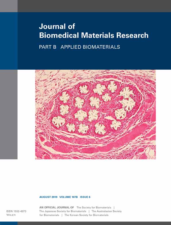
Cartilage/bone interface fabricated under perfusion: Spatially organized commitment of adipose-derived stem cells without medium supplementation
Walter Baumgartner
Division of Plastic and Hand Surgery, University Hospital Zürich, ZKF, Zürich, Switzerland
Search for more papers by this authorLukas Otto
Division of Plastic and Hand Surgery, University Hospital Zürich, ZKF, Zürich, Switzerland
Search for more papers by this authorSamuel C. Hess
Institute for Chemical- and Bioengineering, Department of Chemistry and Applied Biosciences, ETH Zürich, Zürich, Switzerland
Search for more papers by this authorWendelin J. Stark
Institute for Chemical- and Bioengineering, Department of Chemistry and Applied Biosciences, ETH Zürich, Zürich, Switzerland
Search for more papers by this authorSonja Märsmann
Division of Plastic and Hand Surgery, University Hospital Zürich, ZKF, Zürich, Switzerland
Division of Trauma Surgery, University Hospital Zürich, ZKF, Zürich, Switzerland
Search for more papers by this authorGabriella Meier Bürgisser
Division of Plastic and Hand Surgery, University Hospital Zürich, ZKF, Zürich, Switzerland
Search for more papers by this authorMaurizio Calcagni
Division of Plastic and Hand Surgery, University Hospital Zürich, ZKF, Zürich, Switzerland
Search for more papers by this authorPaolo Cinelli
Division of Trauma Surgery, University Hospital Zürich, ZKF, Zürich, Switzerland
Search for more papers by this authorCorresponding Author
Johanna Buschmann
Division of Plastic and Hand Surgery, University Hospital Zürich, ZKF, Zürich, Switzerland
Correspondence to: Johanna Buschmann; e-mail: [email protected]Search for more papers by this authorWalter Baumgartner
Division of Plastic and Hand Surgery, University Hospital Zürich, ZKF, Zürich, Switzerland
Search for more papers by this authorLukas Otto
Division of Plastic and Hand Surgery, University Hospital Zürich, ZKF, Zürich, Switzerland
Search for more papers by this authorSamuel C. Hess
Institute for Chemical- and Bioengineering, Department of Chemistry and Applied Biosciences, ETH Zürich, Zürich, Switzerland
Search for more papers by this authorWendelin J. Stark
Institute for Chemical- and Bioengineering, Department of Chemistry and Applied Biosciences, ETH Zürich, Zürich, Switzerland
Search for more papers by this authorSonja Märsmann
Division of Plastic and Hand Surgery, University Hospital Zürich, ZKF, Zürich, Switzerland
Division of Trauma Surgery, University Hospital Zürich, ZKF, Zürich, Switzerland
Search for more papers by this authorGabriella Meier Bürgisser
Division of Plastic and Hand Surgery, University Hospital Zürich, ZKF, Zürich, Switzerland
Search for more papers by this authorMaurizio Calcagni
Division of Plastic and Hand Surgery, University Hospital Zürich, ZKF, Zürich, Switzerland
Search for more papers by this authorPaolo Cinelli
Division of Trauma Surgery, University Hospital Zürich, ZKF, Zürich, Switzerland
Search for more papers by this authorCorresponding Author
Johanna Buschmann
Division of Plastic and Hand Surgery, University Hospital Zürich, ZKF, Zürich, Switzerland
Correspondence to: Johanna Buschmann; e-mail: [email protected]Search for more papers by this authorAbstract
Tissue engineering of an osteochondral interface demands for a gradual transition of chondrocyte- to osteoblast-prevailing tissue. If stem cells are used as a single cell source, an appropriate cue to trigger the desired differentiation is the use of composite materials with different amounts of calcium phosphate. Electrospun meshes of poly-lactic-co-glycolic acid and amorphous calcium phosphate nanoparticles (PLGA/aCaP) in weight ratios of 100:0; 90:10, 80:20, and 70:30 were seeded with human adipose-derived stem cells (ASCs) and cultured in DMEM without chemical supplementation. After 2 weeks of static cultivation, they were either further cultivated statically for another 2 weeks (group 1), or placed in a Bose® bioreactor with a flow rate per area of 0.16 mL cm−2 min−1 (group 2). Markers for stem cell criteria, chondrogenesis, osteogenesis, adipogenesis and angiogenesis were analyzed by quantitative real-time PCR. Cell distribution, Sox9 protein expression and proteoglycans were assessed by histology. In group 2 (perfusion culture), chondrogenic Sox9 was upregulated toward the cartilage-mimicking side compared to pure PLGA. On the bone-mimicking side, Sox9 experienced a downregulation, which was confirmed on the protein level. Vice versa, expression of osteocalcin was upregulated on the bone-mimicking side, while it was unchanged on the cartilage-mimicking side. In group 1 (static culture), CD31 was upregulated in the presence of aCaP compared to pure PLGA, whereas Sox9 and osteocalcin expression were not affected. aCaP nanoparticles incorporated in electrospun PLGA drive the differentiation behavior of human ASCs in a dose-dependent manner. Discrete gradients of aCaP may act as promising osteochondral interfaces. © 2018 Wiley Periodicals, Inc. J Biomed Mater Res Part B: Appl Biomater 107B: 1833–1843, 2019.
CONFLICT OF INTEREST
All authors disclose any potential sources of conflict of interest.
Supporting Information
| Filename | Description |
|---|---|
| jbmb34276-sup-0001-Supinfo.docxWord 2007 document , 70.4 KB | Appendix S1: Supporting Information |
| jbmb34276-sup-0002-TableS1.docxWord 2007 document , 13.9 KB | Table S1 The sequences of forward and reverse primers. |
Please note: The publisher is not responsible for the content or functionality of any supporting information supplied by the authors. Any queries (other than missing content) should be directed to the corresponding author for the article.
REFERENCES
- 1Di Luca A, Van Blitterswijk C, Moroni L. The osteochondral interface as a gradient tissue: From development to the fabrication of gradient scaffolds for regenerative medicine. Birth Defects Res C Embryo Today Rev 2015; 105(1): 34–52.
- 2Khorshidi S, Solouk A, Mirzadeh H, Mazinani S, Sharifi S, Ramakrishna S. A review of key challenge s of electrospun scaffolds for tissue-engineering applications. J Tissue Eng Regen Med 2015; 10(9): 715–738.
- 3Zhang W, Lian Q, Li D, Wang K, Hao D, Bian W, Jin Z. The effect of interface microstructure on interfacial shear strength for osteochondral scaffolds based on biomimetic design and 3D printing. Mater Sci Eng C 2015; 46: 10–15.
- 4Chen K, Shi P, Teh TK, Toh SL, Goh JCH. In vitro generation of a multilayered osteochondral construct with an osteochondral interface using rabbit bone marrow stromal cells and a silk peptide-based scaffold. J Tissue Eng Regen Med 2016; 10(4): 284–293.
- 5Christakiran MJ, Reardon PJ, Konwarh R, Knowles JC, Mandal BB. Mimicking hierarchical complexity of the osteochondral Interface using electrospun silk bioactive glass composites. ACS Appl Mater Interfaces 2017; 9(9): 8000–8013.
- 6Guo J, Li C, Ling S, Huang W, Chen Y, Kaplan DL. Multiscale design and synthesis of biomimetic gradient protein/biosilica composites for interfacial tissue engineering. Biomaterials 2017; 145: 44–55.
- 7Mellor LF, Mohiti-Asli M, Williams J, Kannan A, Dent MR, Guilak F, Loboa EG, Mellor LF. Extracellular calcium modulates chondrogenic and osteogenic differentiation of human adipose-derived stem cells: A novel approach for osteochondral tissue engineering using a single stem cell source. Tissue Eng Part A 2015; 21(17–18): 2323–2333.
- 8Datta P, Ozbolat V, Ayan B, Dhawan A, Ozbolat IT. Bone tissue bioprinting for craniofacial reconstruction. Biotechnol Bioeng 2017; 114(11): 2424–2431.
- 9Mathieu PS, Loboa EG. Cytoskeletal and focal adhesion influences on mesenchymal stem cell shape, mechanical properties, and differentiation down osteogenic, adipogenic, and chondrogenic pathways. Tissue Eng Part B Rev 2012; 18(6): 436–444.
- 10Komura T, Kato K, Konagaya S, Nakaji-Hirabayashi T, Iwata H. Optimization of surface-immobilized extracellular matrices for the proliferation of neural progenitor cells derived from induced pluripotent stem cells. Biotechnol Bioeng 2015; 112(11): 2388–2396.
- 11Wang Q, Huang H, Wei K, Zhao Y. Time-dependent combinatory effects of active mechanical loading and passive topographical cues on cell orientation. Biotechnol Bioeng 2016; 113(10): 2191–2201.
- 12Cook CA, Huri PY, Ginn BP, Gilbert-Honick J, Somers SM, Temple JP, Mao HQ, Grayson WL. Characterization of a novel bioreactor system for 3D cellular mechanobiology studies. Biotechnol Bioeng 2016; 113(8): 1825–1837.
- 13Yeatts AB, Both SK, Yang W, Alghamdi HS, Yang F, Fisher JP, Jansen JA. In vivo bone regeneration using tubular perfusion system bioreactor cultured nanofibrous scaffolds. Tissue Eng Part A 2014; 20(1–2): 139–146.
- 14Jagodzinski M, Mohiti-Asli M, Williams J, Kannan A, Dent MR, Guilak F, Loboa EG. Effects of cyclic longitudinal mechanical strain and dexamethasone on osteogenic differentiation of human bone marrow stromal cells. Eur. Cell. Mater. 2004; 7: 35–41.
- 15Angele P, Schumann D, Angele M, Kinner B, Englert C, Hente R, Füchtmeier B, Nerlich M, Neumann C, Kujat R. Cyclic, mechanical compression enhances chondrogenesis of mesenchymal progenitor cells in tissue engineering scaffolds. Biorheology 2004; 41(3–4): 335–346.
- 16Pelaez D, Huang C-YC, Cheung HS. Cyclic compression maintains viability and induces chondrogenesis of human mesenchymal stem cells in fibrin gel scaffolds. Stem Cells Dev 2009; 18(1): 93–102.
- 17Park JS, Chu JS, Cheng C, Chen F, Chen D, Li S. Differential effects of equiaxial and uniaxial strain on mesenchymal stem cells. Biotechnol Bioeng 2004; 88(3): 359–368.
- 18Hess SC, Stark WJ, Mohn D, Cohrs N, Märsmann S, Calcagni M, Cinelli P, Buschmann J. Gene expression in human adipose-derived stem cells: comparison of 2D films, 3D electrospun meshes or co-cultured scaffolds with two-way paracrine effects. Eur. Cell. Mater. 2017; 34: 232–248.
- 19Miron RJ, Sculean A, Shuang Y, Bosshardt DD, Gruber R, Buser D, Chandad F, Zhang Y. Osteoinductive potential of a novel biphasic calcium phosphate bone graft in comparison with autographs, xenografts, and DFDBA. Clin Oral Implants Res 2016; 27(6): 668–675.
- 20Fedorovich NE, Leeuwenburgh SC, van der Helm YJ, Alblas J, Dhert WJ. The osteoinductive potential of printable, cell-laden hydrogel-ceramic composites. J Biomed Mater Res A 2012; 100A(9): 2412–2420.
- 21Dadhich P, Das B, Pal P, Srivas PK, Dutta J, Ray S, Dhara S. A simple approach for an eggshell-based 3D-printed osteoinductive multiphasic calcium phosphate scaffold. ACS Appl Mater Interfaces 2016; 8(19): 11910–11924.
- 22Zhang JW, Dalbay MT, Luo X, Vrij E, Barbieri D, Moroni L, de Bruijn JD, van Blitterswijk JP, Knight MM, Yuan H. Topography of calcium phosphate ceramics regulates primary cilia length and TGF receptor recruitment associated with osteogenesis. Acta Biomater. 2017; 57: 487–497.
- 23McCullen SD, Zhu Y, Bernacki SH, Narayan RJ, Pourdeyhimi B, Gorga RE, Loboa EG. Electrospun composite poly(L-lactic acid)/tricalcium phosphate scaffolds induce proliferation and osteogenic differentiation of human adipose-derived stem cells. Biomed Mater 2009; 4(3): 1–9.
- 24Li XM, Liu H, Niu X, Fan Y, Feng Q, Cui FZ, Watari F. Osteogenic differentiation of human adipose-derived stem cells induced by osteoinductive calcium phosphate ceramics. J Biomed Mater Res Part B Appl Biomater 2011; 97B(1): 10–19.
- 25Stavenschi E, Labour MN, Hoey DA. Oscillatory fluid flow induces the osteogenic lineage commitment of mesenchymal stem cells: the, effect of shear stress magnitude, frequency, and duration. J Biomech 2017; 55(1): 99–106.
- 26Guo T, Yu L, Lim CG, Goodley AS, Xiao X, Placone JK, Ferlin KM, Nguyen BN, Hsieh AH, Fisher JP. Effect of dynamic culture and periodic compression on human mesenchymal stem cell proliferation and chondrogenesis. Ann Biomed Eng 2016; 44(7): 2103–2113.
- 27Liu YX, Kongsuphol P, Gourikutty SBN, Ramadan Q. Human adipocyte differentiation and characterization in a perfusion-based cell culture device. Biomed Microdevices 2017; 19(3): 1–10.
- 28Wang H, Riha GM, Yan S, Li M, Chai H, Yang H, Yao Q, Chen C. Shear stress induces endothelial differentiation from a murine embryonic mesenchymal progenitor cell line. Arterioscler Thromb Vasc Biol 2005; 25(9): 1817–1823.
- 29Buschmann J, Gao S, Härter L, Hemmi S, Welti M, Werner CM, Calcagni M, Cinelli P, Wanner GA. Yield and proliferation rate of adipose-derived stem cells as a function of age, BMI and harvest site: Increasing the yield by using adherent and supernatant fractions? Cytotherapy 2013; 15(1): 1098–1105.
- 30Zuk PA, Zhu M, Mizuno H, Huang J, Futrell JW, Katz AJ, Benhaim P, Lorenz HP, Hedrick MH. Multilineage cells from human adipose tissue: implications for cell-based therapies. Tissue Eng 2001; 7(2): 211–228.
- 31Gronthos S, Franklin DM, Leddy HA, Robey PG, Storms RW, Gimble JM. Surface protein characterization of human adipose tissue-derived stromal cells. J Cell Physiol 2001; 189(1): 54–63.
- 32Buschmann J, Härter L, Gao S, Hemmi S, Welti M, Hild N, Schneider OD, Stark WJ, Lindenblatt N, Werner CM, Wanner GA, Calcagni M. Tissue engineered bone grafts based on biomimetic nanocomposite PLGA/amorphous calcium phosphate scaffold and human adipose-derived stem cells. Injury 2012; 43(10): 1689–1697.
- 33Gao SP, Calcagni M, Welti M, Hemmi S, Hild N, Stark WJ, Bürgisser GM, Wanner GA, Cinelli P, Buschmann J. Proliferation of ASC-derived endothelial cells in a 3D electrospun mesh: Impact of bone-biomimetic nanocomposite and co-culture with ASC-derived osteoblasts. Injury 2014; 45(6): 974–980.
- 34Loher S, Stark WJ, Maciejewski M, Baiker A, Pratsinis SE, Reichardt D, Maspero F, Krumeich F, Günther D. Fluoro-apatite and calcium phosphate nanoparticles by flame synthesis. Chem Mater 2005; 17(1): 36–42.
- 35Schneider OD, Loher S, Brunner TJ, Uebersax L, Simonet M, Grass RN, Merkle HP, Stark S. Cotton wool-like nanocomposite biomaterials prepared by electrospinning: in vitro bioactivity and osteogenic differentiation of human mesenchymal stem cells. J Biomed Mater Res Part B-Appl Biomater 2008; 84B(2): 350–362.
- 36Baumgartner W, Welti M, Hild N, Hess SC, Stark WJ, Bürgisser GM, Giovanoli P, Buschmann J. Tissue mechanics of piled critical size biomimetic and biominerizable nanocomposites: Formation of bioreactor-induced stem cell gradients under perfusion and compression. J Mech Behav Biomed Mater 2015; 47(0): 124–134.
- 37Dominici M, Le Blanc K, Mueller I, Slaper-Cortenbach I, Krause D, Deans R, Keating A, Prockop DJ, Horwitz E, Dominici M. Minimal criteria for defining multipotent mesenchymal stromal cells. The International Society for Cellular Therapy position statement. Cytotherapy 2006; 8(4): 315–317.
- 38Abdel-Sayed P, Darwiche SE, Kettenberger U, Pioletti DP. The role of energy dissipation of polymeric scaffolds in the mechanobiological modulation of chondrogenic expression. Biomaterials 2014; 35(6): 1890–1897.
- 39Lam J, Lu S, Lee EJ, Trachtenberg JE, Meretoja VV, Dahlin RL, van den Beucken JJ, Tabata Y, Wong ME, Jansen JA, Mikos AG, Kasper FK. Osteochondral defect repair using bilayered hydrogels encapsulating both chondrogenically and osteogenically pre-differentiated mesenchymal stem cells in a rabbit model. Osteoarthr Cartil 2014; 22(9): 1291–1300.
- 40Camarero-Espinosa S, Cooper-White J. Tailoring biomaterial scaffolds for osteochondral repair. Int J Pharm 2017; 523(2): 476–489.
- 41Zhang T, Lin S, Shao X, Shi S, Zhang Q, Xue C, Lin Y, Zhu B, Cai X. Regulating osteogenesis and adipogenesis in adipose-derived stem cells by controlling underlying substrate stiffness. J Cell Physiol 2017; 233(4): 3418–3428.
- 42Chen Q, Shou P, Zheng C, Jiang M, Cao G, Yang Q, Cao J, Xie N, Velletri T, Zhang X, Xu C, Zhang L, Yang H, Hou J, Wang Y, Shi Y. Fate decision of mesenchymal stem cells: adipocytes or osteoblasts? Cell Death Differ 2016; 23(7): 1128–1139.
- 43Hoshiba T, Kawazoe N, Chen GP. The balance of osteogenic and adipogenic differentiation in human mesenchymal stem cells by matrices that mimic stepwise tissue development. Biomaterials 2012; 33(7): 2025–2031.
- 44Gu H, Huang Z, Yin X, Zhang J, Gong L, Chen J, Rong K, Xu J, Lu L, Cui L. Role of c-Jun N-terminal kinase in the osteogenic and adipogenic differentiation of human adipose-derived mesenchymal stem cells. Exp Cell Res 2015; 339(1): 112–121.
- 45Atashi F, Modarressi A, Pepper MS. The role of reactive oxygen species in mesenchymal stem cell adipogenic and osteogenic differentiation: A review. Stem Cells Dev 2015; 24(10): 1150–1163.
- 46Tang J, Peng R, Ding J. The regulation of stem cell differentiation by cell-cell contact on micropatterned material surfaces. Biomaterials 2010; 31(9): 2470–2476.
- 47Solorio LD, Phillips LM, McMillan A, Cheng CW, Dang PN, Samorezov JE, Yu X, Murphy WL, Alsberg E. Spatially organized differentiation of mesenchymal stem cells within biphasic microparticle-incorporated high cell density osteochondral tissues. Adv Healthc Mater 2015; 4(15): 2306–2313.
- 48Liu WY, Lipner J, Xie J, Manning CN, Thomopoulos S, Xia Y. Nanofiber scaffolds with gradients in mineral content for spatial control of osteogenesis. ACS Appl Mater Interfaces 2014; 6(4): 2842–2849.
- 49Song FL, Jiang D, Wang T, Wang Y, Lou Y, Zhang Y, Ma H, Kang Y. Mechanical stress regulates osteogenesis and adipogenesis of rat mesenchymal stem cells through PI3K/Akt/GSK-3 beta/beta-catenin signaling pathway. Biomed Res Int 2017; 4: 1–10.
- 50Engler AJ, Sen S, Sweeney HL, Discher DE. Matrix elasticity directs stem cell lineage specification. Cell 2006; 126(4): 677–689.
- 51Teong B, Wu SC, Chang CM, Chen JW, Chen HT, Chen CH, Chang JK, Ho ML. The stiffness of a crosslinked hyaluronan hydrogel affects its chondro-induction activity on hADSCs. J Biomed Mater Res B Appl Biomater 2017; 106(2): 808–816.
- 52Mullen CA, Vaughan T, Billiar KL, McNamara LM. The effect of substrate stiffness, thickness, and cross-linking density on osteogenic cell behavior. Biophys J 2015; 108(7): 1604–1612.
- 53Bodle JC, Loboa EG. Concise review: Primary cilia: Control centers for stem cell lineage specification and potential targets for cell-based therapies. Stem Cells 2016; 34(6): 1445–1454.
- 54Sadowska JM, Götz H, Baranowski A, Heid F, Rommens PM, Hofmann A. In vitro response of mesenchymal stem cells to biomimetic hydroxyapatite substrates: A new strategy to assess the effect of ion exchange. Acta Biomater 2018; 76:319–332.
- 55Baumgartner W, Schneider I, Hess SC, Stark WJ, Märsmann S, Brunelli M, Calcagni M, Cinelli P, Buschmann J. Cyclic uniaxial compression of human stem cells seeded on a bone biomimetic nanocomposite decreases anti-osteogenic commitment evoked by shear stress. J Mech Behav Biomed Mater 2018; 83: 84–93.
- 56Chai YC, Roberts SJ, Schrooten J, Luyten FP. Probing the osteoinductive effect of calcium phosphate by using an in vitro biomimetic model. Tissue Eng. Part A 2011; 17(7–8): 1083–1097.
- 57Wang L, Fan H, Zhang ZY, Lou AJ, Pei GX, Jiang S, Mu TW, Qin JJ, Chen SY, Jin D. Osteogenesis and angiogenesis of tissue-engineered bone constructed by prevascularized beta-tricalcium phosphate scaffold and mesenchymal stem cells. Biomaterials 2010; 31(36): 9452–9461.
- 58Xiao W, Liu Y-M, Ren K-G, Shi F, Li Y, Zhi W, Weng J, Qu S-X. Evaluation of vascularization of porous calcium phosphate by Chick Chorioallantoic membrane model ex vivo. J Inorg Mater 2017; 32(6): 649–654.
- 59Bragdon B, Lam S, Aly S, Femia A, Clark A, Hussein A, Morgan EF, Gerstenfeld LC. Earliest phases of chondrogenesis are dependent upon angiogenesis during ectopic bone formation in mice. Bone 2017; 101: 49–61.
- 60Ritz U, Götz H, Baranowski A, Heid F, Rommens PM, Hofmann A. Influence of different calcium phosphate ceramics on growth and differentiation of cells in osteoblast-endothelial co-cultures. J Biomed Mater Res Part B-Appl Biomater 2017; 105(7): 1950–1962.


