Volume 15, Issue 5 e202100357
RESEARCH ARTICLE

In-vivo two-photon visualization and quantitative detection of redox state of cancer

Wei Wang

Wei Wang

Key Laboratory of Optoelectronic Science and Technology for Medicine of Ministry of Education, Fujian Provincial Key Laboratory of Photonics Technology, Fujian Normal University, Fuzhou, P. R. China

Search for more papers by this author
Chenlu Wang

Chenlu Wang

MOE Key Laboratory for Analytical Science of Food Safety and Biology College of Chemistry, Fuzhou University, Fuzhou, P. R. China

Search for more papers by this author
Guoming Liu

Guoming Liu

Department of Orthopaedics, Jinshan Hospital of Fujian Provincial Hospital, Fuzhou, P. R. China

Search for more papers by this author
Long Jin

Long Jin

Department of Pathology, Shengli Clinical Medical College, Fujian Medical University, Fujian Provincial Hospital, Fuzhou, P. R. China

Search for more papers by this author
Zexi Lin

Zexi Lin

Key Laboratory of Optoelectronic Science and Technology for Medicine of Ministry of Education, Fujian Provincial Key Laboratory of Photonics Technology, Fujian Normal University, Fuzhou, P. R. China

Search for more papers by this author
Lisheng Lin

Lisheng Lin

Key Laboratory of Optoelectronic Science and Technology for Medicine of Ministry of Education, Fujian Provincial Key Laboratory of Photonics Technology, Fujian Normal University, Fuzhou, P. R. China

Search for more papers by this author
Ying Wu

Ying Wu

MOE Key Laboratory for Analytical Science of Food Safety and Biology College of Chemistry, Fuzhou University, Fuzhou, P. R. China

Search for more papers by this author
Jianxin Chen

Jianxin Chen

Key Laboratory of Optoelectronic Science and Technology for Medicine of Ministry of Education, Fujian Provincial Key Laboratory of Photonics Technology, Fujian Normal University, Fuzhou, P. R. China

Search for more papers by this author
Hongxin Lin

Corresponding Author

Hongxin Lin

Key Laboratory of Optoelectronic Science and Technology for Medicine of Ministry of Education, Fujian Provincial Key Laboratory of Photonics Technology, Fujian Normal University, Fuzhou, P. R. China

Correspondence

Hongxin Lin, Key Laboratory of Optoelectronic Science and Technology for Medicine of Ministry of Education, Fujian Provincial Key Laboratory of Photonics Technology, Fujian Normal University, Fuzhou 350007, P. R. China.

Email: [email protected]

Jibin Song, MOE Key Laboratory for Analytical Science of Food Safety and Biology College of Chemistry, Fuzhou University, Fuzhou 350108, P. R. China.

Email: [email protected]

Search for more papers by this author
Jibin Song

Corresponding Author

Jibin Song

MOE Key Laboratory for Analytical Science of Food Safety and Biology College of Chemistry, Fuzhou University, Fuzhou, P. R. China

Correspondence

Hongxin Lin, Key Laboratory of Optoelectronic Science and Technology for Medicine of Ministry of Education, Fujian Provincial Key Laboratory of Photonics Technology, Fujian Normal University, Fuzhou 350007, P. R. China.

Email: [email protected]

Jibin Song, MOE Key Laboratory for Analytical Science of Food Safety and Biology College of Chemistry, Fuzhou University, Fuzhou 350108, P. R. China.

Email: [email protected]

Search for more papers by this author
First published: 08 January 2022
Citations: 4

Wei Wang, Chenlu Wang and Guoming Liu contributed equally to this work.

Funding information: Natural Science Foundation of Fujian Province, Grant/Award Numbers: 2021J01392, 2020J01155; Health and Education Commission of Fujian Province, Grant/Award Number: 2019-WJ-20; National Natural Science Foundation of China, Grant/Award Numbers: 21874024, 82001806, 82001875, U21A20377

Abstract

Glutathione (GSH), the most common and abundant antioxidant in the body, is particularly concentrated in cancer cells (2–10 mM). This concentration is approximately 1000 times that of normal cells, making GSH a specific tumor marker. Overexpression of GSH is critical for mapping the redox state of cancer cells. However, there are few probes and detection methods responsive to GSH that can quantitatively visualize GSH in vivo in two-photon excitation fluorescence (TPEF) imaging mode. The experimental results show that TPEF-GSH could not only target GSH in tumors, but also establish the quantitative relationship between TPEF signal and GSH concentration. We explored the optimal two-photon excitation wavelength of TPEF-GSH, the optimal cell incubation duration with TPEF-GSH, the best imaging time point for GSH in cells, and the quantitative relationship between the TPEF signal and the changes in GSH concentrations. In zebrafish embryo and zebrafish experiments, the ratiometric value of TPEF-GSH increased with the decrease of GSH concentration. Microinjection and co-incubation were used to verify whether the ratiometric value could quantify endogenous GSH in tumor-bearing zebrafish, and the obtained GSH levels were 4.66 mM and 5.16 mM, respectively. The ratio TPEF probe could accurately visualize and quantify GSH in vivo, reflecting the redox status of the tumor. The design of the ratiometric molecular probe provides a reliable strategy for the development of TPEF nanoprobe in vivo. In this article, a new GSH sensitive molecular probe, TPEF-GSH, has been developed with good specificity and sensitivity. TPEF-GSH was successfully used to image cancer cells in vitro and tumor-bearing zebrafish in vivo, and to further detect GSH levels.image

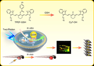
CONFLICT OF INTEREST

The authors declare no potential conflict of interest.

DATA AVAILABILITY STATEMENT

Data openly available in a public repository that issues datasets with DOIs.

The full text of this article hosted at iucr.org is unavailable due to technical difficulties.