Volume 11, Issue 12 e201800186
FULL ARTICLE

Label-free non-linear microscopy to measure myelin outcome in a rodent model of Charcot-Marie-Tooth diseases

Helene Hajjar

Helene Hajjar

Institut des Neurosciences de Montpellier, Université de Montpellier, Montpellier, France

Search for more papers by this author
Hassan Boukhaddaoui

Hassan Boukhaddaoui

Institut des Neurosciences de Montpellier, Université de Montpellier, Montpellier, France

Montpellier Ressources Imagerie (MRI), Montpellier, France

Search for more papers by this author
Amel Rizgui

Amel Rizgui

Institut des Neurosciences de Montpellier, Université de Montpellier, Montpellier, France

Search for more papers by this author
Chamroeun Sar

Chamroeun Sar

Institut des Neurosciences de Montpellier, Université de Montpellier, Montpellier, France

Montpellier Ressources Imagerie (MRI), Montpellier, France

Search for more papers by this author
Jade Berthelot

Jade Berthelot

Institut des Neurosciences de Montpellier, Université de Montpellier, Montpellier, France

Search for more papers by this author
Claire Perrin-Tricaud

Claire Perrin-Tricaud

Institut des Neurosciences de Montpellier, Université de Montpellier, Montpellier, France

Search for more papers by this author
Herve Rigneault

Herve Rigneault

CNRS, École Centrale Marseille, Institut Fresnel, Aix-Marseille Université, Marseille, France

Search for more papers by this author
Nicolas Tricaud

Corresponding Author

Nicolas Tricaud

Institut des Neurosciences de Montpellier, Université de Montpellier, Montpellier, France

Correspondence

Tricaud Nicolas, Institut des Neurosciences de Montpellier, Université de Montpellier, 80 Rue A. Fliche, 34090 Montpellier, France.

Email: [email protected]

Search for more papers by this author
First published: 09 August 2018
Citations: 13
Funding information European Research Council, Grant/Award Number: FP7-IDEAS-ERC 311610; Agence Nationale de la Rechercher
Hajjar Helene and Boukhaddaoui Hassan contributed equally to this study.

Abstract

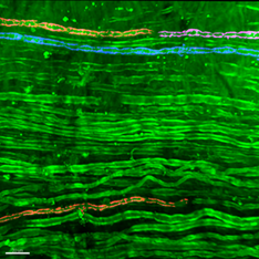
Myelin sheath produced by Schwann cells covers and nurtures axons to speed up nerve conduction in peripheral nerves. Demyelinating peripheral neuropathies result from the loss of this myelin sheath and so far, no treatment exists to prevent Schwann cell demyelination. One major hurdle to design a therapy for demyelination is the lack of reliable measures to evaluate the outcome of the treatment on peripheral myelin in patients but also in living animal models. Non-linear microscopy techniques which include second harmonic generation (SHG), third harmonic generation (THG) and coherent anti-stokes Raman scattering (CARS) were used to image myelin ex vivo and in vivo in the sciatic nerve of healthy and demyelinating mice and rats. SHG did not label myelin and THG required too much light power to be compatible with live imaging. CARS is the most reliable of these techniques for in vivo imaging and it allows for the analysis and quantification of myelin defects in a rat model of CMT1A disease. This microscopic technique therefore constitutes a promising, reliable and robust readout tool in the development of new treatments for demyelinating peripheral neuropathies.
image

The full text of this article hosted at iucr.org is unavailable due to technical difficulties.