Volume 10, Issue 3 pp. 377-384
Full Article

Colocalization of cellular nanostructure using confocal fluorescence and partial wave spectroscopy

John E. Chandler

John E. Chandler

Biomedical Engineering Department, Northwestern University, 2145 Sheridan Rd, Evanston, IL 60208 USA

Search for more papers by this author
Yolanda Stypula-Cyrus

Yolanda Stypula-Cyrus

Biomedical Engineering Department, Northwestern University, 2145 Sheridan Rd, Evanston, IL 60208 USA

Search for more papers by this author
Luay Almassalha

Luay Almassalha

Biomedical Engineering Department, Northwestern University, 2145 Sheridan Rd, Evanston, IL 60208 USA

Search for more papers by this author
Greta Bauer

Greta Bauer

Biomedical Engineering Department, Northwestern University, 2145 Sheridan Rd, Evanston, IL 60208 USA

Search for more papers by this author
Leah Bowen

Leah Bowen

Biomedical Engineering Department, Northwestern University, 2145 Sheridan Rd, Evanston, IL 60208 USA

Search for more papers by this author
Hariharan Subramanian

Hariharan Subramanian

Biomedical Engineering Department, Northwestern University, 2145 Sheridan Rd, Evanston, IL 60208 USA

Search for more papers by this author
Igal Szleifer

Igal Szleifer

Biomedical Engineering Department, Northwestern University, 2145 Sheridan Rd, Evanston, IL 60208 USA

Search for more papers by this author
Vadim Backman

Corresponding Author

Vadim Backman

Biomedical Engineering Department, Northwestern University, 2145 Sheridan Rd, Evanston, IL 60208 USA

Corresponding author: e-mail: [email protected], Phone: 8474913536, Fax: 8474914928

Search for more papers by this author
First published: 25 April 2016
Citations: 2

Abstract

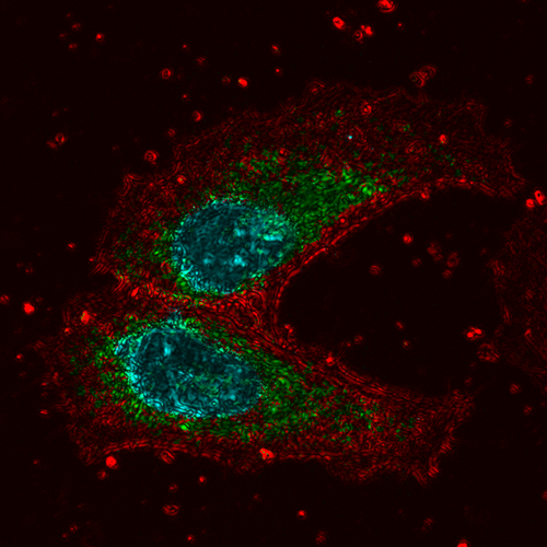
A new multimodal confocal microscope has been developed, which includes a parallel Partial Wave Spectroscopic (PWS) microscopy path. This combination of modalities allows molecular-specific sensing of nanoscale intracellular structure using fluorescent labels. Combining molecular specificity and sensitivity to nanoscale structure allows localization of nanostructural intracellular changes, which is critical for understanding the mechanisms of diseases such as cancer. To demonstrate the capabilities of this multimodal instrument, we imaged HeLa cells treated with valinomycin, a potassium ionophore that uncouples oxidative phosphorylation. Colocalization of fluorescence images of the nuclei (Hoechst 33342) and mitochondria (anti-mitochondria conjugated to Alexa Fluor 488) with PWS measurements allowed us to detect a significant decrease in nuclear nanoscale heterogeneity (Σ), while no significant change in Σ was observed at mitochondrial sites. In addition, application of the new multimodal imaging approach was demonstrated on human buccal samples prepared using a cancer screening protocol. These images demonstrate that nanoscale intracellular structure can be studied in healthy and diseased cells at molecular-specific sites.

The full text of this article hosted at iucr.org is unavailable due to technical difficulties.