High resolution imaging of the fibrous microstructure in bovine common carotid artery using optical polarization tractography
Leila Azinfar
Department of Bioengineering, University of Missouri, Columbia, MO 65211 USA
Search for more papers by this authorMohammadreza Ravanfar
Department of Bioengineering, University of Missouri, Columbia, MO 65211 USA
Search for more papers by this authorYuanbo Wang
Department of Bioengineering, University of Missouri, Columbia, MO 65211 USA
Search for more papers by this authorKeqing Zhang
Department of Molecular Microbiology & Immunology, University of Missouri, Columbia, MO 65211 USA
Search for more papers by this authorDongsheng Duan
Department of Molecular Microbiology & Immunology, University of Missouri, Columbia, MO 65211 USA
Search for more papers by this authorCorresponding Author
Gang Yao
Department of Bioengineering, University of Missouri, Columbia, MO 65211 USA
Corresponding author: e-mail: [email protected]
Search for more papers by this authorLeila Azinfar
Department of Bioengineering, University of Missouri, Columbia, MO 65211 USA
Search for more papers by this authorMohammadreza Ravanfar
Department of Bioengineering, University of Missouri, Columbia, MO 65211 USA
Search for more papers by this authorYuanbo Wang
Department of Bioengineering, University of Missouri, Columbia, MO 65211 USA
Search for more papers by this authorKeqing Zhang
Department of Molecular Microbiology & Immunology, University of Missouri, Columbia, MO 65211 USA
Search for more papers by this authorDongsheng Duan
Department of Molecular Microbiology & Immunology, University of Missouri, Columbia, MO 65211 USA
Search for more papers by this authorCorresponding Author
Gang Yao
Department of Bioengineering, University of Missouri, Columbia, MO 65211 USA
Corresponding author: e-mail: [email protected]
Search for more papers by this authorAbstract
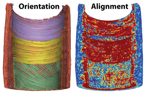
The biomechanical properties of artery are primarily determined by the fibrous structures in the vessel wall. Many vascular diseases are associated with alternations in the orientation and alignment of the fibrous structure in the arterial wall. Knowledge on the structural features of the artery wall is crucial to our understanding of the biology of vascular diseases and the development of novel therapies. Optical coherence tomography (OCT) and polarization-sensitive OCT have shown great promise in imaging blood vessels due to their high resolution, fast acquisition, good imaging depth, and large field of view. However, the feasibility of using OCT based methods for imaging fiber orientation and distribution in the arterial wall has not been investigated. Here we show that the optical polarization tractography (OPT), a technology developed from Jones matrix OCT, can reveal the fiber orientation and alignment in the bovine common carotid artery. The fiber orientation and alignment data obtained in OPT provided a robust contrast marker to clearly resolve the intima and media boundary of the carotid artery wall.
References
- 1G. A. Holzapfel, T .C. Gasser, and R. W. Ogden, J. Elasticity 61, 1–48 (2000).
- 2T. C. Gasser, R. W. Ogden, and G. A. Holzapfel, J. R. Soc. Interface 3, 15–35 (2006).
- 3A. J. Schriefl, G. Zeindlinger, D. M. Pierce, P. Regitnig, and G. A. Holzapfel, J. R. Soc. Interface 9, 1275–1286 (2012).
- 4M. Volpe, J. Hum. Hypertens. 19, 93–102 (2005).
- 5H. C. Stary, D. H. Blankenhorn, A. B. Chandler, S. Glagov, W. Insull, Jr., M. Richardson, M. E. Rosenfeld, S. A. Schaffer, C. J. Schwartz, W. D. Wagner, and R. W. Wissler, Circulation 85, 391–405 (1992).
- 6C. Pagiatakis, R. Galaz, J. C. Tardif, and R. Mongrain, Med. Biol. Eng. Comput. 53, 545–555 (2015).
- 7I. Hariton, G. de Botton, T. C. Gasser, and G. A. Holzapfel, Biomech. Model. Mechanobiol. 6, 163–175 (2007).
- 8A. Karimi, M. Navidbakhsh, and A. Shojaei, Tissue Cell 47, 152–158 (2015).
- 9R. B. Dodson, M. R. Morgan, C. Galambos, K. S. Hunter, and S. H. Abman, Am. J. Physiol. Lung Cell. Mol. Physiol. 307, L822–L828 (2014).
- 10R. B. Dodson, P. J. Rozance, E. Reina-Romo, V. L. Ferguson, and K. S. Hunter, J. Biomech. 46, 956–963 (2013).
- 11P. B. Canham, H. M. Finlay, and D. R. Boughner, Cardiovasc. Res. 34, 557–567 (1997).
- 12A. V. Kamenskiy, I. I. Pipinos, J. N. MacTaggart, S. A. Kazmi, and Y. A. Dzenis, J. Biomech. Eng. 133, 111008 (2011).
- 13P. Farand, A. Garon, and G. E. Plante, Microvasc. Res. 73, 95–99 (2007).
- 14P. Smith, Lab. Invest. 35, 525–529 (1976).
- 15P. B. Canham, H. M. Finlay, J. G. Dixon, D. R. Boughner, and A. Chen, Cardiovasc. Res. 23, 973–982 (1989).
- 16P. B. Canham, E. A. Talman, H. M. Finlay, and J. G. Dixon, Connect. Tissue Res. 26, 121–134 (1991).
- 17P. B. Canham, P. Whittaker, S. E. Barwick, and M. E. Schwab, Can. J. Physiol. Pharmacol. 70, 296–305 (1992).
- 18H. M. Finlay, P. Whittaker, and P. B. Canham, Stroke 29, 1595–1601 (1998).
- 19A. J. Rowe, H. M. Finlay, and P. B. Canham, J. Vasc. Res. 40, 406–415 (2003).
- 20A. Zoumi, X. Lu, G. S. Kassab, and B. J. Tromberg, Biophys. J. 87, 2778–2786 (2004).
- 21T. Boulesteix, A. M. Pena, N. Pagès, G. Godeau, M. P. Sauviat, E. Beaurepaire, and M. C. Schanne-Klein, Cytometry Part A 69, 20–26 (2006).
- 22M. Vielreicher, S. Schürmann, R. Detsch, M. A. Schmidt, A. Buttgereit, A. Boccaccini, and O. Friedrich, J. R. Soc. Interface 10, 20130263 (2013).
- 23S. Ghazanfari, A. Driessen-Mol, G. J. Strijkers, F. P. T. Baaijens, and C. V. C. Bouten, PLoS ONE 10, 1–15 (2015).
- 24I. K. Jang, B. E. Bouma, D. H. Kang, S. J. Park, S. W. Park, K. B. Seung, K. B. Choi, M. Shishkov, K. Schlendorf, E. Pomerantsev, S. L. Houser, H. T. Aretz, and G. J. Tearney, J. Am. Coll. Cardiol. 39, 604–609 (2002).
- 25M. J. Suter, G. J. Tearney, W. Y. Oh, and B. E. Bouma, IEEE J. Sel. Top Quantum Electron 16, 706–714 (2010).
- 26M. E. Brezinski, G. J. Tearney, B. E. Bouma, J. A. Izatt, M. R. Hee, E. A. Swanson, J. F. Southern, and J. G. Fujimoto, Circulation 93, 1206–1213 (1996).
- 27G. J. Tearney, I.-K. Jang, and B. E. Bouma, J. Biomed. Opt. 11, 021002 (2006).
- 28G. Guagliumi and V. Sirbu, Catheter. Cardiovasc. Interv. 72, 237–247 (2008).
- 29S. K. Nadkarni, B. E. Bouma, J. de Boer, and G. J. Tearney, Laser. med. sci. 24, 439–445 (2009).
- 30W.-C. Kuo, N.-K. Chou, C. Chou, C.-M. Lai, H.-J. Huang, S.-S. Wang, and J.-J. Shyu, Appl. Opt. 46, 2520–2527 (2007).
- 31N. Gladkova, S. Nemirova, E. Shakhov, E. Sharabrin, N. Prodanets, L. Snopova, E. Kiseleva, E. Sergeeva, M. Y. Kirillin, and E. Gubarkova, Sovrem. Tehnol. Med 5, 45–55 (2013).
- 32W. Y. Oh, S. H. Yun, B. J. Vakoc, M. Shishkov, A. E. Desjardins, B. H. Park, J. F. De Boer, G. J. Tearney, and B. E. Bouma, Opt. Express 16, 1096–1103 (2008).
- 33S. D. Giattina, B. K. Courtney, P. R. Herz, M. Harman, S. Shortkroff, D. L. Stamper, B. Liu, J. G. Fujimoto, and M. E. Brezinski, Int. J. Cardiol. 107, 400–409 (2006).
- 34S. K. Nadkarni, M. C. Pierce, B. H. Park, J. F. de Boer, P. Whittaker, B. E. Bouma, J. E. Bressner, E. Halpern, S. L. Houser, and G. J. Tearney, J. Am. Coll. Cardiol. 49, 1474–1481 (2007).
- 35M. E. Brezinski, Int. J. Cardiol. 107, 154–165 (2006).
- 36C. Fan and G. Yao, J. Biomed. Opt. 17, 110501–110501 (2012).
- 37C. Fan and G. Yao, Opt. Lett. 37, 1415–1417 (2012).
- 38C. Fan and G. Yao, Opt. Express 20, 22360–22371 (2012).
- 39Y. Wang and G. Yao, Biomed. Opt. Express 4, 2540–2545 (2013).
- 40Y. Wang, K. Zhang, N. Wasala, X. Yao, D. Duan, and G. Yao, Biomed. Opt. Express 5, 2843–2855 (2014).
- 41Y. Wang, K. Zhang, N. Wasala, D. Duan, and G. Yao, Biomed. Opt. Express 6, 347–352 (2015).
- 42N. I. Fisher, Statistical Analysis of Circular Data (Cambridge University Press, 1995), pp. 59–102.
- 43C. Fan and G. Yao, Biomed. Opt. Express 4, 460–465 (2013).
- 44A. J. Schriefl, M. J. Collins, D. M. Pierce, G. A. Holzapfel, L. E. Niklason, and J. D. Humphrey, Thromb. Res. 130, e139–e146 (2012).
- 45R. C. Buck, Am. J. Anat. 156, 1–13 (1979).
- 46L. H. Timmins, Q. Wu, A. T. Yeh, J. E. Moore, and S. E. Greenwald, Am. J. Physiol. Heart Circ. Physiol. 298, H1537–H1545 (2010).
- 47J. M. Clark and S. Glagov, Arteriosclerosis 5, 19–34 (1985).
- 48B. Vakoc, S. Yun, J. De Boer, G. Tearney, and B. Bouma, Opt. Express 13, 5483–5493 (2005).
- 49A. J. Schriefl, H. Wolinski, P. Regitnig, S. D. Kohlwein, and G. A. Holzapfel, J. R. Soc. Interface 10, 20120760 (2013).
- 50S. Polzer, T. C. Gasser, K. Novak, V. Man, M. Tichy, P. Skacel, and J. Bursa, Acta Biomater. 14, 133–145 (2015).
- 51A. Garcia, E. Pena, A. Laborda, F. Lostale, M. A. De Gregorio, M. Doblare, and M. A. Martinez, Med. Eng. Phys. 33, 665–676 (2011).
- 52N. Lippok, M. Villiger, C. Jun, and B. E. Bouma, Opt. Lett. 40, 2025–2028 (2015).
- 53M. J. Marques, S. Rivet, A. Bradu, and A. Podoleanu, Opt. Lett. 40, 3858–3861 (2015).


