Imaging of subchondral bone by optical coherence tomography upon optical clearing of articular cartilage
Corresponding Author
Alexander Bykov
Optoelectronics and Measurement Techniques Laboratory, University of Oulu, P.O. Box 4500, 90014 Oulu, Finland
ITMO University, 49 Kronverksky pr., Saint-Petersburg, 197101 Russia
Interdisciplinary Laboratory of Biophotonics, Tomsk State University, Tomsk, 634050 Russia
Corresponding author: e-mail: [email protected]
Search for more papers by this authorTapio Hautala
Optoelectronics and Measurement Techniques Laboratory, University of Oulu, P.O. Box 4500, 90014 Oulu, Finland
Search for more papers by this authorMatti Kinnunen
Optoelectronics and Measurement Techniques Laboratory, University of Oulu, P.O. Box 4500, 90014 Oulu, Finland
Search for more papers by this authorAlexey Popov
Optoelectronics and Measurement Techniques Laboratory, University of Oulu, P.O. Box 4500, 90014 Oulu, Finland
ITMO University, 49 Kronverksky pr., Saint-Petersburg, 197101 Russia
Interdisciplinary Laboratory of Biophotonics, Tomsk State University, Tomsk, 634050 Russia
Search for more papers by this authorSakari Karhula
Department of Medical Technology, Institute of Biomedicine, University of Oulu, P.O. Box 5000, 90014 Oulu, Finland
Medical Research Center, University of Oulu and Oulu University Hospital, P.O. Box 50, 90029 Oulu, Finland
Search for more papers by this authorSimo Saarakkala
Department of Medical Technology, Institute of Biomedicine, University of Oulu, P.O. Box 5000, 90014 Oulu, Finland
Medical Research Center, University of Oulu and Oulu University Hospital, P.O. Box 50, 90029 Oulu, Finland
Department of Diagnostic Radiology, Oulu University Hospital, P.O. Box 50, 90029 Oulu, Finland
Search for more papers by this authorMiika T. Nieminen
Medical Research Center, University of Oulu and Oulu University Hospital, P.O. Box 50, 90029 Oulu, Finland
Department of Diagnostic Radiology, Oulu University Hospital, P.O. Box 50, 90029 Oulu, Finland
Department of Radiology, University of Oulu, P.O. Box 5000, 90014 Oulu, Finland
Search for more papers by this authorValery Tuchin
Optoelectronics and Measurement Techniques Laboratory, University of Oulu, P.O. Box 4500, 90014 Oulu, Finland
Research-Educational Institute of Optics and Biophotonics, Saratov State University, 410012 Saratov, Russia
Institute of Precise Mechanics and Control of Russian Academy of Sciences, Russian Academy of Sciences, 410028 Saratov, Russia
Search for more papers by this authorIgor Meglinski
Optoelectronics and Measurement Techniques Laboratory, University of Oulu, P.O. Box 4500, 90014 Oulu, Finland
ITMO University, 49 Kronverksky pr., Saint-Petersburg, 197101 Russia
Interdisciplinary Laboratory of Biophotonics, Tomsk State University, Tomsk, 634050 Russia
Search for more papers by this authorCorresponding Author
Alexander Bykov
Optoelectronics and Measurement Techniques Laboratory, University of Oulu, P.O. Box 4500, 90014 Oulu, Finland
ITMO University, 49 Kronverksky pr., Saint-Petersburg, 197101 Russia
Interdisciplinary Laboratory of Biophotonics, Tomsk State University, Tomsk, 634050 Russia
Corresponding author: e-mail: [email protected]
Search for more papers by this authorTapio Hautala
Optoelectronics and Measurement Techniques Laboratory, University of Oulu, P.O. Box 4500, 90014 Oulu, Finland
Search for more papers by this authorMatti Kinnunen
Optoelectronics and Measurement Techniques Laboratory, University of Oulu, P.O. Box 4500, 90014 Oulu, Finland
Search for more papers by this authorAlexey Popov
Optoelectronics and Measurement Techniques Laboratory, University of Oulu, P.O. Box 4500, 90014 Oulu, Finland
ITMO University, 49 Kronverksky pr., Saint-Petersburg, 197101 Russia
Interdisciplinary Laboratory of Biophotonics, Tomsk State University, Tomsk, 634050 Russia
Search for more papers by this authorSakari Karhula
Department of Medical Technology, Institute of Biomedicine, University of Oulu, P.O. Box 5000, 90014 Oulu, Finland
Medical Research Center, University of Oulu and Oulu University Hospital, P.O. Box 50, 90029 Oulu, Finland
Search for more papers by this authorSimo Saarakkala
Department of Medical Technology, Institute of Biomedicine, University of Oulu, P.O. Box 5000, 90014 Oulu, Finland
Medical Research Center, University of Oulu and Oulu University Hospital, P.O. Box 50, 90029 Oulu, Finland
Department of Diagnostic Radiology, Oulu University Hospital, P.O. Box 50, 90029 Oulu, Finland
Search for more papers by this authorMiika T. Nieminen
Medical Research Center, University of Oulu and Oulu University Hospital, P.O. Box 50, 90029 Oulu, Finland
Department of Diagnostic Radiology, Oulu University Hospital, P.O. Box 50, 90029 Oulu, Finland
Department of Radiology, University of Oulu, P.O. Box 5000, 90014 Oulu, Finland
Search for more papers by this authorValery Tuchin
Optoelectronics and Measurement Techniques Laboratory, University of Oulu, P.O. Box 4500, 90014 Oulu, Finland
Research-Educational Institute of Optics and Biophotonics, Saratov State University, 410012 Saratov, Russia
Institute of Precise Mechanics and Control of Russian Academy of Sciences, Russian Academy of Sciences, 410028 Saratov, Russia
Search for more papers by this authorIgor Meglinski
Optoelectronics and Measurement Techniques Laboratory, University of Oulu, P.O. Box 4500, 90014 Oulu, Finland
ITMO University, 49 Kronverksky pr., Saint-Petersburg, 197101 Russia
Interdisciplinary Laboratory of Biophotonics, Tomsk State University, Tomsk, 634050 Russia
Search for more papers by this authorAbstract
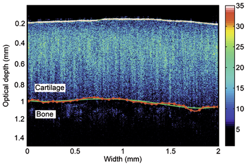
Optical clearing is an effective method to reduce light scattering of biological tissues that provides significant enhancement of light penetration into the biological tissues making non-invasive diagnosis more feasible. In current report Optical Coherence Tomography (OCT) in conjunction with optical clearing is applied for assessment of deep cartilage layers and cartilage-bone interface. The solution of Iohexol in water has been used as an optical clearing agent. The cartilage-bone boundary becomes visible after 15 min of optical clearing that enabling non-invasive estimation of its roughness: Sa = 10 ± 1 µm. The results show that for 0.9 mm thick cartilage optical clearing is stopped after 50 min with an increase of refractive index from 1.386 ± 0.008 to 1.510 ± 0.009. Current approach enables more reliable detection of arthroscopically inaccessible regions, including cartilage-bone boundary and subchondral bone, and potentially improves accuracy of the osteoarthritis diagnosis.
Supporting Information
| Filename | Description |
|---|---|
| jbio201500130-sup-0001-author-biographies.pdfPDF document, 202.3 KB | Author Biographies |
Please note: The publisher is not responsible for the content or functionality of any supporting information supplied by the authors. Any queries (other than missing content) should be directed to the corresponding author for the article.
References
- 1G. Li, J. Yin, J. Gao, T. S. Cheng, N. J. Pavlos, C. Zhang, and M. H. Zheng, Arthritis. Res. Ther. 15, 223–234 (2013).
- 2V. V. Tuchin, Coherent-Domain Optical Methods: Biomedical Diagnostics, Environmental Monitoring and Material Science, V. 2 (Second edition. Berlin, Heidelberg, N.Y.; Springer-Verlag, 2013).
- 3A. Doronin and I. Meglinski, Laser Photon. Rev. 7, 797–800 (2013).
- 4R. Reif and R. K. Wang, Quant. Imaging Med. Surg. 2, 207–212 (2012).
- 5T. Kamali, A. Doronin, T. Rattanapak, S. Hook, and I. Meglinski, Laser Phys. Lett. 6, 607–610 (2012).
- 6T. Rattanapak, J. Birchall, K. Young, M. Ishii, I. Meglinski, T. Rades, and S. Hook, J. Control. Release 172, 894–903 (2013).
- 7A. F. Pena, J. Devine, A. Doronin, and I. Meglinski, Opt. Lett. 38, 2629–2631 (2013).
- 8A. F. Pena, A. Doronin, V. V. Tuchin, and I. Meglinski, J. Biomed. Opt. 19, 086001 (2014).
- 9P. O. Bagnaninchi, Y. Yang, M. Bonesi, G. Maffulli, C. Phelan, I. Meglinski, A. El Haj, and N. Maffulli, Phys. Med. Biol. 55, 3777–3787 (2010).
- 10N. Ugryumova, D. P. Attenburrow, C. P. Winlove, and S. J. Matcher, J. Phys. D: Appl. Phys. 38, 2612–2619 (2005).
- 11G. I. Petrov, A. Doronin, H. T. Whelan, I. Meglinski, and V. V. Yakovlev, Biomed. Opt. Expr. 3 (9), 2154–2161 (2012).
- 12A. Alex, B. Považay, B. Hofer, S. Popov, C. Glittenberg, S. Binder, and W. Drexler, J. Biomed. Opt. 15 (2), 026025 (2010).
- 13N. C. R. te Moller, H. Brommer, J. Liukkonen, T. Virn, M. Timonen, P. H. Puhakka, J. S. Jurvelin, P. R. van Weeren, and J. Tyrss, Vet. J. 197, 589–595 (2013).
- 14Y. T. Pan, Z. G. Li, T. Q. Xie, and C. R. Chu, J. Biomed. Opt. 8, 648–654 (2003).
- 15X. D. Li, S. Martin, C. Pitris, R. Ghanta, D. L. Stamper, M. Harman, J. G. Fujimoto, and M. E. Brezinski, Arthr. Res. Ther. 7, R318–R323 (2005).
- 16S. J. Matcher, J. Appl. Phys. 105, 102041 (2009).
- 17M. C. Mansfield, C. P. Winlove, J. Moger, and S. J. Matcher, J. Biomed. Opt. 13, 044020 (2008).
- 18S. Saarakkala, S. Z. Wang, Y. P. Huang, and Y. P. Zheng, Phys. Med. Biol. 54 (22), 6837–6852 (2009).
- 19D. Zhu, K. V. Larin, Q. Luo, and V. V. Tuchin, Laser Photon. Rev. 7, 732–757 (2013).
- 20S. G. Proskurin and I. Meglinski, Laser Phys. Lett. 4, 824–826 (2007).
- 21M. Bonesi, S. Proskurin, and I. Meglinski, Laser Phys. 20, 891–899 (2010).
- 22J. Wang, Y. Zhang, T. H. Xu, Q. M. Luo, and D. Zhu, Laser Phys. Lett. 9 (6), 469–473 (2012).
- 23Y. Liu, X. Yang, D. Zhu, R. Shi, and Q. Luo, Opt. Lett. 38 (20), 4236–4239 (2013).
- 24V. V. Yakovlev, H. F. Zhang, G. D. Noojin, M. L. Denton, R. J. Thomas, and M. O. Scully, Proc. Natl. Ac. Sci. 107 (47), 20335–20339 (2010).
- 25R. Arora, G. I. Petrov, V. V. Yakovlev, and M. O. Scully, Anal. Chem. 86, 1445–1451 (2014).
- 26K. V. Larin, M. G. Goshn, A. N. Bashkatov, E. A. Genina, N. A. Trunina, and V. V. Tuchin, J. Select Top. Quant. Electron. 18, 1244–1259 (2012).
- 27M. G. Ghosn, V. V. Tuchin, and K. V. Larin, Opt. Lett. 31, 2314–2316 (2006).
- 28M. Kinnunen, A. V. Bykov, J. Tuorila, T. Haapalainen, A. V. Karmenyan, and V. V. Tuchin, J. Biomed. Opt. 19 (7), 071409 (2014).
- 29P. D. Agrba, M. Yu. Kirillin, A. I. Abelevich, E. V. Zagaynova, and V. A. Kamensky, Opt. Spectrosc. 107 (6), 853–858 (2009).
- 30S. J. Madsen, E. A. Chu, and B. J. F. Wong, IEEE J. Select Top. Quant. Electron. 5 (4), 1127–1133 (1999).
- 31M. Szarko, K. Muldrew, and J. E. A. Bertram, BMC Musculoskeletal Disorders 11, 231 (2010).
- 32D. E. T. Shepherd and B. B. Seedhom, Ann. Rheum. Dis. 58, 27–34 (1999).
- 33K. Wang, J. Wu, and T. B. Kirk, J. Biomed. Opt. 18, 105003 (2013).
- 34S.-Z. Wang, Y.-P. Huang, and Q. Wang, Connect. Tiss. Res. 51, 36–47 (2010).
- 35K. Wang, J. Wu, R. E. Day, and T. B. Kirk, J. Microsc. 248, 281–291 (2012).
- 36J. M. Herrmann, C. Pitris, B. E. Bouma, S. A. Boppart, C. A. Jesser, D. L. Stamper, J. G. Fujimoto, and M. E. Brezinski, J. Rheumatol. 26, 627–625 (1999).
- 37V. C. Mow and R. Huiskes, Basic Orthopaedic Biomechanics and Mechano-biology, 3rd ed. (Lippincott Williams & Wilkins, Philadelphia).
- 38K. M. Meek, S. Dennis, and Sh. Khan, Biophys. J. 85, 2205–2212 (2003).
- 39J. H. Gladstone and
T. P. Dale,
Phil. Trans. Royal Soc. London
153,
317–343
(1864).
10.1098/rstl.1863.0014 Google Scholar


