Excessive Plasmin Compromises Hepatic Sinusoidal Vascular Integrity After Acetaminophen Overdose
Potential conflict of interest: Nothing to report.
Supported by the National Institutes of Health (P30GM114731, to C.T.G. and F.L.) and by Oklahoma Medical Research Foundation institutional funds (to C.T.G.).
Abstract
The serine protease plasmin degrades extracellular matrix (ECM) components both directly and indirectly through activation of matrix metalloproteinases. Excessive plasmin activity and subsequent ECM degradation cause hepatic sinusoidal fragility and hemorrhage in developing embryos. We report here that excessive plasmin activity in a murine acetaminophen (APAP) overdose model likewise compromises hepatic sinusoidal vascular integrity in adult animals. We found that hepatic plasmin activity is up-regulated significantly at 6 hours after APAP overdose. This plasmin up-regulation precedes both degradation of the ECM component fibronectin around liver vasculature and bleeding from centrilobular sinusoids. Importantly, administration of the pharmacological plasmin inhibitor tranexamic acid or genetic reduction of plasminogen, the circulating zymogen of plasmin, ameliorates APAP-induced hepatic fibronectin degradation and sinusoidal bleeding. Conclusion: These studies demonstrate that reduction of plasmin stabilizes hepatic sinusoidal vascular integrity after APAP overdose. (Hepatology 2018; 00:1-13).
The circulating zymogen plasminogen (Plg), which is primarily synthesized in the liver, is converted to the serine protease plasmin by widely expressed Plg activators.1 Plasmin is a potent thrombolytic protease that dissolves fibrin blood clots and targets extracellular matrix (ECM) components for degradation. It directly cleaves some ECM components, such as laminin and fibronectin, and it indirectly cleaves additional ECM components by activating matrix metalloproteinases (MMPs).2, 3
We previously reported that excessive plasmin activation and subsequent ECM degradation cause embryonic vascular fragility.4, 5 Embryonic hepatic sinusoidal blood vessels are particularly prone to rupture when their underlying laminin-rich ECM is degraded by plasmin.5 However, because laminin is down-regulated in perisinusoidal spaces after birth,6 it is unclear whether excessive plasmin activity can cause vascular fragility in an adult liver.
Acetaminophen (APAP) overdose is the leading cause of drug-induced liver injury in the United States.7 In addition to centrilobular hepatocyte necrosis, APAP overdose also causes centrilobular bleeding8, and some reports indicate that vascular damage precedes hepatocyte damage in mouse models of APAP overdose.9, 10 Intriguingly, plasmin activation exacerbates APAP-induced hepatic injury in mice. Increased plasmin activation through administration of recombinant tissue plasminogen activator (tPA) elevates APAP-induced liver injury and bleeding in wild-type (WT) mice.11 Conversely, both pharmacological plasmin inhibition and genetic deletion of Plg reduce hepatic damage and bleeding after APAP overdose.11 Moreover, mice with genetic deletion of plasminogen activator inhibitor-1 (PAI-1), which presumably have excessive plasmin activity, suffer massive intrahepatic bleeding after APAP overdose.11, 12 Altogether, these published data suggest a causative relationship between plasmin activation and hepatic vascular damage following APAP overdose. However, a systematic and mechanistic analysis of this putative relationship is missing from the literature and, therefore, is the subject of this study.
Materials and Methods
MICE
Male C57Bl/6J mice, 8-10 weeks old, from Jackson Laboratories (Bar Harbor, ME), with an average weight of 25 g, were used in this study. Mice were socially housed in autoclaved, ventilated microisolator cages with autoclaved corncob bedding and mouse igloos from Bio-Serv (Flemington, NJ) for enrichment in a room with a 14/10-hour light/dark cycle. They were provided chlorinated reverse-osmosis water delivered in pouches and irradiated PicoLab Rodent Diet 20 (catalog no. 5053) from LabDiet (St. Louis, MO). Mice were fasted for approximately 16 hours (overnight) prior to treatment with 300 mg/kg APAP (catalog no. A5000; Sigma-Aldrich, St. Louis, MO) dissolved in warm sterile saline solution (catalog no. 0409-7983-61; Hospira, Lake Forest, IL) and delivered by intraperitoneal injection. Food was returned immediately after treatment with APAP or vehicle. When indicated, mice were additionally treated with 600 mg/kg tranexamic acid (TA; Spectrum Chemicals & Laboratory Products, New Brunswick, NJ) delivered by intraperitoneal injection at 2 hours after APAPoverdose. Male littermate WT (Plg+/+) and Plg-deficient (Plg+/-, Plg–/–) mice, 8-11 weeks old, on a C57Bl/6J background, were used for genetic studies and housed together prior to and during the experiments. Plg-deficient mice13 were genotyped by PCR (65°C annealing temperature) using a forward primer (5′-AATCCCAAGCTGAGTAAAGACAGTGG-3′) and a reverse primer (5′-TTAGGTAATGGAGACACGGAGGTGTT-3′), which produce a 256-bp amplicon from a WT Plg allele, in combination with a forward primer (5′-GACATGGATGGCTGAACCGTCTCTGT-3′) and a reverse primer (5′-ACAAGCAAAACCAAATTAAGGGCCA-3′), which produce a 495-bp amplicon from a knockout Plg allele. All experimental animal protocols were approved by the Institutional Animal Care and Use Committee at the Oklahoma Medical Research Foundation.
HEPATOTOXICITY ASSESSMENT
Blood (300 μL) was drawn from the heart immediately after euthanasia and centrifuged at 7,000g for 5 minutes at 20°C. Serum was collected and subjected toalanine aminotransferase (ALT) measurement using a Catalyst One Chemistry Analyzer from IDEXX Laboratories, Inc. (Westbrook, ME).
HISTOLOGICAL STAINING
Prior to euthanasia, anesthetized animals were perfused with phosphate-buffered saline (PBS) and then perfused with 4% paraformaldehyde at a rate of 5 mL/minute using a 101U/R peristaltic pump from Watson-Marlow Fluid Technology Group (Wilmington, MA). Median liver lobes were subjected to additional immersion fixation in 4% paraformaldehyde overnight and embedded in paraffin blocks. Liver samples from Plg+/+, Plg+/–, and Plg–/– mice were collected without perfusion but immersion-fixed in 4% paraformaldehyde overnight prior to embedding; we did not see dramatic differences in histological blood detection using this technique versus perfusion fixation. Histological sections were cut at 7 μm thickness using an HM 355S microtome from Microm International (Walldorf, Germany). Sections were mounted on UltraClear Microscope slides from Denville Scientific Inc. (Holliston, MA) and allowed to dry overnight prior to hematoxylin and eosin staining, which was performed as described.14 The extent of sinusoidal hemorrhage was determined by measuring the hemorrhage area relative to the entire histological section after rendering red blood cells to black and other cells to white using Fiji software.15
MICROSCOPY AND IMAGE ACQUISITION
Brightfield histological images were obtained with an Eclipse 80i microscope from Nikon (Melville, NY) using ×4 (numerical aperture [NA] 0.13) and ×20 (NA 0.5) objectives and a Nikon DS-Fi1 camera. Fluorescence images were obtained with a Nikon Eclipse Ti-E inverted microscope using a ×40 (NA 0.6) objective, an Andor Zyla 4.2 camera, and an X-cite 120LED light source. NIS-Elements AR3.0 (Nikon) software was used for brightfield image acquisition and assembly, and NIS-Elements AR4.6 (Nikon) software was used for fluorescence image acquisition and assembly.
PLASMIN ACTIVITY ASSAY
Total liver (100 mg) was homogenized in radio immunoprecipitation assay (RIPA) buffer (50 mM trishydroxymethylaminomethane, 1% Triton X-100, 1.1% sodium deoxycholate, 0.1% sodium dodecyl sulfate, and 150 mM NaCl) without protease inhibitors. Lysates were centrifuged at 15,000g for 15 minutes at 4°C. The supernatant was collected and analyzed for plasmin activity. Protein concentration was determined using the Pierce BCA Protein Assay Kit and a NanoDrop 2000 from Thermo Fisher Scientific (Waltham, MA). Plasmin activity was measured using 0.1 mmol/L of a plasmin-specific fluorogenic substrate, Boc-Glu-Lys-Lys-MCA (catalog no. MEK-3105-v; Peptides International Inc., Louisville, KY). Fluorescence from the liver homogenate was compared against a standard curve generated with human plasmin (catalog no. P1867; Sigma-Aldrich). A FLUOstar Omega microplate reader (excitation 355 nm, emission 460 nm) from BMG LABTECH (Ortenberg, Germany) was used for measuring fluorescence.
IN SITU ZYMOGRAPHY
The upper half of left liver lobes was cryosectioned without prior fixation. Sections (12 μm) were overlaid with in situ zymography solution consisting of 1% UltraPure low–melting point agarose (Thermo Fisher Scientific), 10 mmol/L Boc-Glu-Lys-Lys-MCA, or 1 mg/mL quenched BODIPY FL casein from the EnzChek Protease Assay Kit (Thermo Fisher Scientific). Overlaid sections were coverslipped and incubated for 30 minutes at 37°C. Substrate cleavage was detected by fluorescence microscopy.
IMMUNOBLOT
Liver tissue (100 mg) was homogenized in RIPA buffer with Protease Inhibitor Cocktail (catalog no. P8340; Thermo Fisher Scientific). Lysates were centrifuged at 10,000g for 15 minutes at 4°C. The supernatant was collected and analyzed. Protein concentration was determined as described above for the plasmin activity assay. Plg from human plasma (Sigma-Aldrich) was used as a positive control sample for Plg/plasmin detection. Protein (30 μg) was electrophoresed on a 9% sodium dodecyl sulfate-polyacrylamide gel electrophoresis gel and then transferred to a polyvinylidene fluoride membrane that was blocked in 5% nonfat dry milk–trishydroxymethylaminomethane-buffered saline Tween 20 (TBST) for 1 hour. Primary antibodies (diluted in 5% milk–TBST) were incubated at 4°C overnight with gentle agitation, and membranes were then washed 3 times (10 minutes each) in TBST. Horseradish peroxidase–conjugated secondary antibodies (diluted in 5% milk–TBST) were applied at room temperature for 1 hour with gentle agitation, and membranes were then washed 5 times (15 minutes each) in TBST. Secondary antibodies were detected using ECL Western Blotting Detection Reagents (catalog no. RPN2209) from GE Healthcare (Chicago, IL). Primary antibodies used for immunoblotting: Plg/plasmin (1:1,000, no. AMPG-9130) from Haematologic Technologies (Essex Junction, VT), fibronectin (1:1,000, no. Ab2413) from Abcam (Cambridge, UK), glyceraldehyde 3-phosphate dehydrogenase (GAPDH; 1:10,000, no. G9545) from Sigma-Aldrich.
REAL-TIME QUANTITATIVE PCR
Total RNA was extracted from 100 mg of liver samples using the RNeasy Mini kit (no. 74106) from Qiagen (Germantown, MD). Complementary DNA was prepared using the iSCRIPT Reverse Transcriptase Kit from Bio-Rad (Hercules, CA), and quantitative real-time PCR was performed using 2 × SYBR green quantitative real-time PCR master mix (Thermo Fisher Scientific) and the CFX96 Real-Time System (Bio-Rad) with gene-specific primers (Supporting Table S1).
TRANSMISSION ELECTRON MICROSCOPY AND SCANNING ELECTRON MICROSCOPY
Prior to euthanasia, anesthetized animals were perfused with PBS and then perfused with fixative (2.5% glutaraldehyde and 2% paraformaldehyde in 0.1 M sodium cacodylate buffer) at a rate of 5 mL/minute using a 101U/R peristaltic pump from Watson-Marlow Fluid Technology Group. For transmission electron microscopy (TEM), liver samples were further fixed by immersion in the same fixative for 1 hour, followed by postfixation in 1% osmium tetroxide (90 minutes) and 1% tannic acid (1 hour). Samples were subsequently dehydrated in a graded ethanol series and embedded in epoxy resin from Electron Microscopy Sciences (Hatfield, PA). Ultrathin sections (70 nm) were cut using an EM UC7 ultramicrotome from Leica (Wetzlar, Germany) equipped with a diamond knife. Sections were stained with uranyl acetate and lead citrate before being viewed with an H-7600 electron microscope from Hitachi (Tokyo, Japan) equipped with a 4-megapixel digital monochrome camera and AMT-EM image acquisition software from Advanced Microscopy Techniques (Woburn, MA). For scanning electron microscopy (SEM), liver tissue pieces were excised after perfusion fixation and further fixed by immersion in the same fixative overnight. Liver tissue was then sliced into 1-mm-thick sections with a vibratome, washed with sodium cacodylate buffer, and then postfixed with 1% osmium tetroxide in 0.1 M cacodylate buffer for 90 minutes. After washing with cacodylate buffer, tissue slices were dehydrated in a graded ethanol series. The sections were further dehydrated in hexamethyldisilazane and allowed to air-dry overnight. Tissue sections were then sputter-coated with Au/Pd particles using a Med-010 Sputter Coater from Balzers (Schaumburg, IL) and imaged with a Quanta 600 SEM from FEI (Hillsboro, OR) at an accelerating voltage of 20 kV.
IMMUNOFLUORESCENCE
The lower half of left liver lobes was cryosectioned without prior fixation. Sections (7 μm) were taken from the freezer before staining and warmed at room temperature for 3 minutes. Sections were fixed with 4% paraformaldehyde for 15 minutes and permeabilized with 0.1% Triton X-100 for 15 minutes at room temperature, washed 3 times (5 minutes each) with PBS, then blocked in blocking solution (5% normal goat serum/5% normal donkey serum/0.3% bovine serum albumin/0.1% Triton X-100 in PBS) for 1 hour at room temperature. Sections were incubated in primary antibody diluted in blocking solution overnight at 4°C, washed 3 times (3 minutes each) with PBS, then incubated for 1 hour at room temperature in secondary antibody at a 1:500 concentration in blocking solution along with 10 μg/mL Hoechst stain (catalog no. 89139-124) from VWR International (Irving, TX). Primary antibodies used for immunofluorescence: fibronectin (1:100, no. Ab2413; Abcam), collagen I (1:100, no. Ab34710; Abcam), collagen IV (1:100, no. Ab6586; Abcam), laminin (1:100, no. Ab11575; Abcam), cytokeratin 19 (1:1, clone TROMA-III) from Developmental Studies Hybridoma Bank (Iowa City, IA).
Results
APAP-INDUCED HEPATIC SINUSOIDAL BLEEDING IS PLASMIN-DEPENDENT
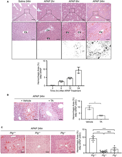
Administration of 300 mg/kg APAP to C57Bl/6J male mice, 8-10 weeks old, following 16 hours of fasting causes increasing centrilobular hepatocyte necrosis (Fig. 1A) and elevated serum ALT measurements (Supporting Fig. S1A) over 24 hours. Bleeding likewise occurs from centrilobular sinusoidal blood vessels by 6 hours after APAP overdose, and it increases in severity by 24 hours (Fig. 1A). Because plasmin contributes to APAP-induced hepatic injury11, 12 and to embryonic hepatic sinusoidal fragility,5 we sought to test the role of plasmin in APAP-induced hepatic bleeding. We first used the pharmacological plasmin inhibitor TA, which we administered 2 hours after APAP overdose. In our hands, TA treatment did not alter serum ALT levels measured 24 hours after APAP overdose (Supporting Fig. S1B). However, centrilobular sinusoidal bleeding at 24 hours after APAP overdose was significantly diminished in TA-treated animals (Fig. 1B). We also took a genetic approach to reduce plasmin in the APAP overdose model by using male mice deficient for the plasmin zymogen Plg (Plg+/– and Plg–/–). Once again, we saw no alteration in serum ALT levels at 24 hours after APAP treatment between littermate WT and Plg+/- or Plg–/– mice (Supporting Fig. S1C). However, as with TA treatment, centrilobular sinusoidal hemorrhage was significantly reduced in Plg+/– and Plg–/– mice versus WT littermates at 24 hours after APAP overdose (Fig. 1C). These pharmacological and genetic data indicate that plasmin contributes to APAP-induced sinusoidal hemorrhage.
PLASMIN ACTIVITY IS ELEVATED BY 6 HOURS AFTER APAP OVERDOSE
In order to determine the level of plasmin activity following APAP overdose, we used a synthetic plasmin substrate that fluoresces upon cleavage.16 With this substrate, we found plasmin activity to be significantly and specifically elevated 6 hours after APAP overdose in whole-liver lysates (Fig. 2A). Notably, this corresponds to the time point at which we see centrilobular sinusoidal bleeding beginning to accumulate after APAP overdose (Fig. 1A). When used for in situ zymography analysis on fresh liver sections, this same synthetic plasmin substrate likewise revealed increased plasmin activity surrounding blood vessels at 6 hours after APAP overdose (Fig. 2B). Immunoblotting of liver lysates with an antibody that recognizes both Plg and plasmin confirmed that plasmin is elevated at 6 hours after APAP overdose (Fig. 2C).
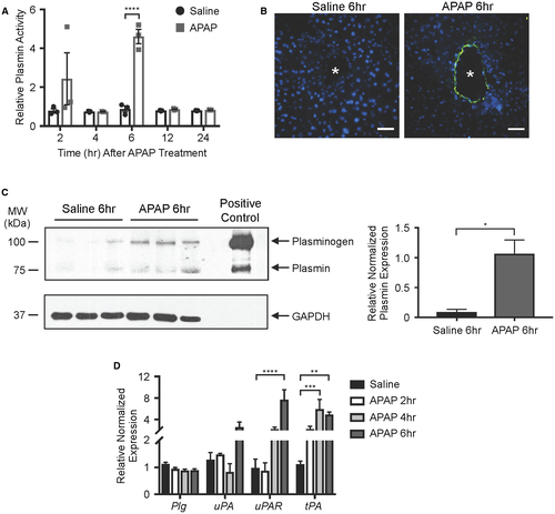
Plasmin is activated from its circulating zymogen Plg by tPA or by urokinase plasminogen activator (uPA) and its receptor (uPAR). In order to address whether expression of Plg or the Plg activators was impacted by APAP overdose, we performed quantitative real-time PCR on total liver samples. We found tPA to be significantly up-regulated by 4 hours after APAP overdose (Fig. 2D), which precedes the 6-hour time point at which our multiple assays showed elevated plasmin activity (Fig. 2A-C). Therefore, an APAP-mediated increase in hepatic tPA expression may contribute to the elevated plasmin activity seen 6 hours after APAP overdose.
SINUSOIDAL ENDOTHELIAL CELLS HAVE CAVERNOUS GAPS BY 6 HOURS AFTER APAP OVERDOSE
To assess sinusoidal endothelial cell morphology at the time of peak APAP-induced plasmin elevation and at the start of APAP-induced sinusoidal bleeding, we processed liver samples for TEM and SEM at 6 hours after APAP overdose. We observed relatively normal sinusoidal endothelial cell morphology at 6 hours after APAP overdose by TEM, unlike the surrounding hepatocytes that showed evidence of necrosis, including swollen organelles and extreme vacuolization (Fig. 3A). However, we did notice a consistent increase in sinusoidal diameter (Supporting Fig. S2A,B) and in the space of Disse between sinusoids and hepatocytes (Supporting Fig. S2A,C) at 6 hours after APAP overdose by TEM. Interestingly, TA treatment did not ameliorate APAP-induced dilation in sinusoids or the space of Disse after APAP overdose (Supporting Fig. S2). We also saw small fenestrations and larger gaps in sinusoidal endothelial surfaces by SEM after saline and APAP treatments (Fig. 3B). However, the larger gaps appeared more cavernous with less underlying meshwork—which may represent ECM—and were seen near extravascular red blood cells after APAP treatment. Additional TA treatment eliminated the extravascular red blood cells and restored some of the meshwork under the larger gaps in the endothelial cell surfaces, although the space of Disse was still visibly enlarged. Altogether, these electron microscopic data indicate that dilated sinusoids and an expanded space of Disse are not plasmin-dependent and do not contribute to APAP-induced sinusoidal bleeding. However, large endothelial gaps with sparse underlying meshwork do correlate with plasmin elevation and may facilitate sinusoidal bleeding after APAP overdose.

FIBRONECTIN IS SIGNIFICANTLY DIMINISHED AROUND HEPATIC VASCULATURE AFTER APAP OVERDOSE
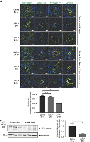
Because excessive plasmin-mediated ECM degradation can compromise embryonic hepatic sinusoidal integrity,5 we immunostained for four major hepatic ECM components following APAP overdose: fibronectin, collagen I, collagen IV, and laminin. At 2 hours after APAP overdose, before we detected significant plasmin elevation (Fig. 2A), we saw no difference in immunostaining for these ECM components compared to saline-treated controls. However, fibronectin, which is abundantly expressed along the sinusoids and can be directly cleaved by plasmin,17 had significantly decreased staining in both centrilobular and periportal regions at 24 hours after APAP overdose (Fig. 4A). Immunoblotting of total liver lysates further confirmed the reduction of fibronectin that occurs by 24 hours after APAP overdose (Fig.4B).
REDUCTION OF PLASMIN RESCUES APAP-INDUCED PERIVASCULAR FIBRONECTIN DEGRADATION
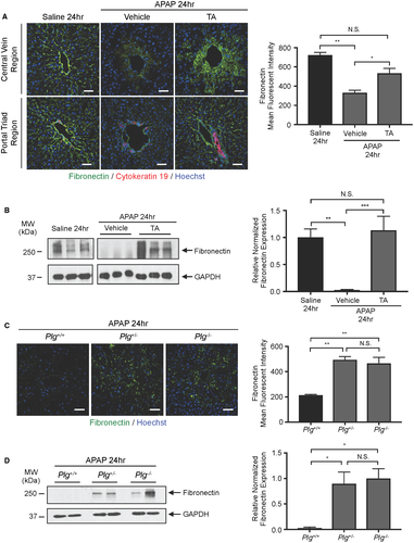
Because plasmin can cleave fibronectin directly or indirectly through activation of MMPs, we sought to determine whether APAP-induced fibronectin diminution is plasmin-dependent. We found that fibronectin immunostaining was significantly rescued in TA-treated animals at 24 hours after APAP overdose (Fig. 5A). We also confirmed that TA treatment rescued fibronectin expression in total liver lysates by immunoblotting at 24 hours after APAP overdose (Fig. 5B). Moreover, fibronectin immunostaining (Fig. 5C) and immunoblotting (Fig. 5D) were also rescued in Plg+/– and Plg–/– livers compared to those from WT littermate controls at 24 hours after APAP overdose. Therefore, plasmin reduction rescues perisinusoidal fibronectin as well as sinusoidal bleeding after APAP overdose, which suggests that fibronectin plays an important role in maintaining sinusoidal integrity in the adult liver.
Discussion
Studies have reported the ability of plasmin to exacerbate APAP-induced liver injury,11, 12 but the specific and mechanistic contribution of plasmin to APAP-induced hepatic vascular damage has been understudied. We considered this to be a relevant question because we recently found that excessive plasmin activity compromises embryonic sinusoidal vascular integrity by promoting hepatic ECM degradation.5 However, we could not predict whether plasmin would cause similar sinusoidal fragility in adult mice because hepatic ECM composition changes after birth.6 We now report that hepatic plasmin activity is significantly up-regulated within 6 hours in a murine APAP overdose model. Moreover, plasmin activity correlates with perisinusoidal fibronectin degradation and bleeding in this model. Therefore, excessive plasmin activity compromises hepatic sinusoidal integrity in both embryos and adult mice, despite the different composition of perisinusoidal ECM before and after birth.
Our TEM analyses at 6 hours after APAP overdose, the time point at which we saw a peak in plasmin activity levels, did not reveal striking morphological abnormalities in centrilobular sinusoidal endothelial cells. Specifically, we did not see evidence of cellular oncosis and vacuolization in sinusoidal endothelial cells, like we did in adjacent hepatocytes. However, we did observe dilated centrilobular sinusoids and an expanded space of Disse in our sections, which is consistent with previous reports.9, 18 Interestingly, these phenotypes were not rescued after pharmacological plasmin inhibition with TA, although TA treatment did reduce perisinusoidal fibronectin degradation and centrilobular sinusoidal bleeding. These data indicate that plasmin does not contribute to sinusoidal dilation or space of Disse expansion after APAP overdose. However, we believe the expanded space of Disse may be responsible for the diffuse fibronectin staining seen in TA-treated mice at 24 hours after APAP overdose (Fig. 5A). Presumably the perisinusoidal fibronectin that is normally degraded by plasmin after APAP overdose can fill the expanded space of Disse when plasmin activity is reduced with TA treatment. Nevertheless, even with this diffuse distribution pattern, the rescue of fibronectin expression still correlates with reduced centrilobular bleeding in TA-treated mice after APAP overdose. Because our SEM data likewise indicate that a meshwork layer reminiscent of ECM is visibly reduced underlying large gaps in sinusoidal endothelial cells after APAP overdose but is rescued after additional TA treatment, we believe that fibronectin-rich ECM presents a physical barrier that impedes red blood cell extravasation.
Our study indicates that fibronectin is reduced around both periportal and centrilobular sinusoids after APAP overdose (Fig. 4A). However, unlike centrilobular sinusoids, periportal sinusoids do not bleed after APAP overdose. The cytochrome P-450 enzyme CYP-4502E1, which metabolizes APAP to its toxic intermediate N-acetyl-p-benzoquinone imine (NAPQI) that induces cellular damage, is primarily expressed in centrilobular hepatocytes.19 This distribution likely causes the characteristic centrilobular hepatocyte necrosis seen after APAP overdose. Based on the centrilobular sinusoidal bleeding observed after APAP overdose, we believe necrotic hepatocytes contribute to this bleeding, perhaps by compromising the structural support that hepatocytes normally lend to sinusoidal vessels. Nevertheless, our data also indicate that fibronectin restoration after plasmin reduction is sufficient to prevent centrilobular sinusoidal bleeding, even in the presence of necrotic hepatocytes. Therefore, we conclude that fibronectin-rich ECM plays a more critical role than hepatocytes in maintaining centrilobular sinusoidal integrity. However, further studies will be needed to determine whether fibronectin provides important prointegrity signaling cues to sinusoidal endothelial cells in addition to scaffolding and barrier supports that maintain sinusoidal integrity.
Despite the fact that reduction of plasmin activity with either pharmacological TA treatment or genetic Plg ablation reduced APAP-induced fibronectin degradation and perisinusoidal bleeding in our study, we did not find that these plasmin inhibition approaches reduced histological evidence of hepatocyte necrosis or serum ALT levels. Our data indicate that plasmin activity and fibronectin degradation do not contribute to APAP-induced hepatocyte damage like NAPQI does. However, these data also stand in contrast to a previous report, in which serum ALT levels and centrilobular necrotic hepatocyte areas were significantly decreased by 24 hours after APAP overdose in mice treated with TA or in Plg–/– mice.11 The cause of this discrepancy at the 24-hour time point is unclear; perhaps it can be attributed to diet or gut microbiota or the number of times that the respective genetic models were crossed onto the C57Bl/6J background. Nevertheless, we propose that it would be interesting to design future studies investigating whether maintenance of sinusoidal integrity with TA treatment helpsto accelerate liver recovery beyond 24 hours after APAP overdose. In light of the report that knockout of PAI-1 delays liver recovery and regeneration processes after APAP overdose,12 it is reasonable to hypothesize that plasmin-mediated sinusoidal hemorrhage impairs tissue repair in the APAP overdose model. A systematic analysis of the efficacy of TA treatment in preventing sinusoidal bleeding and promoting subsequent tissue repair when administered at varying times after APAP overdose is warranted and could reveal new and therapeutically beneficial options for combating APAP-induced liver damage.
Acknowledgment
We thank Vijay Muthukumar, Shruti Desai, and Jun Xie for assistance with mouse colony maintenance and Griffin lab members for helpful discussions. We also thank Francis Castellino (Notre Dame) for sharing Plg+/– mice and Lisa Whitworth (Oklahoma State University Microscopy Laboratory) for assistance with SEM.
REFERENCES
Abbreviations
-
- ALT
-
- alanine aminotransferase
-
- APAP
-
- acetaminophen
-
- ECM
-
- extracellular matrix
-
- GAPDH
-
- glyceraldehyde 3-phosphate dehydrogenase
-
- NA
-
- numerical aperture
-
- PBS
-
- phosphate-buffered saline
-
- Plg
-
- plasminogen
-
- SEM
-
- scanning electron microscopy
-
- TA
-
- tranexamic acid
-
- TBST
-
- trishydroxymethylaminomethane-buffered saline Tween 20
-
- TEM
-
- transmission electron microscopy
-
- tPA
-
- tissue plasminogen activator
-
- uPA
-
- urokinase plasminogen activator
-
- WT
-
- wild type


