Hepatitis C virus–induced CCL5 secretion from macrophages activates hepatic stellate cells
Potential conflict of interest: Nothing to report.
Supported by the National Institutes of Health (R01 DK081817) and Saint Louis University Liver Center Seed Grants.
Abstract
Hepatitis C virus (HCV)–mediated chronic liver disease is a serious health problem around the world and often causes fibrosis/cirrhosis and hepatocellular carcinoma. The mechanism of liver disease progression during HCV infection is still unclear, although inflammation is believed to be an important player in disease pathogenesis. We previously reported that macrophages including Kupffer cells exposed to HCV induce proinflammatory cytokines. These secreted cytokines may activate hepatic stellate cells (HSCs) toward fibrosis. In this study, we examined crosstalk between macrophages and HSCs following HCV infection. Primary human HSCs and immortalized HSCs (LX2 cells) were incubated with conditioned medium derived from HCV-exposed human macrophages. Expression of inflammasome and fibrosis-related genes in these cells was examined, with increased expression of inflammatory (NLR family pyrin domain containing 3, interleukins 1β and 6, and cysteine-cysteine chemokine ligand 5 [CCL5]) and profibrogenic (transforming growth factor β1, collagen type 4 alpha 1, matrix metalloproteinase 2, and alpha-smooth muscle actin) markers. Further investigation suggested that CCL5, secreted from HCV-exposed macrophages, activates inflammasome and fibrosis markers in HSCs and that neutralizing antibody to CCL5 inhibited activation. Conclusion: Together, our results demonstrate that human macrophages exposed to HCV induce CCL5 secretion, which plays a significant role in hepatic inflammation and fibrosis. (Hepatology 2017;66:746–757).
Abbreviations
-
- CCL5
-
- cysteine-cysteine chemokine ligand 5
-
- CM
-
- conditioned medium
-
- COL4A1
-
- collagen type 4 alpha 1
-
- ERK
-
- extracellular signal–regulated kinase
-
- GAPDH
-
- glyceraldehyde 3-phosphate dehydrogenase
-
- HCV
-
- hepatitis C virus
-
- HSC
-
- hepatic stellate cell
-
- IL
-
- interleukin
-
- KC
-
- Kupffer cell
-
- MMP2
-
- matrix metalloproteinase 2
-
- NF-κB
-
- nuclear factor kappa B
-
- NLRP3
-
- NLR family pyrin domain containing 3
-
- α-SMA
-
- alpha-smooth muscle actin
-
- TGF
-
- transforming growth factor
-
- TNF
-
- tumor necrosis factor
The World Health Organization estimates that approximately 150 million people have chronic hepatitis C virus (HCV) infection worldwide.1 A significant number of chronically infected people will develop a potentially life-threatening problem, such as liver fibrosis, cirrhosis, and hepatocellular carcinoma.2 Approximately 700,000 people die each year from HCV-related liver diseases.3 Advanced hepatic fibrosis including cirrhosis is the primary lesion in most cases of hepatocellular carcinoma.4 Sustained inflammatory stimuli cause activation of hepatic stellate cells (HSCs), leading to the storage of considerable extracellular matrixes in the liver and to fibrosis.5 Therefore, understanding the mechanisms of fibrosis following HCV infection is important for prevention and control of liver disease progression.
Different cell types in the liver make a network and relate to hepatic fibrogenesis regulation.6 We and others have shown that macrophages/Kupffer cells (KCs), exposed to HCV, induce interleukin-1β (IL-1β)/IL-18 expression.7-10 However, the mechanism by which HCV induces proinflammatory cytokines in macrophages and its consequence on other liver cell types, such as HSCs, is poorly understood. KCs are resident macrophages of the liver and the largest population of innate immune cells. Liver damage, including induction of fibrosis, may at least in part be attributed to cytokines produced by KCs. The onset of fibrosis reflects a complicated interplay among cytokines, and the cells secrete cytokines and chemokines. In this study, we examined the relationship between conditioned medium (CM) from HCV-exposed macrophages and HSCs. We observed that CM from HCV-exposed macrophages induces inflammasome and fibrosis marker genes. Further study suggested that cysteine-cysteine chemokine ligand 5 (CCL5) is one of the players in HSC inflammation and activation.
Materials and Methods
CELLS AND VIRUSES
Human monocytic THP-1 cells were cultured in Roswell Park Memorial Institute 1640 medium (Sigma-Aldrich, St. Louis, MO) containing l-glutamine, 2-mercaptoethanol (50 μM), 25 mM 4-(2-hydroxyethyl)-1-piperazine ethanesulfonic acid, and 10% fetal bovine serum and maintained at 37°C in a 5% CO2 atmosphere. For macrophage differentiation, THP-1 cells were seeded at a density of 1 × 10⁵ cells/mL and treated with phorbol 12-myristate 13-acetate (100 ng/mL) at 37°C overnight. The cells were then kept in fresh complete medium for 2 days before exposure to virus. Unless mentioned otherwise, we used phorbol 12-myristate 13-acetate-differentiated THP-1 cells throughout our studies as described.7 Human KCs with 98% purity (Thermo Fisher Scientific) were procured and maintained in Dulbecco's modified Eagle's medium supplemented with 10% human AB serum and 1% antibiotics. The immortalized human HSC line LX2 was obtained from Dr. Scott Friedman (Mount Sinai School of Medicine, New York, NY). The cells were cultured in Dulbecco's modified Eagle's medium supplemented with 2% fetal bovine serum and penicillin–streptomycin at 37°C in 5% CO2. Primary human HSCs were procured from ScienCell, maintained in Stellate Cell Medium (ScienCell), and cultured in poly-l-lysine-coated flasks.
For infection, cells were incubated with HCV genotype 2a (clone JFH1; multiplicity of infection of 0.1) in a minimum volume of medium. Phorbol 12-myristate 13-acetate–treated THP-1 cells and KCs were incubated with HCV for 4 hours. After incubation, cells were washed with medium without serum three times and maintained for 24 hours in respective maintenance medium. Culture supernatants were collected, subjected to centrifugation at 10,000g for 10 minutes at 4°C, and kept frozen at –80°C until use (HCV-MΦ-CM/HCV-KC-CM). Culture supernatant from mock-infected THP1 macrophages or KCs served as a control CM. LX2 cells or primary human HSCs were incubated with CM from control or HCV-infected macrophages at indicated time points. Cells were lysed and processed for either RNA isolation or protein lysate preparation.
RNA ISOLATION AND QUANTITATIVE RT-PCR
Total RNA was isolated using TRIZOL reagent (Life Technologies). RNA was quantified using a NanoDrop ND-1000 spectrophotometer (Thermo Fisher Scientific). Complementary DNA was synthesized using random hexamers and a Superscript III reverse transcriptase kit (Invitrogen, CA). Real-time PCR was performed with complementary DNA for quantitation using the TaqMan gene expression PCR master mix and 6-carboxyfluorescein-minor groove binder probes for NLR family pyrin domain containing 3 (NLRP3; assay Hs00918082_m1), IL-1β (assay Hs01555410_m1), ACTA2 (assay Hs00426835_g1), tissue inhibitor of metalloproteinase 1 (assay Hs00171558_m1), collagen type 4 alpha 1 (COL4A1; assay Hs00266237_m1), transforming growth factor β1 (TGFβ1; assay Hs00998133_m1), IL-6 (assay Hs00985639_m1), CCL5 (assay Hs00982282_m1), tumor necrosis factor-alpha (TNF-α; assay Hs01113624_g1), and matrix metalloproteinase 2 (MMP2; assay Hs01548727_m1). A 6-carboxyfluorescein-minor groove binder probe for 18S ribosomal RNA (assay Hs03928985_g1) was used as an endogenous control. The reactions were performed at 50°C for 2 minutes and 95°C for 10 minutes, cycled at 95°C for 15 seconds and 60°C for 1 minute for 40 cycles, and held at 25°C for 2 minutes in a 7500 real-time PCR system (Applied Biosystems). Relative gene expression levels were normalized to the 18S ribosomal RNA level using the 2–ΔΔCT formula (ΔΔCT = ΔCT sample − ΔCT untreated control).
WESTERN BLOT ANALYSIS
Control or HCV-MΦ-CM-treated LX2 cells were lysed using a sodium dodecyl sulfate–polyacrylamide gel electrophoresis sample loading buffer. Proteins were subjected to electrophoresis on a polyacrylamide gel and transferred onto a nitrocellulose membrane. The membrane was probed with specific antibody. Proteins were detected using an enhanced chemiluminescent western blot substrate (Pierce, IL). Membranes were reprobed with antibody to glyceraldehyde 3-phosphate dehydrogenase (GAPDH; Cell Signaling) as an internal control for normalization of protein load. All western blot experiments were performed at least three times for reproducibility. ImageJ software (National Institutes of Health) was used for densitometric scanning of western blot images. The membrane was probed with antibodies to phospho-nuclear factor kappa B (NF-κB) p65 (Ser536) (93H1), NF-κB p65 (D14E12), phospho-IκBα (14D4), IκBα (L35A5), phospho-p44/42 mitogen-activated protein kinase (extracellular signal–regulated kinase 1/2 [Erk1/2]) (Thr202/Tyr204) (D13.14.4E), p44/42 mitogen-activated protein kinase (L34F12), GAPDH (Cell Signaling), and α-actin (1A4):Sc-32251 (Santa Cruz).
LUCIFERASE ASSAY
LX2 cells (3 × 10⁴/well) were seeded and transfected with pNF-κB Luc reporter plasmid (a minimal promoter with three NF-κB binding sites linked with a luciferase reporter gene)11 using jetPRIME trasnsfection reagent (Polyplus). After 24 hours of transfection, cells were treated with control or HCV-MΦ-CM for another 24 hours. Cells were harvested in 1× passive lysis buffer (Promega), and luciferase activity was determined using GloMax (Promega).
INFLAMMATION ANTIBODY ARRAY
CM from control or HCV-infected macrophages was analyzed for the presence of 40 human cytokine proteins using an inflammation membrane antibody array (Abcam; ab134003) following the manufacturer's instructions. The intensity of each cytokine or chemokine spot was determined by ImageJ software and represented as relative expression compared to control CM.
NEUTRALIZATION ASSAY
Neutralizing antibody to IL-1β, TNF-α, CCL5, or normal goat immunoglobulin G control was procured (R&D Systems, Minneapolis, MN) and used (2 μg/mL) for the neutralization assay. Antibodies were incubated with CM for 1 hour at 37°C before addition to LX2 cells.
STATISTICAL ANALYSIS
The results are presented as means ± standard deviations. Data were analyzed by Student t test with a two-tailed distribution. P < 0.05 was considered statistically significant.
Results
CM FROM HCV-EXPOSED MACROPHAGES PROMOTES PROINFLAMMATORY MARKER GENE EXPRESSION IN HUMAN HSCs
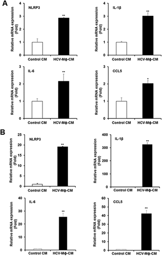
LX2 cells were incubated for 48 hours with CM from THP1 macrophages incubated with HCV (HCV-MΦ-CM) and analyzed for changes in the expression levels of various known proinflammatory cytokine genes compared to CM from THP1 macrophages (control CM). Our results suggested enhanced expression (2-fold to 3-fold) of NLRP3, IL-1β, IL-6, and CCL5 genes in LX2 cells (Fig. 1A). Next, we verified the expression of these genes in primary HSCs. We observed a significantly higher level (>20-fold) of expression of these proinflammatory cytokine genes in primary HSCs following incubation with CM from macrophages (Fig. 1B). Interestingly, the expression level of IL-1β was highest (>300-fold) compared to control. NLRP3 inflammasome plays an important role in inflammation and fibrosis during nonalcoholic steatohepatitis and alcoholic steatohepatitis development.12 However, the specific contribution of persistent NLRP3 inflammasome activation in HSCs during HCV infection remains to be understood.
FIBROGENIC MAKERS ARE ELEVATED IN HSCs FOLLOWING EXPOSURE OF CM FROM HCV-INCUBATED MACROPHAGES
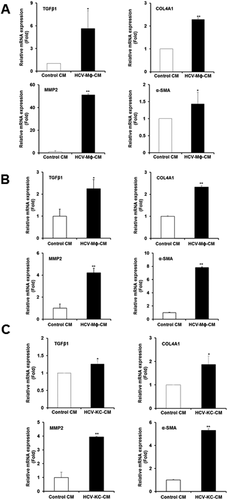
We examined the modulation of fibrosis markers in LX2 cells incubated with HCV-MΦ-CM compared to control CM. Our results demonstrated activation of fibrosis markers in HCV-MΦ-CM-incubated LX2 cells (Fig. 2A). The results suggested higher TGFβ1 (∼6-fold), COL4A1 (>2-fold), MMP-2 (>50-fold), and alpha-smooth muscle actin (α-SMA; ∼1.5-fold). Similarly, we examined for changes in primary human HSCs and observed a significant up-regulation of profibrogenic markers TGFβ1 (>2.5-fold), COL4A1 (>2.5-fold), MMP2 (>4-fold), and α-SMA (>8-fold) in primary HSCs incubated with HCV-MΦ-CM compared to control CM (Fig. 2B). Other collagen markers such as COL1A1 and COL1A2 are also increased in primary HSCs incubated with HCV-MΦ-CM compared to control CM (data not shown). In order to determine the modulation of HSCs by liver-resident macrophages, we incubated KCs with HCV. Primary HSCs were exposed to CM from control or HCV-exposed KCs. Our results demonstrated a significant up-regulation of profibrogenic markers TGFβ1 (<1.5-fold), COL4A1 (∼2-fold), MMP2 (>4-fold), and α-SMA (>5-fold) in primary HSCs exposed to HCV KC CM compared to control CM (Fig. 2C). Together, our results indicated that soluble mediators from HCV-exposed macrophages when exposed to HSCs exert proinflammatory and profibrogenic effects on HSCs. Interestingly, the effect was higher in the generation of MMP2 and α-SMA compared to the other two cytokine genes (COL4A1 and TGFβ1).
CM FROM HCV-EXPOSED MACROPHAGES ACTIVATES NF-κB IN LX2 CELLS
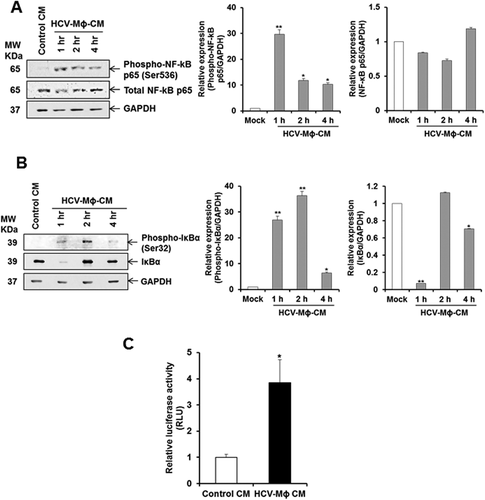
Several signaling pathways regulate the expression of proinflammatory cytokines, of which the NF-κB pathway is the most important.13 To understand the intracellular mechanisms responsible for HSC modulation by HCV-MΦ-CM, the NF-κB signaling pathway was examined. We observed an increase of phosphorylated NF-κB in LX2 cells following incubation with CM from HCV-exposed macrophages (Fig. 3A). In the canonical pathway, NF-κB/Rel proteins are bound and inhibited by IκB proteins. Proinflammatory cytokines, lipopolysaccharide, growth factors, and antigen receptors activate an IKK complex (IKKβ, IKKα, and NEMO), which phosphorylates IκB proteins. Phosphorylation of IκB leads to its ubiquitination and proteasomal degradation, freeing NF-κB/Rel complexes.14 We therefore examined the phospho-IκBα and IκBα expression in LX2 cells. We observed up-regulation of phospho-IκBα and significant reduction in total IκBα level (Fig. 3B). The degradation of IκBα with concomitant increase in phosphorylated NF-κB p65 in LX2 cells suggested activation of this pathway. We also examined the NF-κB-dependent luciferase reporter activity in LX2 cells in the presence of control CM or HCV-MΦ-CM. There was a significant induction of NF-κB-dependent luciferase activity in LX2 cells incubated with HCV-MΦ-CM compared to control CM (Fig. 3C). Together, our results demonstrated that the NF-κB pathway is active in HSCs in response to CM from HCV-exposed macrophages.
IDENTIFICATION OF FIBROGENIC ACTIVATOR FROM CM OF HCV-EXPOSED MACROPHAGES
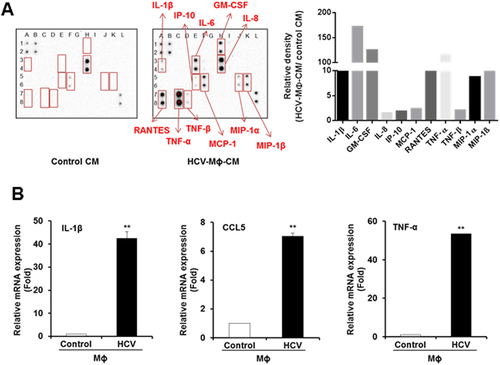
In order to identify soluble mediator in CM from HCV-exposed macrophages, we performed a human inflammation array using control and CM from HCV-exposed macrophages. We observed a significant induction of proinflammatory cytokines and chemokines (Fig. 4). Levels of IL-1β; IL-6; granulocyte-macrophage colony-stimulating factor; IL-8; interferon-inducible protein 10; monocyte chemoattractant protein 1; regulated upon activation, normal T cell expressed, and secreted (CCL5); TNF-α; TNF-β; and macrophage inflammatory proteins 1α and 1β were significantly higher in CM from HCV-exposed macrophages compared to control CM (Fig. 4A). Based on the information in the literature as well as our previous results, we focused initially on IL-1β, TNF-α, and CCL5 to examine their role in hepatic fibrosis. In HCV-infected patients, serum IL-1β, TNF-α, and CCL5 levels were higher compared to healthy individuals.15-17 We confirmed the expression of these molecules in HCV-exposed macrophages at the mRNA level (Fig. 4B).
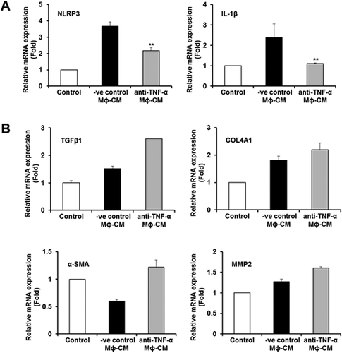
Several studies have indicated that IL-1β contributes to the development of nonalcoholic steatohepatitis and fibrosis.18 We observed higher-level expression of IL-1β in HSCs after incubation with CM from HCV-exposed macrophages (Fig. 1). We next incubated CM from HCV-exposed macrophages with different doses of IL-1β neutralizing antibody and further incubated with HSCs. We did not observe blockade in inflammasome or fibrosis marker gene expression (data not shown). TNF-α has also been implicated in the promotion of HSC survival and proliferation.18-20 We incubated different concentrations of TNF-α neutralizing antibody with CM from HCV-exposed macrophages, following exposure to HSCs. The CM from HCV-exposed macrophages treated with different concentrations of negative control antibody was used in parallel as control. Interestingly, we observed a significant reduction in expression of NLRP3 and IL-1β genes after TNF-α neutralization (Fig. 5A) but not the fibrosis marker genes (Fig. 5B), suggesting that TNF-α mediated induction of the inflammasome pathway in HSCs.
CCL5 SECRETED FROM HCV-EXPOSED MACROPHAGES INDUCES INFLAMMASOME AND FIBROSIS MARKERS IN HSCs
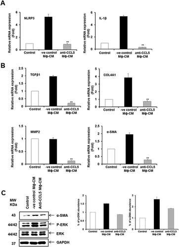
CCL5 is up-regulated in patients with advanced fibrosis or cirrhosis and correlated with higher inflammation scores and greater liver damage during chronic HBV or HCV infection.15-17, 21 CCL5 plays a role in hepatic activation in mice when treated with carbon tetrachloride for reactive phenomenon of liver functions.18 Our inflammation array data demonstrated HCV-induced CCL5 secretion in macrophages. To examine the intercellular communication between macrophages and HSCs during HCV infection, we evaluated the role of CCL5 by neutralization assay. The negative control antibody was used in parallel as control. When LX2 cells were incubated with CCL5 neutralized CM from HCV-exposed macrophages, we observed a significant down-regulation of inflammatory (NLRP3 and IL-1β) and profibrogenic (TGFβ1, COL4A1, MMP2, and α-SMA) genes compared to the negative control antibody (Fig. 6A,B). We also observed that CCL5-mediated α-SMA expression is reduced when neutralizing antibody to CCL5 was used (Fig. 6C). CCL5 induces HSC activation through phosphorylation of ERK22 and phosphorylation of AKT.23 We observed up-regulation of ERK phosphorylation compared to negative control, and phosphorylation of ERK was down-regulated in HSCs when HCV-MΦ-CM was incubated with anti-CCL5 neutralizing antibody (Fig. 6C). However, we did not observe modulation of the Akt signaling pathway (data not shown).
Discussion
We have demonstrated that CM from HCV-exposed macrophages activates human HSCs. We have also shown that HCV induces secretion of CCL5 from macrophages, which in turn activates inflammatory molecules and fibrosis markers in HSCs. This indirect pathway is thought to be mediated by HSC activation through KC-derived CCL5 following HCV exposure. Chronic HCV infection promotes secretion of cytokines and chemokines by modulating promoter activity in hepatic cell lines23 and by circulatory and resident liver macrophages through the NF-κB signaling pathway.7 In diseased liver, most extracellular matrix components are produced by HSCs,24 and accumulation of these causes liver fibrosis. Cytokines and chemokines stimulate key biological processes in HSCs, such as activation, proliferation, and migration.22 We have shown recently that exosomes carrying microRNA 19a from HCV-infected hepatocytes induce HSC fibrosis markers by modulating the SOCS–STAT3 axis.25 We also examined whether residual HCV from macrophage CM activates HSC. However, we could not detect HCV RNA in HSCs incubated with HCV-MΦ-CM. Therefore, residual HCV activating HSCs is unlikely. Treatment of chronic HCV infection with direct-acting antiviral agents is associated with high rates of sustained virologic response (>90%). Direct-acting antiviral agents can reduce the viral load and may thus be effective in reverting liver pathogenesis at an early stage of disease. We postulate that HCV infection activates HSCs using multiple pathways, resulting in the development and progression of fibrosis.
Resident macrophages are major regulators of inflammation and fibrogenesis by regulating the crosstalk between HSCs and immune system cells to achieve a cellular response.22, 23 Macrophages produce cytokines and chemokines that directly activate fibroblasts and recruit inflammatory cells.24 We previously demonstrated that HCV exposure to macrophages, including KCs, induces inflammasome formation.7 In this study, we observed a significant induction of proinflammatory cytokines and chemokines. Although TNF-α enhances inflammasome markers, it did not exert an effect on activation of fibrosis marker genes. Interestingly, IL-1β did not play a role in activation of HSCs in our experimental system.
CCL5 is secreted by macrophages through activation of the Nod-like receptor proteins Nod1 and Nod2, and NF-κB signaling is key in the Nod-dependent stimulation of the CCL5 promoter.26 We previously reported that HCV induces mature IL-1β/IL-18 in cell-free supernatants through the NF-κB signaling pathway in macrophages.24 Therefore, it is possible that HCV-exposed macrophages secrete CCL5 through the NF-κB signaling pathway. Clinically, CCL5 is up-regulated in serum and livers of HCV-infected individuals.23 The CCL5 mRNA level correlates with the HCV RNA load and histological activity index, including score of fibrosis, intralobular degeneration, and focal necrosis, periportal with or without bridging necrosis and portal inflammation. CCL5 and its receptors, CCR5 and CCR1, are important players in liver fibrosis in the carbon tetrachloride mice model27, 28 and in HSC proliferation and migration in primary human HSCs.20 Our data demonstrate that HSCs incubated with CCL5 neutralized CM of HCV-exposed macrophages and displayed a significant down-regulation of inflammatory and profibrotic marker genes compared to negative control. Our study further indicated that ERK phosphorylation was up-regulated in HSCs incubated with CM from HCV-exposed macrophages. Other inflammatory molecules may also have a role in the activation of fibrosis and will be investigated in the future.
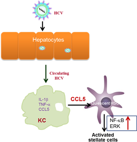
HCV induces CCL5 secretion in human macrophages, which plays a role in hepatic inflammation and fibrosis. Interestingly, our data suggested that HCV-MΦ-CM leads to significant reduction in IκBα level in LX2 cells. The degradation of IκBα with a concomitant increase in phosphorylated NF-κB p65 in LX2 cells suggests involvement of the NF-κB pathway. We also observed a significant induction of NF-κB-dependent luciferase activity in LX2 cells in the presence of HCV-MΦ-CM. NF-κB has been implicated in the activation of cytokines.29, 30 In the human immunodeficiency virus and HCV coinfection model, human immunodeficiency virus and HCV cooperatively promote hepatic fibrogenesis through the NF-κB dependent pathway.31 Based on our results, we propose a mechanism (Fig. 7) that suggests the onset of fibrosis during HCV infection. NF-κB plays an important role in the regulation of proinflammatory cytokines, and we observed that NF-κB signaling is activated for IL-1β/IL-18 transcription in HCV-exposed macrophages.7 NF-κB signaling is also activated in HSCs treated with HCV-MΦ-CM but not fibrosis markers. On the other hand, CCL5 is known to activate fibrosis through the ERK pathway.22 We demonstrated that CCL5 indeed activates fibrosis markers through the ERK pathway and is reduced following the use of neutralizing antibody to CCL5. Thus, the NF-κB and ERK signaling pathways are likely to be independently involved in HSC activation. Understanding the in-depth mechanism will help in the development of future therapeutic strategies against HCV-associated fibrosis.
Acknowledgment
We thank Scott Friedman for providing LX2 cells.
REFERENCES
Author names in bold designate shared co-first authorship.


