How to conduct EEG recordings—A video-based educational resource
[Correction added on 25 January 2024, after first online publication: The copyright line was changed.]
Abstract
Content available: Video
In this multimedia teaching material, we address the following learning objective of the ILAE's competency-based curriculum1: “Demonstrate knowledge on how to conduct EEG recordings, including technical requirements (e.g., mounting electrodes, using filters, amplifiers, electrode arrays, etc.).”
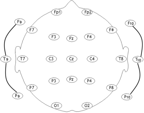
Standardized EEG electrode positioning is paramount both in the clinical and research realms. The classical 10–20 system, initially developed by Jasper and colleagues in the 1950s,2 has been widely used as it allows a consistent and replicable method of recording EEG.3 It was not until 2017 that the International Federation of Clinical Neurophysiology (IFCN) proposed an updated standardized EEG electrode array.4
This 25-electrode array4 includes inferior temporal electrodes thus covering the anterior and basal areas of the temporal lobes—which are the most frequent source of epileptogenicity (Figure 1). Electrodes are placed in accordance with 20% and 10% positions of standardized measurements from skull anatomical landmarks (Figure 2). We illustrate this topic by presenting two educational videos: a manikin demonstration (Video 1) and a live demonstration (Video 2).


ACKNOWLEDGMENTS
We thank Dr. Bernard Chang, Dr. Mouhsin Shafi, and Mr. Daniel Sweeney for their invaluable assistance in this educational project.
CONFLICT OF INTEREST STATEMENT
F. A. Nascimento is an Associate Editor for Epileptic Disorders. S. Beniczky is an Editor-in-Chief for Epileptic Disorders. M. Salazar, J. Colonetti, and D. Schomer report no disclosures relevant to this article.
REFERENCES
Test yourself
-
How many EEG electrodes does the standardized EEG electrode array proposed by the International Federation of Clinical Neurophysiology (IFCN) include?
- 16
- 18
- 22
- 25
-
What additional coverage has been included in the standardized EEG electrode array proposed by the International Federation of Clinical Neurophysiology (IFCN)?
- Inferior temporal
- Superior frontal
- Inferior occipital
- Superior parietal
Answers may be found in the supporting information.


