Unveiling the uncommon: Gas-containing renal stones—A unique case study
Key Clinical Message
Gas-containing renal stones are a rare condition. There is an association between renal stones containing gas, urinary tract infection, and renal fusion anomalies, so it is essential to know the radiographic features for prompt diagnosis and treatment.
1 INTRODUCTION
Gas-containing renal stones, documented in only 12 cases before this,1 remain a medical rarity, often associated with recurrent urinary tract infections (UTIs).2 This contribution sheds light on the thirteenth reported case, exploring clinical nuances, imaging nuances, and tailored management approaches.
2 CASE PRESENTATION
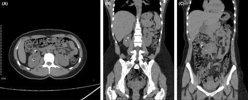
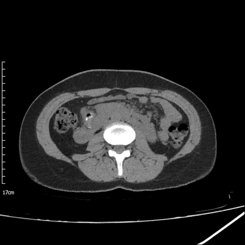
A 31-year-old woman with a history of three times UTIs, who has undergone two times percutaneous nephrolithotomies (PCNL) and one transurethral lithotomy (TUL) due to previous renal stones, and without a history of diabetes mellitus, vesicourethral reflux or hypertension, came at the emergency department with right flank pain and macroscopic hematuria. Plain computed tomography (CT) revealed numerous stones containing gas in all calyces and pelvis with maximum dimensions of 22 × 14 mm in the right kidney accompanied by horseshoe kidneys (Figures 1A–C and 2). Urine analysis also showed a significant white blood cell count, and moderate bacteria and urine culture results showed Escherichia coli growth.


3 MANAGEMENT
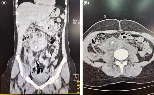
The patient initially received conservative management with antibiotics and hydration. Imaging studies, including enhanced CT, validated the stone's location. Given the unusual nature of the Case, a multidisciplinary approach involving urologists, nephrologists, and infectious disease specialists is needed to determine the most appropriate course of action. The absence of fat stranding and free fluid in perinephric space further supported the gas-containing stones diagnosis. Ultimately, the patient went under PCNL and a subsequent follow-up CT scan was conducted to monitor the patient's condition (Figure 3A,B). A preoperative urine analysis showed moderate bacteriuria with a positive urine culture for E. coli. A repeat urine analysis was performed one week after surgery, which revealed significant bacteriuria, although the urine culture was negative.

4 CONCLUSION
Gas-containing renal stones present an uncommon diagnostic and management challenge. This case report contributes to the sparse literature on this subject, emphasizing the need to consider this rare condition in the differential diagnosis of renal stones. Future investigations should delve into the underlying renal fusion anomalies, and metabolic and infectious factors contributing to gas-containing stone formation, guiding optimal therapeutic strategies.3
5 DISCUSSION
Gas-containing renal stones present a diagnostic conundrum, necessitating differentiation from emphysematous pyelonephritis.3 UTIs play a pivotal role in the development of these stones, evident in most of the reported cases, including this one.2 Bacteria within stones metabolize generating gas and in most cases E. coli was responsible.4 The presence of gas inside the stones indicates a combination of two pathologies, called tissue metabolism and metabolic activity.4 Imaging features, showing distinct gas pockets within the stone, underscore the uniqueness of gas-containing stones compared with other renal pathologies.
Regarding prognosis, follow-up CT-scan assessments post-surgery indicated a favorable outcome. The size of the largest stone reduced significantly from 22 × 12 mm to 12 × 6 mm, and the overall number of stones also decreased (Figure 3A,B). Notably, the stones were no longer gas-containing, suggesting effective surgical management and resolution of the underlying pathology.
AUTHOR CONTRIBUTIONS
Fahimeh Zeinalkhani: Conceptualization; supervision. Peyman Kamali Hakim: Conceptualization; resources. Mahdiyeh Movahedi: Formal analysis; software; visualization; writing – review and editing. Fatemeh Zare Bozorgabadi: Data curation; methodology. Maryam Movahedi: Project administration; writing – original draft; writing – review and editing.
ACKNOWLEDGMENTS
We would like to thank the patient for permitting us to publish this article.
FUNDING INFORMATION
None.
CONFLICT OF INTEREST STATEMENT
The authors declare no conflict of interest.
CONSENT
Written informed consent was obtained from the patient to publish this report in accordance with the journal's patient consent policy.
Open Research
DATA AVAILABILITY STATEMENT
The data that support the findings of this study are available from the corresponding author, upon reasonable request.


