Right sided posterior diaphragmatic hernia with liver incarceration: A case report
Key clinical Message
Right sided posterior diaphragmatic hernias are a rare diagnosis, especially in adult populations. This patient presented with right thoracic pain for 20 years before investigation. Imaging has provided an accurate diagnosis in this case. Repair can be done safely laparoscopically.
1 CASE PRESENTATION
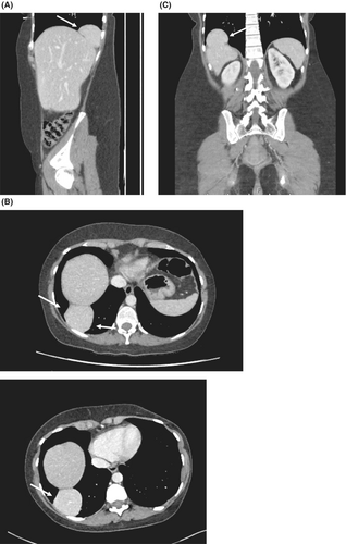
A 48-year-old woman presented to the outpatient clinic complaining of persistent pain in the right thoracic base, which had been present since she underwent elective cholecystectomy 19 years earlier. Despite this discomfort, further imaging was not pursued until she visited the emergency room due to a urinary tract infection. Initial blood tests showed elevated white blood cell count (10,220/mm3), elevated C-reactive protein (85.6 mg/L). Liver function tests were all withing normal range. Urinalysis and blood cultures were both positive for Escherichia Coli. Subsequent thoracoabdominal computed tomography revealed acute left pyelonephritis as well as a right diaphragmatic hernia with intrathoracic liver segment VII protrusion (Figure 1A–C). She was treated with Ciprofloxacine 500 mg twice daily for the urinary tract infection. On outpatient clinic follow-up a month later, no further urinary tract infection was detected. She was referred to our department for management of the right sided diaphragmatic hernia.

In our clinic, she still complained of right upper quadrant pain. No prior traumatic event was described by the patient. Prior history shows a laparoscopic cholecystectomy 19 years earlier as well as an open appendectomy in 1990. Repeat thoracoabdominal computed tomography was performed that showed no evolution of the right sided diaphragmatic hernia. Since persistent pain was described by the patient, we recommended a laparoscopic hernia repair.
2 MANAGEMENT
Under general anesthesia, patient was placed in the French position. Upon achieving pneumoperitoneum, we placed a 12 mm trocar on the right midclavicular line, 3 cm below the 12th rib. Two 5 mm trocars were then inserted on either side of the first trocar, followed by another 5 mm trocar on the left flank. Liver mobilization began at the falciform ligament, extending to the right lateral aspect of the right suprahepatic vein. The right coronary ligament was divided until reaching the right triangular ligament. Adhesiolysis was performed on the liver's inferior aspect, with the hepaticoduodenal ligament dissected laterally toward the right triangular ligament. The herniated liver segment was reduced, revealing a 5 × 2 cm right posterior diaphragmatic hernia with access to the right pleural cavity. Closure of the defect was achieved using two running sutures of nonabsorbable V-loc (™ Medtronic) 3–0, without mesh placement. The postoperative course was uneventful, and patient was discharged on postoperative day 2.
Patient was seen again in the outpatient clinic 1 month postoperatively and was pain free. She did not present any thoracic or right upper quadrant discomfort for the first time in nearly 20 years.
3 DISCUSSION
Posterolateral diaphragmatic hernias, most commonly called Bochdalek hernias, represent 80%–90% of all diaphragmatic hernias. They can be either congenital or acquired. When congenital, they appear 85% of all cases on the left side, as the right pleuroperitoneal canal obliterates faster than the left one, as well as being “protected” by the liver.1 Adult onset is rare as its incidence is estimated at 0.17%, with commonly herniated organs including the colon, small bowel, liver, and right kidney.2 Acquired defects are mostly posttraumatic. Usually asymptomatic if no abdominal viscera is involved, diaphragmatic hernias are usually found incidentally. Adult onset present with vague symptoms such as diffuse abdominal pain, chest pain or even shortness of breath. Clinical examination is often unremarkable, and imaging using computerized tomography is usually sufficient for the diagnosis.1, 2 In this patient, it is unknown if the defect was acquired or congenital. No prior imaging is available for review.
Given its rarity, there is no consensus on the optimal management of diaphragmatic hernias. A systematic review encompassing 41 studies described a variety of surgical approaches, including laparotomy, laparoscopy, robotic repair, thoracoscopy, thoracotomy, or a combination thereof.3 We present a case of right sided diaphragmatic hernia treated laparoscopically with no mesh reinforcement of the primary closure. Patient showed significant improvement in her symptoms.
We believe that the abdominal laparoscopic approach is safe, feasible, and yields satisfying results. Further studies are required to assess if mesh placement is required.
AUTHOR CONTRIBUTIONS
Boitsios Alexi: Conceptualization; investigation; writing – original draft; writing – review and editing. Lacroix Sacha: Writing – review and editing. Jehaes François: Writing – review and editing. Monami Benoit: Writing – review and editing.
FUNDING INFORMATION
No funding was received for this article.
CONFLICT OF INTEREST STATEMENT
The authors state that they have no conflicts of interest.
Open Research
DATA AVAILABILITY STATEMENT
Data sharing not applicable to this article as no datasets were generated or analysed during the current study.
Written informed consent was obtained from the patient to publish this report in accordance with the journal's patient consent policy.


