Diagnostic and response assessment FDG PET-CT in neurolymphomatosis
Key Clinical Message
FDG PET-CT is a useful imaging tool in the diagnosis and response assessment of neurolymphomatosis, especially in cases of otherwise unexplained neuropathy following conventional diagnostic work-up including lumbar puncture, CT, and MRI. The use of a novel PET reconstruction algorithm improves image quality and lesion detection through increased signal-to-noise ratio.
A 66-year-old woman with stage IVB diffuse large B-cell lymphoma (DLBCL) with bone marrow involvement but no other sites of extranodal disease, as confirmed on a contrast-enhanced CT of the neck, chest, abdomen and pelvis and lumbar puncture, was treated to a complete response with six cycles of R-CHOP (rituximab, cyclophosphamide, doxorubicin, vincristine, and prednisone), two additional rituximab infusions, and four cycles of intrathecal methotrexate for central nervous system prophylaxis. One month following completion of treatment, the patient developed a painful progressive sensorimotor neuropathy in both arms and legs. Biochemical tests including serum B12, folate, and thyroid function tests were normal. Myeloid-associated glycoprotein antibody and paraneoplastic antibody assays including anti-Hu, Yo, Ri, and Ma were negative. A repeat staging CT was normal as was a MRI of her brain and whole spine. CSF analysis showed no evidence of lymphoma and only a mildly elevated CSF protein of 0.52 g/L (normal range 0.15–0.45 g/L) with normal CSF glucose. A trial of high-dose steroids had no effect. Nerve conduction studies showed severe multifocal sensory and motor axonal loss with lymphomatous nerve infiltration or paraneoplastic vasculitis as possible causes (rather than demyelination or vincristine-related neuropathy).
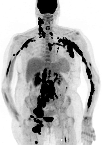
A baseline FDG PET-CT scan prior to commencing chemotherapy (Fig. 1) was acquired using a three-dimensional mode time-of-flight (ToF) Discovery 690 PET-CT system (GE Healthcare) with PET images reconstructed using a novel Bayesian penalized likelihood PET reconstruction algorithm, an iterative technique recently introduced by GE Healthcare (Q.Clear, GE Healthcare, Milwaukee, WI, USA); the previous standard PET reconstruction was a ToF-ordered subset expectation maximization (OSEM) with two iterations, 24 subsets, and a 6.4-mm Gaussian filter (VPFX, GE Healthcare). PET-CT imaging revealed extensive intensely FDG avid (peri)neural linear uptake along both arms (left > right) without corresponding structural abnormality, in addition to further linear FDG uptake along the left fifth intercostal nerve (Q.clear = SUVmax 20.3, ToF OSEM = SUVmax 8.3), both L3 nerves (left L3; Q.clear = SUVmax 28.5, ToF OSEM = SUVmax 16.0), and left sciatic nerve (Q.clear = SUVmax 13.6, ToF OSEM = SUVmax 5.4). There was new bulky intensely FDG avid nodal disease above and beneath the diaphragm. Overall appearances were consistent with widespread FDG avid neurolymphomatosis and recurrent nodal disease above and beneath the diaphragm.

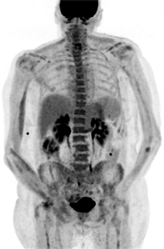
She was commenced on salvage treatment with rituximab, ifosfamide, carboplatin, and etoposide (R-ICE) chemotherapy. Response assessment FDG PET-CT imaging after two cycles of chemotherapy (Fig. 2) revealed an excellent response to treatment with complete resolution of the widespread (peri)neural FDG uptake. There was some clinical improvement, with the patient being able to walk with assistance having been previously bed bound, but she continued to be troubled by neuropathic pain at the time of discharge.

Neurolymphomatosis (NL) is defined as peripheral or cranial nerve, nerve root, or plexus infiltration by malignant lymphocytes, usually of B-cell non-Hodgkin's lymphoma origin 1; the most common subtype is DLBCL 1, 2. NL is a rare and potentially devastating manifestation of NHL that can occur at diagnosis or relapse; it should be considered in any patient with NHL presenting with unexplained neuropathy. Rarely T-cell NHL 3 or acute leukemia 4 can give rise to NL. Presentation is variable and depends on the neural structures involved. A painful sensorimotor neuropathy is most common. However, pure motor, pure sensory, or any combination of mono or polyneuropathy has been described 1.
FDG PET-CT has proven efficacy in high-grade lymphoma for the detection of FDG avid nodal and extranodal disease with superior sensitivity to MRI in the detection of NL 5, 6. Standard iterative techniques, for example, OSEM, are commonly used for reconstruction of PET data. Multiple iterations are performed with each iteration producing an image closer to the true image. However, the number of iterations is limited by the increasing level of noise associated with each iteration. The novel Bayesian penalized likelihood PET reconstruction algorithm is an iterative technique using a penalty function to act as a noise suppressor. This permits an increased number of iterations without an accompanying increase in noise, thereby resulting in improved image quality through increased signal-to-noise ratio and affording improved lesion detection particularly of small lesions (Fig. 3) when compared with standard reconstruction techniques (Fig. 3) 7, 8. FDG PET-CT also permits accurate assessment of response to treatment, especially of extranodal disease. CSF analysis and bone marrow examination are often unhelpful in these circumstances, and if diagnostic uncertainty remains, FDG PET-CT can help guide biopsy of potentially accessible affected nerves.

Conflict of Interest
None declared.


