Look Outside the Brain: Incidentally Detected Cutis Verticis Gyrata
Funding: The authors received no specific funding for this work.
Guarantor: Shritik Devkota, Department of Radiodiagnosis and Imaging, Anil Baghi Hospital, Punjab, India.
ABSTRACT
Cutis verticis gyrata (CVG) is a rare dermatological condition characterized by thickened and folded scalp skin, often discovered incidentally during neuroimaging for unrelated issues. Clinicians should remain vigilant and consider a multidisciplinary approach to recognize and manage potential comorbidities, emphasizing the importance of a thorough physical examination beyond primary neurological concerns.
1 Case Presentation
A 60-year-old female presented with a three-day history of persistent, diffuse headache, described as a dull, global ache that gradually worsened and was unresponsive to over-the-counter analgesics. She had no associated symptoms, such as nausea, vomiting, or visual disturbances, and no history of migraines or recent trauma. Her medical history included major depressive disorder for which she was receiving antidepressant therapy. The headache affected her daily activities but did not worsen with physical exertion, and she had no significant surgical history.
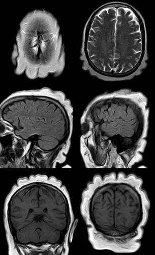
The initial physical examination was unremarkable, with stable vital signs Laboratory and initial imaging studies were inconclusive; therefore, magnetic resonance imaging (MRI) was performed, which revealed findings consistent with those of cutis verticis gyrata (CVG) (Figure 1).

On review of the head-to-toe examination, clinical identification of the condition was missed earlier because of thick scalp hair. The patient was diagnosed with CVG based on clinico-radiologic findings. Other secondary causes of CVG were excluded, and the final diagnosis was CVG with a tension-type headache. The patient was treated with analgesics that provided relief and was asymptomatic at the last follow-up after 3 months.
2 Discussion
CVG is a rare benign cutaneous disorder characterized by convoluted folds and deep furrows of the scalp that mimic the cerebral sulci and gyri. The primary form can be idiopathic or may be associated with neuropsychiatric conditions. Secondary CVG has been linked to conditions such as pachydermoperiostosis, acromegaly, syphilis, scleromyxedema, amyloidosis, leukemia, Ehler-Danlos syndrome, and inflammatory scalp diseases. Chromosomal abnormalities, including Turner, Noonan, and fragile X syndromes, as well as systemic diseases, such as diabetes mellitus and tuberous sclerosis, have also been associated [1, 2]. CVG is rare and predominantly observed in men, with a reported prevalence of approximately 1 in 100,000 males and 0.026 in 100,000 females [2, 3].
The pathogenesis of headache in CVG is multifactorial, involving peripheral neural stimulation and central mechanisms. Thickened scalp folds in CVG may stimulate cervical roots and trigeminal branches, leading to secondary cortical sensitization. Additionally, inflammatory factors, such as elevated TNFα levels, and cervical spine hypermobility causing facet joint erosion may contribute. Psychological stress from the condition's aesthetic impact can further exacerbate headaches [3].
CVG presents with symmetric redundant scalp folds that exhibit deep furrows and convolutions. Hypertrophy and folding of the skin produce an appearance mimicking a cerebral gyrus. Presentation may differ based on whether the CVG is primary or secondary or on the underlying abnormality [2].
MRI reveals diffuse scalp thickening with prominent ridges and furrows, involving the dermis and often the subcutis, maintaining a normal signal intensity. Folds are typically anterior to posterior, except transverse in the occiput, and are symmetric in primary and asymmetric in secondary diseases. Severe cases exhibit a “cogwheel” pattern. MRI is crucial for assessing the brain and orbits in primary non-essential diseases and for detecting underlying causes, such as pituitary adenomas in secondary diseases [1].
Clinical diagnosis is usually straightforward owing to its distinctive appearance. However, accurate diagnosis of underlying or associated disorders is challenging and requires a multidisciplinary approach. Although CVG is benign, its secondary causes and complications require management [2].
We excluded other etiological diagnoses of secondary CVG and headache based on clinical, laboratory, and imaging evaluations. Secondary causes including pachydermoperiostosis, cerebriform intradermal nevus, acromegaly, and amyloidosis were ruled out based on the absence of systemic or dermatological features. Chromosomal conditions, such as Turner syndrome and Ehlers-Danlos syndrome, were excluded because of a lack of characteristic markers [2].
Author Contributions
Shritik Devkota: conceptualization, validation, writing – review and editing. Om Prakash Bhatta: conceptualization, writing – original draft, writing – review and editing.
Acknowledgments
The authors have nothing to report.
Consent
Written informed consent was obtained from the patient to publish this report in accordance with the journal's patient consent policy.
Conflicts of Interest
The authors declare no conflicts of interest.
Open Research
Data Availability Statement
All relevant data supporting this case image are included in the article. No additional datasets were generated or analyzed beyond what is presented in the manuscript.


