Congenital Partial Hypoplasia of the Lower Lip: A Rare Form of 28–29 Tessier Cleft? Report of a Case
Funding: The authors received no specific funding for this work.
ABSTRACT
We aim to describe a case of congenital partial hypoplasia of the lower lip in a 2-year-old child. The presence of a gingival notch, a mucosal bridle, and difficulties with tooth eruption raised the possibility of a modified 28–29 Tessier cleft. However, other hypotheses have been proposed as the anomaly does not conform to standard cleft anatomical findings.
1 Introduction
Congenital clefts of the lower lip and mandible are among the rarest craniofacial malformations, first described by Couronne in 1819. These anomalies typically manifest as midline defects, with less than 100 cases reported in the literature [1, 2]. Paramedian clefts of the lower lip, a subset of these malformations, are even rarer, with only five documented cases to date. The anatomical classification proposed by Tessier in 1976 [3] is widely accepted as the standard for describing craniofacial clefts; however, rare presentations challenge its applicability. In Tessier's classification, cleft lip and/or mandible midline is defined as no. 30, and paramedian lower cleft lip defects are therefore classified as 28–29. The present report details a unique case of congenital partial hypoplasia of the lower lip, characterized by a lateral third defect. This anomaly lacks the hallmark features of standard clefts and does not formally align with Tessier's classification. By detailing the clinical presentation and associated findings, this report aims to expand the understanding of such rare anomalies and explore alternative hypotheses for their embryological origin.
2 Case History
We aim to report the case of a 2-year-old boy who was born with hypoplasia of the lateral third of the lower lip. To the best of our knowledge, this type of anomaly has not been previously reported in the literature.
The patient was initially referred to the craniofacial cleft team at the Grenoble University Hospital Centre at the age of 5 months, due to parental concerns regarding the anomaly. He was the first child of healthy parents with no consanguinity, and the pregnancy was spontaneous, despite the mother having previously experienced a miscarriage. There was a family history of congenital dental anomalies: the mother's aunt had only milk teeth, and the mother of our patient was missing two permanent teeth (34 and 44, FDI World Dental Federation notation). There had been no exposure to teratogens or X-rays during pregnancy. The mother was 30 years old at the time of delivery, and the infant was delivered at term by normal vaginal delivery. The birth weight was 3215 g, with a cranial circumference of 35 mm, and the Apgar score was 10/10. There were no other anomalies.
3 Physical Examination
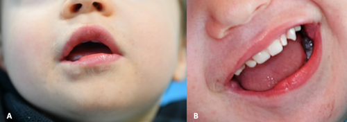
Physical examination revealed hypoplasia of the right lateral third of the lower lip (Figure 1A). Facial palsy was absent and the smile appeared symmetrical. Intra-oral examination revealed a gingival notch, a mucosal bridle, and the median labial frenulum was deviated on the right side (Figure 1B). Lip eversion was possible. No abnormalities were detected on the tongue, oral mucosa, pharynx, or tonsils. However, a cystic lesion was observed on the anterior part of the uvula. Feeding was found to be normal, although the parents noted a tendency to drool due to difficulties in achieving complete lip closure. No abnormalities were detected on the rest of the body, and no circumcisions or amputations of digits or limbs were observed. As the development and growth were normal, a simple follow-up with an annual visit was chosen.

4 Investigations
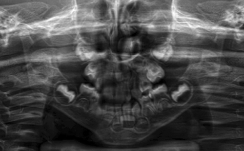
At the age of 27 months, tooth number 82 and 83 were not in position, and tooth number 84 was found to be abnormal (while teeth numbers 72 and 73 were present on the arch, FDI World Dental Federation notation). The orthopantomogram taken for the consultation showed good mandibular continuity but revealed the absence of teeth numbers 42 and 43 (whereas teeth numbers 32 and 33 were visible). This could be related to the known history of missing teeth, but as this anomaly was unilateral, it could also be related to the lip deformity (Figure 2).

Further anomalies were sought, with particular attention paid to those of a cardiologic nature. The results of the cardiologic examination and echocardiography were found to be within the normal range.
5 Treatment
The management of this labial hypoplasia can be discussed, but the less invasive technique remains lipofilling with autologous fat graft or hyaluronic acid. In the meantime, follow-up is required to monitor speech development and the eruption of the teeth.
6 Discussion
The presence of a gingival notch, a mucosal bridle, and difficulty erupting teeth led us to believe that this anomaly may be an incomplete 28–29 Tessier cleft. In the existing literature on paramedian cleft of the lower lip [4-8], no reports were found that described the extent of the cleft to the oral commissure. The defects ranged from localized lower lip hypoplasia associated with bilateral upper lip and palate clefts (Oka et al. 1983) to complete lower lip clefts without mandibular clefts (Hassanpour et al. 2018, Chauvel-Picard et al. 2018, Ghorpade et al. 2023) to incomplete lower lip cleft with mandibular defect (Morritt et al. 2007). In all these cases, the lip defect is localized on a thin part of the lower lip without extension to the commissure. Regarding the anatomical differences between a lower cleft lip defect and this total external third lower lip hypoplasia, we believe that this may be the first congenital partial lower lip hypoplasia reported in the literature.
For Chisholm et al. [9], atrophic skin conditions are caused by abnormalities in the dermis and/or subcutaneous tissue. Although the epidermis may be thin and atrophic, there must be a loss of substance in the subepidermal tissue for the skin to look and feel atrophic. In this case, we can assume that both the orbicularis oris and the depressor labii inferioris are hypoplasia but remain functional (Figure 3A,B).

While much research has been done to understand and explain the embryological mechanism of upper cleft lip, the etiology of paramedian lower cleft lip is still unclear, and there is no consensus on its embryogenesis.
There are several hypotheses. The first is a fusion defect between the mandibular prominences of the first branchial arches. According to Warbrick [10], it is due to three depressions in the mandibular process during the fetal period. He suggested that a disturbance in the embryological formation of the mandibular process could lead to a disturbance in one of these depressions, resulting in paramedian defects.
Secondly, clefts may result from the failure of mesodermal migration and penetrance. In addition, growth centers within the developing mandible may be required for formation. Partial or complete failure of growth center differentiation may contribute to mandibular defects, rather than a simple failure of mandibular protrusions to fuse at the midline [11].
Thirdly, there is a belief that intrauterine trauma may be responsible for cleft lip and palate [6, 12]. In 2018, Chauvel-Picard et al. observed a paramedian cleft of the lower lip in a child whose mother had undergone fetal reduction for a multiple pregnancy. They believed that the introduction of a transabdominal or transvaginal needle could cause craniofacial abnormalities [12]. Burton et al. studied 394 fetuses and infants whose mothers had undergone chorionic villus sampling. Thirteen (3.3%) had major congenital anomalies, including four with missing limbs or parts of limbs and three with cleft lip, with or without cleft palate [13]. In our case, the mother did not experience any trauma or fetal reduction. However, it is important to note that our patient was sucking his thumb on the first and second fetal scans. Prolonged intrauterine lip pressure could explain this anomaly, but it would probably have been seen in many other babies.
Fourthly, two main syndromes could explain this anomaly: Amniotic band syndrome and Van der Woude syndrome. The amniotic band syndrome is known to be responsible for many forms of clefting. It was not considered here because there were no ring strings and no amputations of digits or limbs. Van der Woude syndrome involves paramedian pits or clefts. This syndrome is highly variable and has been reported as lip pits in combination with a cleft. However, the present case has neither the characteristic lip pits nor the cleft. It has therefore been excluded.
A fifth hypothesis could be a vascular origin. Ischemia of the stapedial arterial system, which arises from the second pharyngeal arch and is responsible for the vascularization of the face during embryogenesis, could eventually lead to facial malformation. In 1973, Poswillo attempted to demonstrate this vascular theory in an animal model by injecting triazene into the carotid artery, causing a hematoma in the distribution area of the stapedial artery and eventually leading to the formation of some defects found in the otomandibular syndrome, such as macrostomia and hemifacial microsomia [14]. This theory remained controversial and was criticized in 1995 by Louryan et al. [15].
Our case has many similarities with cases of paramedian inferior cleft lip, but the anatomical analysis of the lip deformity led us to believe that it could not be formally classified as a 28–29 Tessier cleft. Thus, the mechanism may be completely novel.
7 Conclusion
This may be the only report of a congenital partial hypoplasia of the lower lip that does not quite fit the Tessier classification, as it does not have the shape of a common cleft. However, the associated anomalies (mucosal bridle, gingival notch, difficulty in eruption of teeth) can be seen in usual cleft lips and suggest a longer follow-up of this young patient to evaluate speech and dentition development. Surgical lip correction will certainly be performed by minimally invasive lipofilling with autologous fat graft or hyaluronic acid.
Author Contributions
Olina Rios: conceptualization, data curation, methodology, writing – original draft, writing – review and editing. Virginie Lafontaine: methodology, supervision, visualization. Cyril Debortoli: methodology, visualization. Charles Savoldelli: methodology, supervision, visualization. Beatrice Morand: data curation, methodology, project administration, validation, visualization, writing – review and editing.
Acknowledgments
The authors have nothing to declare.
Ethics Statement
Written informed consent was obtained from the parents of the child. This case report was submitted with their consent and with the approval of the Ethics Committee of the Face and Neck Institute.
Consent
Written informed consent was obtained from the patient's parents to publish this report in accordance with the journal's patient consent policy.
Conflicts of Interest
The authors declare no conflicts of interest.
Open Research
Data Availability Statement
The data supporting the findings of the present study are available from the corresponding author upon request.


