Keloids on stretch marks: Case of a Cameroonian lady
Abstract
Keloids are hypertrophic scars that develop as a result of various pathophysiologic mechanisms. We report a case of a 30-year-old Cameroonian woman who presented with keloid-like masses in the abdomen. The onset was postpartum without trauma. After histopathologic confirmation, we concluded a postinflammatory keloid.
1 BACKGROUND
Keloids are benign intradermal tumors most often secondary to trauma. These tumors usually result from imbalance between collagen fiber production and degradation.1 Apart from traumatic keloids, inflammatory and spontaneous keloids are other commonly reported etiologic forms.2 Pruritus caused by these lesions can be at times unbearable, with negative impacts mainly affecting the esthetic and psychogenic spheres of activities of daily living.3 Keloids are very common amongst persons of African descent. Nevertheless, apart from skin color, genetic factors have been implicated in its pathogenesis. Particularly implicated is the TNF-β1 gene, responsible for the formation of extracellular matrix, and hence directly implicated in scar formation.4 Folliculitis, herpes zoster, and varicella are the most common causes of inflammatory keloid.5 Here, we report a case of postinflammatory keloid secondary to stretch marks.
2 CASE PRESENTATION
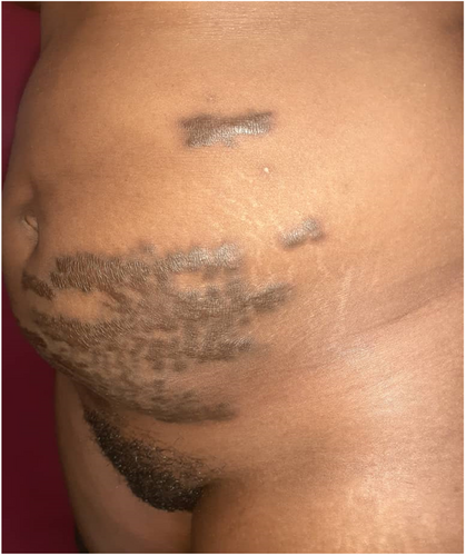
We had a 30-year-old woman who came presenting with pruritic tumor-like masses on the suprapubic area and left flank of her abdomen that appeared 12 months after a successful vaginal delivery. The evolution of the lesions was marked by spreading and extension over the abdomen. She reported that the lesions developed over stretch marks that developed during pregnancy. No history of inflammatory dermatosis nor trauma was identified. Still, no obvious genetic disease, personal, or family history of keloids was identified. Clinically, there were hyperpigmented cracked tumoral plaques with sinuous borders, some limited by an erythematous halo over the suprapubic area and left flank (Figure 1). Amongst these were also found linear atrophic, achromic depressive macular lesions in the distribution of the stretch marks.

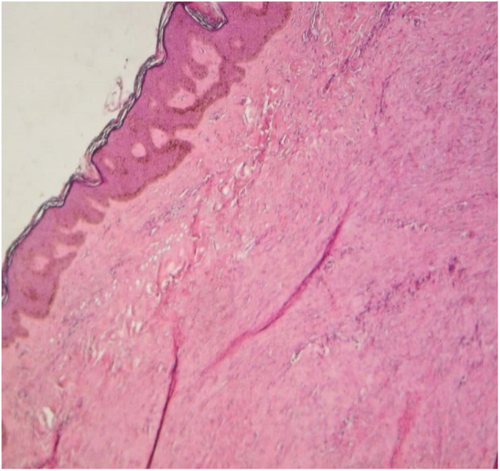
With these lesions, a histopathologic exam was realized after hematoxylin and eosin staining of biopsied tissue and confirmed its keloid nature with few collagen fibers and dermal fibrosis (Figure 2).

3 DISCUSSION
We report a case of postinflammatory keloid that developed on stretch marks that occurred following pregnancy. The diagnosis was made on the basis of the absence of obvious trauma and the distribution of keloids over the stretch marks.
Stretch marks are white atrophic cutaneous lesions that usually begin with inflammation, whose occurrence is favored by pregnancy or obesity.6 Clinically, there exist two phases: an inflammatory and a scaring phase.5 Postinflammatory keloids have been described due to folliculitis, acne, varicella, and herpes zoster.7 Certain authors doubt the spontaneity of keloids.8 If we should adhere to this principle, we think that the keloids presented by our patient developed during the inflammatory phase of stretch mark formation.
The pathophysiology of postinflammatory keloids is that of secondary keloids. The proliferative phase is marked by fibroblast hyperactivity and reduced collagenase activity, culminating in faulty resorption and overproduction of collagen.9 Proinflammatory cytokines levels, particularly TNF alpha, are increased in keloids. Thus, keloids can be considered as a site of an abnormal immune response.10 The inflammation during the inflammatory phase of stretch marks became abnormal and inappropriate in our patient, provoking the development of keloids.
4 CONCLUSION
We have reported to the best of our knowledge the first case of postinflammatory keloids secondary to stretch marks in Africa. The pathophysiology of this clinical entity is not expected to be different from that of other postinflammatory keloids.
AUTHOR CONTRIBUTIONS
Defo Defo conceived the study. Julie Carole Zoua, Alexandra Dominique Ngangue Engome, Coralie Reine Mendouga Menye, Dahlia Noelle Tounouga, and Emmanuel Armand Kouotou drafted the manuscript. Julie Carole Zoua, Alexandra Dominique Ngangue Engome, Coralie Reine Mendouga Menye, Dahlia Noelle Tounouga, and Emmanuel Armand Kouotou proofread and corrected the manuscript. All authors agreed with the final manuscript to be submitted for publication.
ACKNOWLEDGMENT
We thank Dr. Chindo Puis-Mary for participating in the redaction of the final manuscript.
FUNDING INFORMATION
This research did not receive any specific grant from funding any agencies.
CONFLICT OF INTEREST
The authors declare that they have no conflicts of interest with regard to this article.
CONSENT
Written consent was obtained from the patient for the publication of this case and the accompanying images. A copy of the consent should be available for review.
Open Research
DATA AVAILABILITY STATEMENT
Data sharing is not applicable to this article as no datasets were generated or analyzed during the current study.


