Craniolacunia in A neonate; A clinical and CT scan illustrative case report
Funding information
No funding was secured for this study
Abstract
Neural tube defects can be accurately diagnosed prenatally. Every effort must be made to get this and its associations with Craniolacunia right, especially in low-resource settings. This case highlights the importance of three-dimensional CT in diagnosing neonatal skull abnormalities.
1 INTRODUCTION
The etiology of craniolacunia is unclear with varied school of thoughts. Fetuses with this rare condition may have associated neural tube defects (NTDs) and hydrocephalus, which may require neurosurgical interventions. Craniolacunia may be self-limiting without NTDs. This case report emphasizes the importance of three-dimensional CT scan in diagnosing Craniolacunia.
Craniosynostosis results from the fusion of one or more of the cranial sutures prematurely. This can be an isolated defect or as part of a syndrome.1, 2 The etiology of most single-suture synostosis is thought to be idiopathic, even though defective dural-mesenchymal signaling issues and genetic causes are increasingly being identified. However, single-gene or chromosomal disorders are the possible causes of genetically influenced multisuture synostoses.2, 3 There are varieties of skull alterations in most patients with craniosynostosis severest in the syndromic type. These are thought to be caused mainly by high intracranial pressure and from the compensatory growth of the skull after stenosis of some sutures.1-3
Craniosynostosis is an extremely rare condition occurring in 0.4 per 10,000 births, of these, craniolacunia is said to be present in only 10% of the cases.1, 4 Craniolacunia, which is an ossification abnormality presents as a fenestrated fetal skull.5-7 The etiology of craniolacunia is unclear and a subject of much controversy as literature on the subject is sparse and somewhat contradictory.6, 7 It is almost always associated with Chiari malformation, meningomyelocele, and encephalocele but may also be seen in conjunction with aqueductal stenosis and phenylketonuria or even in healthy children.5, 7 It has been shown to develop in utero, with resolution in infancy as a result of skull remodeling.8 Craniolacunia also known as lacunar skull and luckenschadel was first described by West in 1875 and named as “luckenschadel” by Engstler in 1905.5-7 We present a case of a two-week-old baby boy born with meningomyelocele and craniolacunia, in order to document another case of this condition to add to the few reported cases in the literature.6
2 CASE REPORT
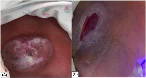
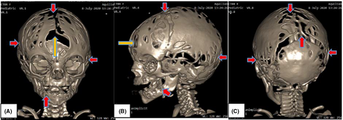
We report a case of a two-week-old baby boy who was born with a defect at the low back and consequently referred to the pediatric department of the Cape Coast Teaching Hospital (CCTH) with the low back defect (meningomyelocele) Figure 1A, without any other complaints. The baby was born through a spontaneous vaginal delivery and cried promptly and spontaneously immediately after delivery. Apgar score after delivery was normal (10 out of 10). The neonate passed meconium immediately after delivery and breastfed appropriately. The immediate postnatal examination revealed normal respiratory and heart sounds, his limbs and face were unremarkable except for the presence of a meningomyelocele at the low back. Neither neurological deficit nor other abnormality was noted. The diagnosis of this defect was likely missed during the routine antenatal ultrasonography. The neurosurgical team repaired the meningomyelocele Figure 1B after thorough investigations including referral to the radiology department of the CCTH for a brain CT scan with 3D reconstruction and bone window, in order to rule out hydrocephalus and encephalocele. The non-contrast CT scan revealed normal brain parenchyma without any ventriculomegaly nor hydrocephalus and normal gyri and sulci as shown in Figure 2A, B, and C below. No extracerebral extension or outpouching of the brain/meninges is seen also shown in Figure 2B. There were diffuse calvarial morphological abnormalities of the inner and outer tables of the skull with areas of discontinuations in the calvarium on the bone window Figure 3A, B, and C. The 3D reconstruction showed multiple lacunae in the calvarium and “lace-like” holes diffusely spread in the calvarium and facial bones giving it a “cobblestone” appearance as shown in Figure 4, which are in keeping with the diagnosis of Craniolacunia. The neurosurgical team is currently managing the patient conservatively and he is doing well.


3 DISCUSSION
Craniolacunia is manifested by numerous, radiolucent, oval, or round defects, which are distinctly separated by dense bony strips giving it a honeycomb-like appearance, on plain skull radiograph.5, 7 The defect is termed as craniolacunia when it involves the inner table of the skull and it's impalpable from outside of the skull. The reverse involving both the inner and outer tables of the skull, making it palpable from outside of the skull is termed as craniofenestra.4, 5, 7 Brain tissue may bulge into the gaps and occasionally the brain may herniate through these defects. The occipital, frontal, and parietal bones are most frequently affected.5-7
The etiology of this type of deformity is still obscure. Darouich et al. reported that some observers believe intracranial hypertension may dampen the intracranial pressure by acting as a protective mechanism and thereby causing this ossification defect, whereas others postulate that the ossification of normal membranous calvarium because of the failure of induction and the leakage of cerebrospinal fluid as a result of neural tube defect may result in poorly distended cerebral ventricles.6 But, according to Zoeller et al., both theories have fallen short in the sense that the former cannot explain why craniolacunia has been demonstrated in infants with low-intracranial pressure, and the latter fails to explain the development of craniolacunia in patients without neural tube defects.7 Also, the fact that this birth defect resolves by 4–6 months of age has been explained with remodeling as a result of either normal expansion of brain tissue or development of hydrocephalus.5-7 Craniolacunia is usually absent in adults since it is primarily a developmental defect of the calvarium in fetuses with neural tube disorders.5
This condition can be diagnosed with physical examination, neuroimaging (CT Scan/MRI), and plain radiography.6, 7 Copper beaten configuration (CBC) on skull radiograph is diagnostic of craniolacunia (luckenschadel).1, 2, 5 Even though, this may be normal and resolve on its own until puberty, it cannot be overlooked as it may be a sign of raised intracranial pressure in order to determine the underlying causes and provide appropriate neurosurgical management/interventions.4-8 In most instances, the radiologic imaging findings are not recognizable by 4 to 6 months of age, but some radiologic changes may be present in utero around seven months of gestation.5 Craniolacunia, which is a defect of the skull in newborns, is usually associated with Chiari II malformation (seen in 80.0%), meningocele, spina bifida, meningomyelocele, and encephalocele in some cases. 2, 4-9
Craniolacunia, which is usually an antenatal X-ray finding is less commonly seen in modern obstetric practices as X-ray exposures are not routinely recommended in pregnancy. Currently, most diagnoses are usually done postnatally. 5, 6, 8 Postnatal ultrasound evaluation usually shows a wavy inner calvarium with an irregular outline on coronal sections, which is more conspicuous posterior to the Sylvian fissure.6, 8 The “lemon sign” which is indicative of severe neural tube defects and related to craniolacunia/luckenschadel is usually seen before 24 weeks of gestation, but this usually tends to resolve in the third trimester.5, 10 As ultrasound has been found to be the most available imaging modality in Ghana, many intrauterine fetal abnormalities like neural tube defects can painstakingly be diagnosed employing this modality.11 The diagnosis of this condition in our case appears to have been missed during routine antennal ultrasonography for fetal anomaly usually done between 18–22 weeks, even though fetal anomaly scans are one of the most important reasons why pregnant women undergo antennal ultrasonography as corroborated by a recent study.12
Taylor et al, reported in a case series the presence of craniolacunia in neonates without neurological deficits or associated abnormalities.13 But unlike those cases, our patient presented with an associated meningomyelocele. No extension of the brain parenchyma from the calvarium was noted, though extensive craniolacunia without any ventriculomegaly nor hydrocephalus were seen on CT scan in our case. The patient has since been enrolled in the neurosurgical clinic for follow-up with periodic transfontanelle ultrasound scan.
4 CONCLUSION
The diagnosis of craniolacunia/luckenschadel in newborns can be made using skull X-rays, CT scan, and neurosonography. The strong association between craniolacunia and spina bifida/meningomyelocele is well established. This case report highlights the importance of three-dimensional CT scan in patients with skull abnormalities.
ACKNOWLEDGMENTS
The authors are appreciative of the mother of the baby for giving us the consent for this paper to be written. Also, we thank the radiographer in charge of the CT scan unit for helping us with the acquisition of these images. Published with written consent of the patient.
CONFLICT OF INTEREST
The authors have no conflict of interest to declare.
AUTHOR CONTRIBUTIONS
All authors contributed significantly to the conception, designing, writing, revising, and approving the manuscript draft for publication.
ETHICAL APPROVAL
A written informed consent was obtained from the mother of the patient for this case to be reported for publication. The patient`s mother was assured of absolute confidentiality and anonymity. Ethical clearance was not required for this study.
Open Research
DATA AVAILABILITY STATEMENT
The data that support the findings of this study are available from the Head of the Research Unit of the Cape Coast Teaching Hospital upon reasonable request. Postal address is as follows: P.O. Box CT I363, Cape Coast, Ghana. E-mail is [email protected].


